. 2024 Oct 15;213(8):1212-1224.
doi: 10.4049/jimmunol.2300844.

MAFB in Macrophages Regulates Prostaglandin E2-Mediated Lipid Mediator Class Switch through ALOX15 in Ischemic Acute Kidney Injury

Affiliations

MAFB in Macrophages Regulates Prostaglandin E2-Mediated Lipid Mediator Class Switch through ALOX15 in Ischemic Acute Kidney Injury

Maho Kanai et al. J Immunol. .

Abstract

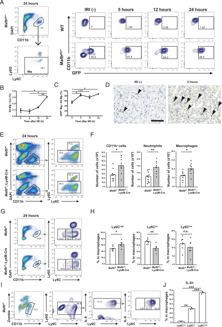
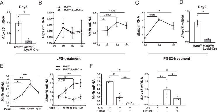
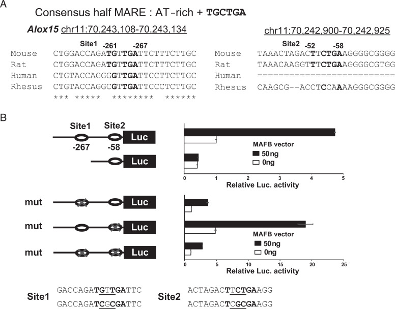
Monocytes and macrophages express the transcription factor MAFB (V-maf musculoaponeurotic fibrosarcoma oncogene homolog B) and protect against ischemic acute kidney injury (AKI). However, the mechanism through which MAFB alleviates AKI in macrophages remains unclear. In this study, we induced AKI in macrophage lineage-specific Mafb-deficient mice (C57BL/6J) using the ischemia-reperfusion injury model to analyze these mechanisms. Our results showed that MAFB regulates the expression of Alox15 (arachidonate 15-lipoxygenase) in macrophages during ischemic AKI. The expression of ALOX15 was significantly decreased at the mRNA and protein levels in macrophages that infiltrated the kidneys of macrophage-specific Mafb-deficient mice at 24 h after ischemia-reperfusion injury. ALOX15 promotes the resolution of inflammation under acute conditions by producing specialized proresolving mediators by oxidizing essential fatty acids. Therefore, MAFB in macrophages promotes the resolution of inflammation in ischemic AKI by regulating the expression of Alox15. Moreover, MAFB expression in macrophages is upregulated via the COX-2/PGE2/EP4 pathway in ischemic AKI. Our in vitro assay showed that MAFB regulates the expression of Alox15 under the COX-2/PGE2/EP4 pathway in macrophages. PGE2 mediates the lipid mediator (LM) class switch from inflammatory LMs to specialized proresolving mediators. Therefore, MAFB plays a key role in the PGE2-mediated LM class switch by regulating the expression of Alox15. Our study identified a previously unknown mechanism by which MAFB in macrophages alleviates ischemic AKI and provides new insights into regulating the LM class switch in acute inflammatory conditions.


Conflict of interest statement

The authors have no financial conflicts of interest.

Figures

FIGURE 1.
FIGURE 1.
Loss of Mafb in macrophages worsens the prognosis of ischemic AKI. (A) Survival curve of Mafbf/f and Mafbf/f::LysM-Cre mice subjected to IRI with 60-min clamp (n = 6 for each group). p <0.01, by log-rank test. (B) Serum BUN and creatinine levels of Mafbf/f and Mafbf/f::LysM-Cre mice subjected to IRI with 30-min clamp (n = 6 for Mafbf/f, n = 5 for Mafbf/f::LysM-Cre). (C) Representative periodic acid–Schiff (PAS)–stained kidney from Mafbf/f and Mafbf/f::LysM-Cre mice at 7 d post-IRI. Scale bar, 100 µm. Arrowheads point to intraluminal debris. (D and E) Quantification of PAS+ intraluminal debris at the corticomedullary junction (D), as well as the acute tubular necrosis (ATN) scores (E) in the kidneys of Mafbf/f and Mafbf/f::LysM-Cre mice at 7 d post-IRI. Intraluminal debris (D) is presented as a percentage area per whole corticomedullary junction in a slide. (F) Fetal liver cells of WT or MAFB p.Leu239Pro (Mafbmt/mt) CD45.2 embryo were transplanted to lethally irradiated WT CD45.1 mice. After the 8-wk recovery period, mice were subjected to renal IRI. (G) Serum blood urea nitrogen (BUN) and creatinine levels of WT or Mafbmt/mt cell–transplanted mice subjected to IRI with a 30-min clamp. n = 5 for each group. (H) Representative PAS-stained kidney from WT or Mafbmt/mt cell–transplanted mice at 7 d post-IRI. Scale bar, 100 µm. Arrowheads point to intraluminal debris. (I) The ATN scores in the kidneys of WT or Mafbmt/mt cell–transplanted mice at 7 d post-IRI. All data are expressed as means ± SEM. *p <0.05, **p <0.01, ***p <0.001, by Welch t test.
FIGURE 2.
FIGURE 2.
MAFB deficiency alters leukocyte infiltration and activation patterns during AKI. (A) Kidney cells of WT or MafbGFP/+ mice at the indicated time points were stained with CD11b, Ly6G, and Ly6C Abs. Ly6GlowLy6C+CD11b+ cells were gated as macrophages, and the percentages of GFP+ macrophages among all macrophages were measured. (B and C) The percentage of all macrophages in total live cells (B) and the percentage of GFP+ macrophages in all macrophages (C) at the indicated time points are plotted with SEM (n = 3 for each group). (D) Immunostaining of IRI (−) or 5 h post-IRI kidney of Mafbf/f mice with anti-MAFB Ab. The image represents a corticomedullary junction of the kidney. Arrowheads point to the MAFB-positive areas. Scale bar, 100 µm. (E) CD11b+ cells were isolated from kidneys of Mafbf/f or Mafbf/f::LysM-Cre mice 24 h after IRI by MACS. Flow cytometric analysis was performed on these CD11b+ cells with anti-CD11b, anti-Ly6G, and anti-Ly6C Abs. Ly6GhighCD11b+cells were gated as neutrophils, and Ly6C+Ly6GlowCD11b+ cells were gated as macrophages. (F) The absolute number of cells in each cell population was calculated from the overall number of CD11b+ cells taken from the kidney tissues and the percent of each cell population (n = 9 for the Mafbf/f group and n = 8 for the Mafbf/f::LysM-Cre group). (G) Kidney cells of Mafbf/f or Mafbf/f::LysM-Cre mice at 24 h post-IRI were stained with CD11b, Ly6G, and Ly6C Abs. Ly6GlowLy6C+CD11b+ cells were gated as macrophages. Numbers indicate the percentage of Ly6Clow, Ly6Cint, and Ly6Chigh macrophages in all macrophages. (H) Percentage of Ly6Clow, Ly6Cint, and Ly6Chigh macrophages in all macrophages (n = 6 for each group). (I) IL-6 expression levels in Ly6Clow, Ly6Cint, and Ly6Chigh macrophages in the kidney of Mafbf/f mice 24 h after IRI were measured by FACS analysis. (J) Percentages of Ly6Clow, Ly6Cint, and Ly6Chigh IL-6+ macrophages in all macrophages (n = 3 for each group). All data are expressed as means ± SEM. *p <0.05, **p <0.01, *** p <0.001, by Welch t test.
FIGURE 3.
FIGURE 3.
MAFB regulates the expression of Alox15 in macrophages infiltrated into the kidney of ischemic AKI. (A) Volcano plot of overall gene expression in macrophages sorted from the kidney of Mafbf/f and Mafbf/f::LysM-Cre mice at 24 h post-IRI. The blue and red dots indicate the downregulated and upregulated genes in the Mafbf/f::LysM-Cre group, respectively. (B) Normalized count of Alox15 mRNA from RNA-seq data (n = 3 for each group). ***False discovery rate<0.001, edgeR. (C) Immunostaining of kidney from Mafbf/f and Mafbf/f::LysM-Cre mice without IRI at 12 and 24 h post-IRI with anti-ALOX15 and anti-F4/80 Ab. Scale bars, 100 µm. (D) The number of ALOX15+F4/80+ cells per field is presented. Seven different fields (original magnification, ×200) were analyzed for each slide. All data are expressed as means ± SEM. **p <0.01, by Welch’s t test. (E) Immunostaining of kidney from WT and MafbGFP/+ mice at 24 h post-IRI with anti-ALOX15 and anti-GFP Ab. Arrowheads point to the ALOX15+GFP+ area. Scale bar, 50 µm.
FIGURE 4.
FIGURE 4.
MAFB regulates Alox15 expression under the COX-2/PGE2/EP4 pathway in macrophages. (A and B) Thioglycollate-elicited PMs were primed with LPS, and Alox15 mRNA levels were measured 3 d after treatment using qRT-PCR (A). Ptgs2 mRNA in the Mafbf/f or Mafbf/f::LysM-Cre group (left) and Mafb mRNA in the Mafbf/f group (right) were measured using qRT-PCR (B). (CE) Thioglycollate-elicited peritoneal macrophages were primed with PGE2. Mafb mRNA in the Mafbf/f group was measured by qRT-PCR (C). The Alox15 mRNA was measured 2 d after treatment (D). In (A)–(D), n = 3 for each group. Thioglycollate-elicited PMs were primed with 10 nM, 100 nM, and 1 µM PGE2 and Mafb mRNA in the Mafbf/f group (left), and Alox15 mRNA levels in each group (right) were measured by RT-qPCR. Data are shown with the expression level at PGE2 (−) as 1. n = 8 for Mafbf/f, n = 5 for Mafbf/f::LysM-Cre (E). (F) LPS-induced Mafb and Alox15 expression was prevented using an EP4 antagonist (L161982) at day 3. All data are expressed as means ± SEM. *p <0.05, **p <0.01, ***p <0.001, by Welch t test. For (F), the p value was adjusted using the Holm method.
FIGURE 5.
FIGURE 5.
MAFB regulates the Alox15 gene through the half-MARE site at −267 bp upstream of the transcriptional initiation site of Alox15. (A) Half of the Maf recognition element (half-MARE) sites were identified in Alox15 gene promoters using the UCSC Genome Browser. The half-MARE site (site 1) in the Alox15 gene promoter (bold) was highly conserved among mammalian species. (B) An MAFB-expressing vector was cotransfected with luciferase reporter constructs of Alox15 promoter containing half-MARE sites or mutant half-MARE sites into RAW264.7 cells. The luciferase activity was analyzed 48 h after transfection. The term “mut” indicates three base mutations in the half-MARE sites. Data are presented as the means ± SEM. n = 3 for each group.
FIGURE 6.
FIGURE 6.
Mechanistic scenario for induction of ALOX15 by MAFB in macrophages. In macrophages infiltrating the kidney after IRI, COX-2–derived PGE2 activates the EP4 receptor, and MAFB expression is induced. Under the COX-2/PGE2/EP4 pathway, MAFB induces the expression of ALOX15. ALOX15 would promote the resolution of inflammation in ischemic AKI by producing specialized proresolving mediators (SPMs) from DHA.

References

    1. Ronco, C., Bellomo R., Kellum J. A.. 2019. Acute kidney injury. Lancet 394: 1949–1964. - PubMed
    1. Goldstein, S. L., Jaber B. L., Faubel S., Chawla L. S.; Acute Kidney Injury Advisory Group of American Society of Nephrology . 2013. AKI transition of care: a potential opportunity to detect and prevent CKD. Clin. J. Am. Soc. Nephrol. 8: 476–483. - PubMed
    1. Huen, S. C., Cantley L. G.. 2017. Macrophages in renal injury and repair. Annu. Rev. Physiol. 79: 449–469. - PubMed
    1. Cao, Q., Harris D. C. H., Wang Y.. 2015. Macrophages in kidney injury, inflammation, and fibrosis. Physiology (Bethesda) 30: 183–194. - PubMed
    1. Kinsey, G. R., Li L., Okusa M. D.. 2008. Inflammation in acute kidney injury. Nephron Exp. Nephrol. 109: e102–e107. - PMC - PubMed