. 2024 Sep 10;121(37):e2404250121.
doi: 10.1073/pnas.2404250121. Epub 2024 Sep 4.

Structure-based design of a soluble human cytomegalovirus glycoprotein B antigen stabilized in a prefusion-like conformation

Affiliations

Structure-based design of a soluble human cytomegalovirus glycoprotein B antigen stabilized in a prefusion-like conformation

Madeline R Sponholtz et al. Proc Natl Acad Sci U S A. .

Abstract

Human cytomegalovirus (HCMV) glycoprotein B (gB) is a class III membrane fusion protein required for viral entry. HCMV vaccine candidates containing gB have demonstrated moderate clinical efficacy, but no HCMV vaccine has been approved. Here, we used structure-based design to identify and characterize amino acid substitutions that stabilize gB in its metastable prefusion conformation. One variant containing two engineered interprotomer disulfide bonds and two cavity-filling substitutions (gB-C7), displayed increased expression and thermostability. A 2.8 Å resolution cryoelectron microscopy structure shows that gB-C7 adopts a prefusion-like conformation, revealing additional structural elements at the membrane-distal apex. Unlike previous observations for several class I viral fusion proteins, mice immunized with postfusion or prefusion-stabilized forms of soluble gB protein displayed similar neutralizing antibody titers, here specifically against an HCMV laboratory strain on fibroblasts. Collectively, these results identify initial strategies to stabilize class III viral fusion proteins and provide tools to probe gB-directed antibody responses.

Keywords: cryo-EM; cytomegalovirus; herpesvirus; vaccine.


Conflict of interest statement

Competing interests statement:M.R.S., P.O.B., A.R.R., J.A.G., R.S.M., L.Z., N.V.J., C.-L.H., and J.S.M. are inventors on a patent application entitled “Prefusion-Stabilized CMV gB Proteins” (PCT/US2023/073369). J.D.C., D.Y., and M.J.B. are employees of Dynavax and hold Dynavax stock. The other authors declare that they have no competing interests.

Figures
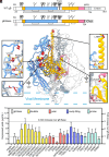
Fig. 1.
Fig. 1.
Exemplary substitutions for HCMV gB stabilization. (A) Schematic of WT Towne strain HCMV gB and ectodomain base construct (gB Base). Native disulfide bonds are shown as connecting black lines. N-linked glycosylation sites are shown as branched lines. The native furin cleavage site is shown as a thick black line. The N-terminal signal sequence (SS) and C-terminal domain (C-term) are shown as white boxes. The first and second regions expected to move during the conformational rearrangement from pre-to-postfusion gB are colored blue and pink, respectively, and the region that does not undergo substantial rearrangement is colored yellow. The MPR and TM domain are shown in gray, with the TM demarcated by dashed blue and black lines. The soluble gB Base construct consists of the first 704 residues of WT gB with substitutions C246S, R457S, and R460S, of which the latter two remove the native furin cleavage site. In gB Base, residue 704 is followed by the foldon (Fd) domain and C-terminal tags. (B) Side view of trimeric prefusion HCMV gB (PDB ID: 7KDP) (23). One protomer is colored as in (A) and shown as a ribbon diagram, one protomer is colored gray and shown as a cartoon trace of the α-carbon backbone, and one protomer is shown as a transparent surface. Exemplary substitutions are shown as spheres with sulfur atoms in yellow, nitrogen atoms in blue, and oxygen atoms in red. The approximate location of the viral membrane is shown as dashed blue lines. Insets show exemplary substitutions as sticks. Anticipated hydrogen bonds are shown as black dashed lines and anticipated disulfide bonds are shown as yellow dashed lines. (C) Absolute and relative expression levels of individual variants determined by quantitative BLI. Variants are colored by substitution type.
Fig. 2.
Fig. 2.
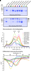
Characterization of single disulfide gB variants. (A) Reducing and non-reducing SDS-PAGE of gB variants. Molecular weight standards are indicated on the left in kDa. White, gray, and black triangles correspond to monomeric, dimeric, and trimeric gB molecular weights, respectively. (B) SEC traces of purified gB variants. The approximate location of the void volume, HMW peak, and LMW peak are identified with black arrows. (C) DSF analysis of gB variant thermostability colored as in (B).
Fig. 3.
Fig. 3.
Characterization of combination gB variants. (A) Absolute and relative expression levels of individual variants determined by quantitative BLI. Dashed lines denote onefold and fivefold increases in expression relative to gB Base. (B) Reducing and non-reducing SDS-PAGE gels of gB variants. Molecular weight standards are indicated at the Left in kDa. White, gray, and black triangles correspond to monomeric, dimeric, and trimeric gB molecular weights, respectively. (C) DSF analysis of gB variant thermostability colored as in (A). (D) Negative-stain EM 2D class averages of six variants.
Fig. 4.
Fig. 4.
Cryo-EM structure of gB-C7 bound to 1G2 and 7H3 Fabs. (A) Side (Top, Left) and Top (Top, Right) views of the composite EM map (global plus local refinement) of HCMV gB-C7 complexed with 1G2 and 7H3 Fabs shown above the gB-C7 complex model (Bottom, Left). One protomer of the model is colored as in Fig. 1A and shown as a ribbon diagram, the second is colored gray and shown as a cartoon tube trace of the α-carbon backbone, and the third is shown as a transparent surface. The Inset (Bottom, Right) shows a zoomed view of the substitutions that comprise design gB-C7 with side chains shown as sticks. (B) Side view of HCMV gB-C7 bound to 1G2 and 7H3 Fabs shown as a ribbon diagram (Top) above a zoomed view of the binding interface between 7H3 and gB-C7 (Bottom). 7H3 HC is pink, 7H3 light chain (LC) is pale pink, and gB-C7 is yellow. In the zoomed interface, key residues are shown as sticks, and hydrogen bonds are shown as black dashed lines. (C) The structure of gB-C7 (yellow) is superimposed with the previously determined structure of prefusion HCMV gB (blue, PDB ID: 7KDP) (23), both shown as cartoon tube traces of the α-carbon backbones. Side and Top views are shown for the superimposition of HCMV gB both as a trimer (Left) and as a single protomer (Right). Shifts in domain arrangement are highlighted with arrows.
Fig. 5.
Fig. 5.
Engineered gB variants are immunogenic in mice. (A) Schematic of mouse immunization. 6-to-8-week-old female BALB/c mice (n = 8/group) were immunized at weeks 0, 3, and 6 with 2.5 μg of gB Base (orange), gB V134C/I653C (red), or gB-C7 (green), all adjuvanted with CpG 1018 plus alum or unadjuvanted gB-C7 (purple). Blood samples were collected from mice 2 wk after the week 6 injection. SM5-1-equivalent antibody concentrations of immunized mouse sera that bind to (B) gB Base or (C) gB-C7. Plots represent ELISA measurements relative to an SM5-1 mFc standard from two experimental replicates. AD169-GFP infection of human MRC-5 fibroblasts. (D) Neutralization by Cytogam is enhanced 3.6-fold in the presence of 12.5% guinea pig complement. Data are averaged across 16 plates with SD shown. Neutralization by mouse sera in the (E) absence and (F) presence of 12.5% guinea pig complement. Plots represent averages from two independent experiments. In violin plots, horizontal lines represent the first quartile, median, and third quartile. Statistical significance was determined by one-way ANOVA followed by Tukey’s HSD test in GraphPad Prism: *P ≤ 0.05, **P ≤ 0.01, ***P ≤ 0.001, and ****P ≤ 0.0001.

References

    1. Gatherer D., et al. , ICTV virus taxonomy profile: Herpesviridae 2021. J. Gen. Virol. 102, 001673 (2021). - PMC - PubMed
    1. Griffiths P., Reeves M., Pathogenesis of human cytomegalovirus in the immunocompromised host. Nat. Rev. Microbiol. 19, 759–773 (2021). - PMC - PubMed
    1. Sapuan S., Theodosiou A. A., Strang B. L., Heath P. T., Jones C. E., A systematic review and meta-analysis of the prevalence of human cytomegalovirus shedding in seropositive pregnant women. Rev. Med. Virol. 32, e2399 (2022). - PMC - PubMed
    1. Manicklal S., Emery V. C., Lazzarotto T., Boppana S. B., Gupta R. K., The “silent” global burden of congenital cytomegalovirus. Clin. Microbiol. Rev. 26, 86–102 (2013). - PMC - PubMed
    1. Anonymous, “Babies born with congenital CMV” in Cytomegalovirus (CMV) and Congenital CMV Infection (Centers for Disease Control and Prevention, 2022).

LinkOut - more resources