. 2024 Nov;11(41):e2401424.
doi: 10.1002/advs.202401424. Epub 2024 Sep 4.

Lysosome-Targeted Bifunctional Therapeutics Induce Autodynamic Cancer Therapy

Affiliations

Lysosome-Targeted Bifunctional Therapeutics Induce Autodynamic Cancer Therapy

Athira Raveendran et al. Adv Sci (Weinh). 2024 Nov.

Abstract

Autodynamic cancer therapy possesses tremendous potential for enhancing therapeutic efficacy by initiating the treatment process autonomously within targeted cells. However, challenges related to biocompatibility and targeted delivery have hindered its clinical translation owing to the induction of adverse effects and cytotoxicity in healthy cells. In this study, a novel approach for auto-initiated dynamic therapy by conjugating zwitterionic near-infrared fluorophores to a cell-penetrating peptide is proposed. This enables efficient cellular uptake and specific targeting of therapy to desired cells while avoiding off-target uptake. The zwitterionic bioconjugate causes cancer-specific toxicity following its internalization into the targeted cells, triggered by specific intracellular conditions in lysosomes. This innovative approach enables selective targeting of lysosomes in malignant cells while minimizing cytotoxic effects on normal cells. By targeting lysosomes, the method overcomes inherent risks and side effects associated with conventional cancer treatments, offering a selective and effective approach to cancer therapy.

Keywords: anticancer agent; cell‐penetrating peptide; fluorescence dye; near‐infrared imaging; targeting therapy.


Conflict of interest statement

The authors declare no conflict of interest.

Figures
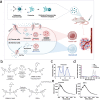
Figure 1
Figure 1
Mechanism of action and synthetic scheme of ZWPro. a) Schematic illustration of cellular uptake through organic anion transporting polypeptides (OATPs)‐mediated endocytosis of the anticancer ZWPro conjugates in cancer (A) and normal cells (B). b) Synthesis and characteristics of ZWPro. ZW800‐1C N‐Hydroxysuccinimide (NHS) ester reacts with the N‐terminal proline of protamine (Pro) to yield ZWPro. c) Fast protein liquid chromatography data shows 3 fraction peaks equivalent to ZWPro, Pro, and ZW. d) Optical spectra of ZW, ZWPro, and Pro. All optical properties were quantified using a 1X phosphate‐buffered saline (PBS) buffer solution. e) Matrix‐assisted laser desorption ionization‐time‐of‐flight‐mass spectrometry data showing Pro, ZW, and ZWPro. The shifted peak of ZWPro indicates successful conjugation.
Figure 2
Figure 2
In vitro cellular evaluations of ZWPro in cancer cells. a) Cytotoxicity assay showing HT29, MCF7, L929, and NIH3T3 cells treated with 20 µM of ZW and ZWPro for 2 and 24 h, respectively, followed by an assessment of cell viability using the water‐soluble tetrazolium assay (n = 5, mean ± s.e.m.). b) Cellular uptake and intracellular endosomal localization of ZW and ZWPro were observed by treating HT29 and L929 cells with 20 µM of ZW and ZWPro. ZWPro was observed in the cells, while ZW was not internalized. The internalization index was determined in 2–9 photographic areas (ns, not significant, *p <  0.05 based on two‐way analysis of variance followed by Bonferroni's multiple comparison test). c) Inhibition assay of cellular uptake of ZWPro to determine the entry mechanisms in HT29 cells. Cultured cells were incubated with bromsulphthalein for 20 min and then incubated with ZWPro (20 µM) in media for 15 min. Data are presented as means ± standard deviations (n = 7–8) **p < 0.01 based on Welch's t‐test. d) Findings from HT29 human colorectal adenocarcinoma cells and L929 fibroblast cell lines co‐stained with the lysosome marker LysoTracker Green. Co‐localization occurs at specific time intervals during imaging. The co‐localizing pixels (%) were calculated by dividing the area of overlap between ZWPro and LysoTracker by the total area of the LysoTracker, respectively. Scale bars: 10 µm.
Figure 3
Figure 3
Annexin V/Propidium iodide (PI) double staining flow cytometric analysis at a) different time intervals or b) different concentrations of ZWPro after ZWPro treatment of HT29 and MCF7 cells for 24 h. c) Apoptosis/necrosis/live cell assay and fluorescence microscopy. HT29 and NIH3T3 cells were treated with 20 µM of ZWPro for 16 and 24 h. Images were acquired after co‐staining with apopxin (apoptosis detection dye, green fluorescence), 7‐AAD (necrosis detecting cell impermeable dye, red fluorescence), and CytoCalcein (live cell detection, blue fluorescence). d) Relative fluorescence intensity was calculated by dividing the area of apoptotic, necrotic, and live cells by the total number of cells, respectively. e) Western blot data show the expression of cathepsin B, TNF‐α, phosphorylated MLKL antibody, and β‐actin as the loading control. The samples were loaded as follows: HT29, HT29 + ZWPro, MCF7, MCF7 + ZWPro, L929, L929 + ZWPro, NIH3T3, and NIH3T3 + ZWPro.
Figure 4
Figure 4
In vivo, tumor growth inhibition and studies in tumor‐induced mice subcutaneously injected with HT29 cells and treated with ZWPro. a) Schematic of the treatment schedule and experimental design of PBS, ZW, and ZWPro. Biodistribution and pharmacokinetic (PK) parameters of targeted ZW and ZWPro in normal mice. PK parameters of ZW and ZWPro (0.3 µmol kg−1) (t1/2α; distribution half‐life, t1/2β; elimination half‐life, AUC; area under the curve, Vd volume of distribution, and urinary excretion (% injected dose, %ID)) were calculated using the software Prism 9. Urinary excretion occurred at 48 h post‐injection (n = 2–3 per group, mean ± s.e.m.). (b) In total, 20 nmol of each compound was injected into BLAB/C nude mice (weights = 20–25 g) and time‐dependent imaging was performed post‐injection. c) Color and NIR fluorescence in vivo images of ZW and ZWPro (0.3 µmol kg−1) were taken at different time intervals. d) Tumor‐to‐background (TBR) ratios of tumors (white dotted circles in (c)) compared with muscles obtained from at each time point; and e) signal‐to‐background (SBR) outcomes were calculated using NIR images of resected organs at 48 h post‐injection. (Exposure time = 100 ms, n = 3, mean ± s.e.m.) The SBR was calculated by the fluorescence intensity of each organ (Or) against the system background (Exposure time = 1000 ms, n = 3, mean ± s.e.m.). Scale bar: 1 cm. f) Tumor reduction assay of the PBS, ZW, and ZWPro. g) Photographs of the dissected tumor tissues. h) Postoperative histopathological examination; H&E stained images and NIR fluorescence microscopy images. Abbreviations used are: He, heart; Lu, lungs; Li, liver; Pa, pancreas; Sp, spleen; Ki, kidneys; In, intestine; Mu, muscle; Tu, tumor. Data are presented as mean ± standard deviation (SD) (Scale bar: 100 µm, = 5, ***p < 0.001, **p < 0.01).

Similar articles

Cited by

References

    1. Lei Z., Zhang X., Zheng X., Liu S., Xie Z., Org. Biomol. Chem. 2018, 16, 8613. - PubMed
    1. Hu Z., Fang C., Li B., Zhang Z., Cao C., Cai M., Su S., Sun X., Shi X., Li C., Zhou T., Zhang Y., Chi C., He P., Xia X., Chen Y., Gambhir S. S., Cheng Z., Tian J., Nat. Biomed. Eng. 2020, 4, 259. - PubMed
    1. Tuguntaev R. G., Hussain A., Fu C., Chen H., Tao Y., Huang Y., Liu L., Liang X.‐J., Guo W., J. Nanobiotechnol. 2022, 20, 236. - PMC - PubMed
    1. Mitchell M. J., Billingsley M. M., Haley R. M., Wechsler M. E., Peppas N. A., Langer R., Nat. Rev. Drug Discovery 2021, 20, 101. - PMC - PubMed
    1. Navya P. N., Kaphle A., Srinivas S. P., Bhargava S. K., Rotello V. M., Daima H. K., Nano Convergence 2019, 6, 23. - PMC - PubMed

LinkOut - more resources