. 2024 Nov 5;33(21):1916-1928.
doi: 10.1093/hmg/ddae128.

A new mouse model for PRPH2 pattern dystrophy exhibits functional compensation prior and subsequent to retinal degeneration

Affiliations

A new mouse model for PRPH2 pattern dystrophy exhibits functional compensation prior and subsequent to retinal degeneration

Breyanna L Cavanaugh et al. Hum Mol Genet. .

Abstract

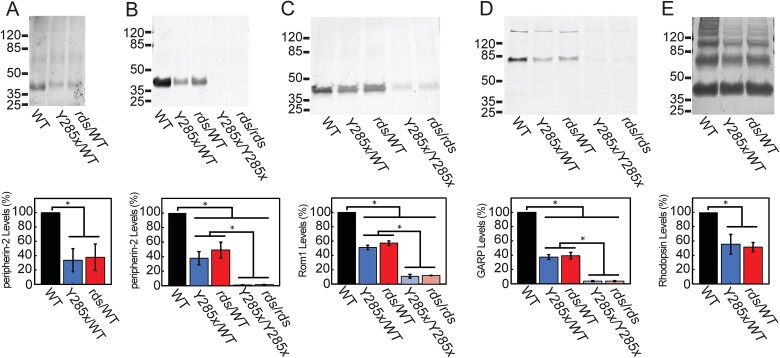
Mutations in PRPH2 are a relatively common cause of sight-robbing inherited retinal degenerations (IRDs). Peripherin-2 (PRPH2) is a photoreceptor-specific tetraspanin protein that structures the disk rim membranes of rod and cone outer segment (OS) organelles, and is required for OS morphogenesis. PRPH2 is noteworthy for its broad spectrum of disease phenotypes; both inter- and intra-familial heterogeneity have been widely observed and this variability in disease expression and penetrance confounds efforts to understand genotype-phenotype correlations and pathophysiology. Here we report the generation and initial characterization of a gene-edited animal model for PRPH2 disease associated with a nonsense mutation (c.1095:C>A, p.Y285X), which is predicted to truncate the peripherin-2 C-terminal domain. Young (P21) Prph2Y285X/WT mice developed near-normal photoreceptor numbers; however, OS membrane architecture was disrupted, OS protein levels were reduced, and in vivo and ex vivo electroretinography (ERG) analyses found that rod and cone photoreceptor function were each severely reduced. Interestingly, ERG studies also revealed that rod-mediated downstream signaling (b-waves) were functionally compensated in the young animals. This resiliency in retinal function was retained at P90, by which time substantial IRD-related photoreceptor loss had occurred. Altogether, the current studies validate a new mouse model for investigating PRPH2 disease pathophysiology, and demonstrate that rod and cone photoreceptor function and structure are each directly and substantially impaired by the Y285X mutation. They also reveal that Prph2 mutations can induce a functional compensation that resembles homeostatic plasticity, which can stabilize rod-derived signaling, and potentially dampen retinal dysfunction during some PRPH2-associated IRDs.

Keywords: PRPH2; homeostatic plasticity; inherited retinal degeneration; peripherin-2; rod and cone photoreceptors.


Figures

Figure 1
Figure 1
Design and generation of Y285X gene edited mice. Prph2Y285X knock-in mice were generated using the CRISPR/Cas9 system in conjunction with pronuclear microinjection into single-cell zygotes. (A) A ssDNA donor template was used to introduce a C>A transversion at c.1095 in murine Prph2 exon 3, mimicking a nonsense mutation associated with human macular disease [20]. Three additional silent mutations were included to introduce a diagnostic Mae III restriction site. Mutations are indicated by asterisks. (B) Founder mice were identified via endpoint PCR and Mae III digest (arrow) and (C) Sanger DNA sequencing. The Y285X mutation was then backcrossed more than seven generations onto the C57BL/6J background. (D) Cartoon shows Protter predicted peripherin-2 transmembrane topology and locations of the Y285 truncation site (red), and the N-terminal (pink) and C-terminal (blue) antibody reactivity sites. Extracellular 2 (EC2) and cytoplasmic c-terminal (CTER) domains are indicated.
Figure 2
Figure 2
Peripherin-2 levels are reduced in Prph2Y285X/WT mice. Protein levels were assessed by western blotting of retinal lysates from P21 mice of the indicated genotypes using primary antibodies against (A) the peripherin-2 N-terminus, (B) the peripherin-2 C-terminus, (C) rom1, (D) GARPs, and (E) rhodopsin. Primary antibody details are provided in Table S1. No truncated peripherin-2 was detected in the Prph2Y285X/WT disease model mice, and full-length peripherin-2 abundance was reduced to less than 50% of WT. Levels of rom1, GARPs, and rhodopsin each dropped to 40%–50% of WT. No peripherin-2 was detected in Prph2Y285X/Y285X homozygotes. Signal intensity data are represented as mean ± SEM (n = 3). Statistically significant differences were calculated using t-tests (*P < 0.05). Together, these data suggest that the Prph2Y285X allele produces an unstable product that can reduce abundance of the full-length protein produced by the WT allele.
Figure 3
Figure 3
The Prph2Y285X/WT disease model develops near-normal numbers of rod photoreceptors. (A) H&E-stained ocular cross-sections of WT and mutant P21 mouse retinas; ONL thickness was measured as an index of photoreceptor number, as described in Methods. (B) Spider plots of ONL thickness for Prph2Y285X/WT (blue symbols/lines), Prph2rds/WT (red symbols/lines), and WT control (black symbols/lines) mice are shown. Photoreceptor numbers in the heterozygous mutant mice (circles) did not differ from WT controls (squares), apart from modest reductions in the superior periphery of Prph2Y285X/WT. Differences did not meet statistical significance of P < 0.05. In contrast, the homozygous mutant mice (triangles) each showed substantial reductions in photoreceptor numbers across the central retina, relative to WT controls. Data are represented as mean ± SEM (n = 3-4). Statistically significant differences were calculated using two-way ANOVA with Tukey’s post hoc test. (C) Left: Maximum projection showing IHC labeling of M- (red) and S-cone (green) OSs in flat-mounted WT mouse retinal explants. Primary antibody details are provided in Table S1. Z-stack images were used to reconstruct 3D volumes acquired from four anatomically-standardized quadrants (superior, S; medial, M; inferior, I). Scale bar = 500 μm. Right: A representative Z-stack volume reconstruction from the medial quadrant; inset shows a higher magnification view. (D) Comparison of cone OS numbers for mutant (blue, red bars) and WT control (black bars) mice by region; medial regions were averaged for each retina. Small, but statistically significant reductions in Prph2Y285X/WT M- and S-cone numbers (blue bars) were seen relative to WT (black bars). By comparison, the Prph2rds/WT retinas were less affected (red bars). Statistically significant differences were calculated using two-way ANOVA (n = 9–11), P < 0.05.
Figure 4
Figure 4
Dysmorphic photoreceptor ultrastructure does not affect OS protein localization in P21 Prph2Y285X/WT mouse retina. Left panels: Typical examples of rod OSs in WT (A) and Prph2Y285X/WT (B and B′) mice. Typical examples of cone OSs in WT (C) and Prph2Y285X/WT mice (D and D′). Typical examples of photoreceptors lacking OSs in Prph2Y285X/Y285X mice (E and E′). Scale bar = 0.5 μm. Right panels: IHC labeling of fixed ocular tissue sections from WT and Prph2Y285X/WT mice. Proteins of interest appear in red. Photoreceptor inner segments (anti-Na+,K+-ATPase) appear in green. Nuclei labeled with Hoechst dye appear in blue. Confocal projection images illustrate that targeting and localization of: Peripherin-2, its interactors rom1 and GARPs, and rhodopsin are not affected in the disease model. Primary antibody details are provided in Table S1. Scale bar = 10 μm.
Figure 5
Figure 5
In vivo ERG finds P21 rod signals severely compromised, but downstream rod-derived signaling preserved. (A) Representative full-field scotopic ERG response traces from WT, Prph2Y285X/WT disease model, and Prph2rds/WT mice to 1.3 log cd.s/m2 light flashes. (B) Scotopic a-wave amplitudes of the Prph2Y285X/WT and Prph2rds/WT mice were reduced (≥ 50%), relative to those of WT. (C) In contrast, scotopic b-wave amplitudes of Prph2Y285X/WT mice were not significantly different than those of WT. Prph2rds/WT mice showed a small, but significant reduction at the highest flash luminance. (D) Prph2Y285X/WT and Prph2rds/WT mutant mice each showed significant increases of b-wave/a-wave ratio at all flash luminances. (E) Representative photopic responses to 1.9 log cd.s/m2 light flashes on a 30 cd/m2 background from WT, Prph2Y285X/WT, and Prph2rds/WT model mice. (F) Photopic b-wave amplitudes of Prph2Y285X/WT mice were not significantly different than those of WT. Prph2rds/WT mice showed a small, but significant reduction at higher flash luminances. Data are represented as mean ± SEM (n = 10–12), statistically significant differences were calculated using one-way ANOVA with Tukey’s post hoc test; (*P < 0.05).
Figure 6
Figure 6
Ex vivo ERG finds P21 rod and cone signaling severely compromised, but downstream rod signaling relatively preserved. (A) Representative transretinal scotopic response traces from WT (top) and Prph2Y285X/WT (bottom) mice to light flash intensities from −6.26 to −0.6 log photons/cm2/s. (B) Left: Scotopic a-wave amplitudes of the Prph2Y285X/WT mutant were reduced (≥ 70%), relative to those of WT. Right: Scotopic b-wave amplitudes from Prph2Y285X/WT retina were reduced less than 30% from WT at most light intensities. (C) Representative photopic responses to light flash intensities of −3.25 to 0.0 log photons/cm2/s (0 log = 2.20 × 10¹⁶ photons/cm2/s) light flashes on a −2.82 log photons/cm2/s background illumination from WT (top) and Prph2Y285X/WT (bottom) mice. (D) Left: Photopic a-wave amplitudes of Prph2Y285X/WT mice were reduced about 50% relative to WT. Right: Photopic b-wave amplitudes of Prph2Y285X/WT mice were lower than WT at all luminances tested, but did not show statistically significant differences. (E) Left: Prph2Y285X/WT mutant mice display b/a-wave ratios that are significantly elevated over WT across the scotopic luminance range. Right: Prph2Y285X/WT mutant mice display b/a-wave ratios that were higher than WT at all luminances tested, but did not show statistically significant differences. Data for (B), (D), and (E), are represented as mean ± SEM (n = 10–12, one-way ANOVA with Tukey’s post hoc test (*P < 0.05).
Figure 7
Figure 7
The basis of functional preservation in P21 Prph2Y285X/WT and Prph2rds/WT mice remains to be determined. (A) Upper panels: Representative H&E-stained ocular cross-sections from Prph2Y285X/WT mice (middle), compared to sections from WT (left) and Prph2rds/WT mice (right). Scale bar = 50 μm. Lower panel: Quantitation of photoreceptor-bipolar cell synaptic layer thickness. No significant differences in OPL thickness were found between WT (black bars) and Prph2Y285X/WT (blue bars) or Prph2rds/WT (red bars). (B) Upper panels: Typical examples of rod spherule synaptic ribbons imaged by conventional TEM. Scale bar = 0.5 μm. Lower panel: Comparisons of ribbon numbers in WT and the Prph2Y285X/WT mice. Data (A and B) are represented as mean ± SEM (n = 3–4), no differences were calculated using one-way ANOVA. (C) IHC double-labeling of fixed ocular vibratome sections with anti-PKC (red) and anti-Na+,K+-ATPase (green) antibodies. Primary antibody details are provided in Table S1. Top: Although bipolar cell length appeared slightly reduced, no change in bipolar cell dendritic structure was observed. Monochrome (center) and enlarged (bottom) images provide enhanced contrast and clarity for bipolar cell dendritic structure. Scale bar = 10 μm.
Figure 8
Figure 8
The Prph2Y285X/WT knock-in mouse is a new model for IRD that retains functional compensation with aging. (A) Spider plots of ONL thickness for P90 Prph2Y285X/WT, Prph2rds/WT, and WT control mice are shown. Each of the mutant lines show significantly reduced photoreceptor numbers across the retina relative to WT controls (n = 4). (B) Bar graphs plotting ONL thicknesses (250 μm superior to the ONH) at P21 and at P90 illustrate a progressive loss of photoreceptors with age for the Prph2Y285X/WT (blue) and Prph2rds/WT (red) mice, relative to WT controls (black). (C) In vivo ERG a-wave amplitudes of P90 Prph2Y285X/WT and Prph2rds/WT mice recorded under scotopic conditions were reduced (≥63%), relative to those of WT. (D) In vivo ERG b-waves recorded under scotopic conditions were reduced (~40%) in each of the mutants relative to WT. (E) P90 Prph2Y285X/WT and Prph2rds/WT mutant mice displayed in vivo b/a-wave ratios that were significantly elevated over WT across the scotopic luminance range. (F) Bar graphs show in vivo scotopic ERG b/a-wave ratios in response to −0.3 log cd.s/m2 flashes. The data illustrates that the functional preservation observed in P21 mutant mice has been retained at P90. Data are represented as mean ± SEM (A and B; n = 4, C, D, E, and F; n = 12–14). Statistically significant differences were calculated using two-way ANOVA (A and B) or one-way ANOVA (C, D, E, and F) with Tukey’s post hoc test (*P < 0.05).

References

    1. Goetz KE, Reeves MJ, Gagadam S. et al. Genetic testing for inherited eye conditions in over 6,000 individuals through the eyeGENE network. Am J Med Genet C Semin Med Genet 2020;184:828–837. - PMC - PubMed
    1. Pontikos N, Arno G, Jurkute N. et al. Genetic basis of inherited retinal disease in a molecularly characterized cohort of more than 3000 families from the United Kingdom. Ophthalmology 2020;127:1384–1394. - PMC - PubMed
    1. Reeves MJ, Goetz KE, Guan B. et al. Genotype-phenotype associations in a large PRPH2-related retinopathy cohort. Hum Mutat 2020;41:1528–1539. - PMC - PubMed
    1. Boon CJ, Den Hollander AI, Hoyng CB. et al. The spectrum of retinal dystrophies caused by mutations in the peripherin/RDS gene. Prog Retin Eye Res 2008;27:213–235. - PubMed
    1. Peeters M, Khan M, Rooijakkers A. et al. PRPH2 mutation update: In silico assessment of 245 reported and 7 novel variants in patients with retinal disease. Hum Mutat 2021;42:1521–1547. - PMC - PubMed

Publication types

MeSH terms