. 2024 Sep 3;12(9):e009236.
doi: 10.1136/jitc-2024-009236.

Targeting IL-33 reprograms the tumor microenvironment and potentiates antitumor response to anti-PD-L1 immunotherapy

Affiliations

Targeting IL-33 reprograms the tumor microenvironment and potentiates antitumor response to anti-PD-L1 immunotherapy

Yanyang Nan et al. J Immunother Cancer. .

Erratum in

Abstract

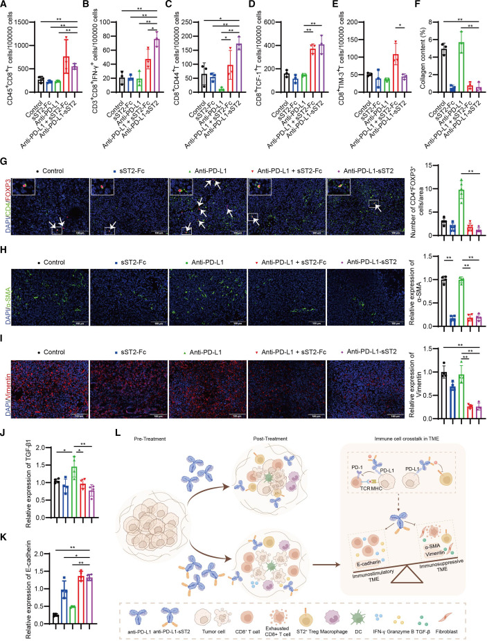
Background: The main challenge against patients with cancer to derive benefits from immune checkpoint inhibitors targeting PD-1/PD-L1 appears to be the immunosuppressive tumor microenvironment (TME), in which IL-33/ST2 signal fulfills critical functions. However, whether IL-33 limits the therapeutic efficacy of anti-PD-L1 remains uncertain.

Methods: Molecular mechanisms of IL-33/ST2 signal on anti-PD-L1 treatment lewis lung carcinoma tumor model were assessed by RNA-seq, ELISA, WB and immunofluorescence (IF). A sST2-Fc fusion protein was constructed for targeting IL-33 and combined with anti-PD-L1 antibody for immunotherapy in colon and lung tumor models. On this basis, bifunctional fusion proteins were generated for PD-L1-targeted blocking of IL-33 in tumors. The underlying mechanisms of dual targeting of IL-33 and PD-L1 revealed by RNA-seq, scRNA-seq, FACS, IF and WB.

Results: After anti-PD-L1 administration, tumor-infiltrating ST2+ regulatory T cells (Tregs) were elevated. Blocking IL-33/ST2 signal with sST2-Fc fusion protein potentiated antitumor efficacy of PD-L1 antibody by enhancing T cell responses in tumor models. Bifunctional fusion protein anti-PD-L1-sST2 exhibited enhanced antitumor efficacy compared with combination therapy, not only inhibited tumor progression and extended the survival, but also provided long-term protective antitumor immunity. Mechanistically, the superior antitumor activity of targeting IL-33 and PD-L1 originated from reducing immunosuppressive factors, such as Tregs and exhausted CD8+ T cells while increasing tumor-infiltrating cytotoxic T lymphocyte cells.

Conclusions: In this study, we demonstrated that IL-33/ST2 was involved in the immunosuppression mechanism of PD-L1 antibody therapy, and blockade by sST2-Fc or anti-PD-L1-sST2 could remodel the inflammatory TME and induce potent antitumor effect, highlighting the potential therapeutic strategies for the tumor treatment by simultaneously targeting IL-33 and PD-L1.

Keywords: Immune Checkpoint Inhibitor; Immune modulatory; Immunotherapy; T regulatory cell - Treg; Tumor microenvironment - TME.


Conflict of interest statement

Competing interests: None declared.

Figures

Figure 1
Figure 1. IL-33/ST2 signal upregulated after anti-PD-L1 treatment. Volcano plots generated from analyzing differential gene expression between control and anti-PD-L1 treatment group (A). IL1RL1 gene level in tumor tissues of LLC tumor-bearing mouse injected with anti-PD-L1 (n=3) (B). ST2 protein level in tumor tissues was analyzed by WB (C). The quantification of ST2 expression in tumor tissues during anti-PD-L1 using ImageJ software (n=4) (D). IL-33 tumor tissue expression was analyzed by ELISA (n=4) (E). The expression of Myd88 and activation of JAK2/STAT3 pathway in tumor tissues treated with anti-PD-L1 (F). The gene correlation among CD274 and IL1RL1 in LUAD was analyzed using GEPIA (G). Representative images of ST2 (green) and FOXP3 (red) labeled in the LLC tumor tissues after anti-PD-L1 treatment (H). Scale bar=100 µm. White arrows point to ST2+FOXP3+ cells. *P < 0.05, **P < 0.01. GEPIA, Gene Expression Profiling Interactive Analysis; LLC, lewis lung carcinoma; LUAD, lung adenocarcinoma.
Figure 2
Figure 2. Dual targeting of PD-L1 and IL-33 enhanced antitumor effect of anti-PD-L1 therapy in mouse models. Treatment scheme (top), tumor growth curves (bottom left), and tumor weight (bottom right) of CT26 (A) or LLC (B) tumor-bearing mice in four groups (n=5). Schematic diagram of treatment protocol (left) and survival curves (right) of metastasis CT26 tumor model (n=5) (C). Scheme (left), H&E-stained lung tissues together with their representative photos (middle), and the quantity of tumor burden (right) from each group with metastasis LLC tumor (n=5) (D). *P < 0.05, **P < 0.01. LLC, lewis lung carcinoma.
Figure 3
Figure 3. Combined blockade of IL-33 and PD-L1 enhanced T cells response. Bulk RNA-seq to evaluate immune landscape in LLC tumors (A–E). Gene expression levels of Prf1, Ifng, and Gzmb in tumor tissue of LLC tumor-bearing mice (n=3) (A). The data were presented in Log2TPM and every group was compared with the combination therapy group. Heat map of genes that under the category of immune signatures (n=3) (B–D). Gene set enrichment analysis enrichment plot of KEGG pathway for genes in Combo-treated tumors, relative to anti-PD-L1-treated tumors (E). Flow cytometry was used to analyze the absolute number of CD4+ (F), CD8+ (G), CD8+GranB+ (H) and CD8+IFN-γ+ (I) cells and CD4+FOXP3+ cells (J) in CT26 tumor tissues (n=3). *P < 0.05, **P < 0.01. LLC, lewis lung carcinoma.
Figure 4
Figure 4. Construction and characterization of bifunctional fusion proteins targeting both PD-L1 and IL-33. Structure of bifunctional fusion proteins (A). Atezolizumab and sST2 were used to targeting PD-L1 and IL-33, respectively, and were fused by a flexible (Gly4Ser)3Gly linker. The purity determination of anti-PD-L1-sST2 (B) and sST2-anti-PD-L1 (C). Affinity of bifunctional fusion proteins for hPD-L1 (D) or mIL-33 (E) proteins measured by SPR. PD-1/PD-L1 blockade bioassay of bifunctional fusion proteins was confirmed (n=2) (F). Naïve CD4+ T cells were incubated with anti-CD3/CD28 and TGF-β1, subjected to the specified treatments for 3 days to determine the frequencies of FOXP3+ T cells (n=3) (G). Effects of bifunctional fusion proteins on LPS-induced production of IL-6 (H) and TNF-α (I) from Raw264.7 in vitro (n=3). * and $ represent the Student’s t-test of each groups compared with control group and IL-33 group, respectively. *P < 0.05, **P < 0.01, $P < 0.05.
Figure 5
Figure 5. Antitumor activity of bifunctional fusion proteins in murine LLC models. Tumor growth (A) and tumor weight (B) of LLC tumor-bearing mice therapied with bifunctional fusion proteins (11.5 mg/kg) or anti-PD-L1 (7.5 mg/kg) and sST2-Fc (6.3 mg/kg) combined therapy (n=5). Schematic overview of the experimental study (C). Mice were intravenously injected with LLC cells (2*105), treated at day 0, 4, 7, and 11 after injection (red arrows) and observed until day 100 (n=8). Then, the survived mice were rechallenged with injection of 5*105 LLC cells and observed for three additional weeks. Survival curves of tumor-bearing mice in five groups (D). H&E-stained lung tissues of mice in rechallenge assay (E). *P < 0.05, **P < 0.01. LLC, lewis lung carcinoma.
Figure 6
Figure 6. Dual targeting of PD-L1 and IL-33 changed T-cell cellular landscapes. Design of scRNA-seq experiment (A). UMAP of all cell types from four groups (B). UMAP plots showing all CD4+ (C) and CD8+ (E) T cells from tumor tissues in four groups. Bar plots exhibiting the proportion of CD4+ (D) and CD8+ (F) T cells within each cluster. Selected significantly enriched pathways in KEGG analyses based on the differentially expressed genes in T cell clusters (G). Gene Ontology in biological function enrichment analysis of the upregulated (H, J) or downregulated (I, K) expressed genes in T cells of anti-PD-L1-sST2 versus atezolizumab group, and anti-PD-L1-sST2 versus combination group, respectively. LLC, lewis lung carcinoma.
Figure 7
Figure 7. Dual targeting of PD-L1 and IL-33 reprograms immuno-inflammatory phenotype TME. Absolute number of CD8+ cells (A), CD3+CD8+IFN-γ+ T cells (B), CD8+CD44+ cells (C), CD8+TCF-1+ cells (D), CD8+TIM-3+ cells (E) within the CD45+ cells from tumor tissues (n=3). The quantification of collagen (n=3) (F). The quantification of CD4+FOXP3+ cells in LLC tumor-bearing mice (n=5) (G). White arrows point to CD4+FOXP3+ cells. The quantification of α-SMA (H), Vimentin (I), TGF-β1 (J), and E-cadherin (K) (n=4). In figure 7G–K, data in groups of control, sST2-Fc, anti-PD-L1, anti-PD-L1 + sST2 Fc, and anti-PD-L1-sST2 are shown in color of blank, blue, green, red, and violet, respectively. Schematic diagram illustrating the potent antitumor impact of anti-PD-L1-sST2 bifunctional fusion protein (l). *P < 0.05, **P < 0.01. LLC, lewis lung carcinoma; TME, tumor microenvironment.

References

    1. van Gulijk M, van Krimpen A, Schetters S, et al. PD-L1 checkpoint blockade promotes regulatory T cell activity that underlies therapy resistance. Sci Immunol. 2023;8:eabn6173. doi: 10.1126/sciimmunol.abn6173. - DOI - PubMed
    1. Wang R, Liu H, He P, et al. Inhibition of PCSK9 enhances the antitumor effect of PD-1 inhibitor in colorectal cancer by promoting the infiltration of CD8+ T cells and the exclusion of Treg cells. Front Immunol. 2022;13:947756. doi: 10.3389/fimmu.2022.947756. - DOI - PMC - PubMed
    1. Wang Y, Zhang X, Xu C, et al. Targeting 4-1BB and PD-L1 induces potent and durable antitumor immunity in B-cell lymphoma. Front Immunol. 2022;13:1004475. doi: 10.3389/fimmu.2022.1004475. - DOI - PMC - PubMed
    1. Abril-Rodriguez G, Torrejon DY, Liu W, et al. PAK4 inhibition improves PD-1 blockade immunotherapy. Nat Cancer. 2020;1:46–58. doi: 10.1038/s43018-019-0003-0. - DOI - PMC - PubMed
    1. Vesely MD, Zhang T, Chen L. Resistance Mechanisms to Anti-PD Cancer Immunotherapy. Annu Rev Immunol. 2022;40:45–74. doi: 10.1146/annurev-immunol-070621-030155. - DOI - PubMed

LinkOut - more resources