Case Reports
. 2024 Sep 19;38(15-16):755-771.
doi: 10.1101/gad.352032.124.

Heterozygous RPA2 variant as a novel genetic cause of telomere biology disorders

Affiliations
Case Reports

Heterozygous RPA2 variant as a novel genetic cause of telomere biology disorders

Rima Kochman et al. Genes Dev. .

Abstract

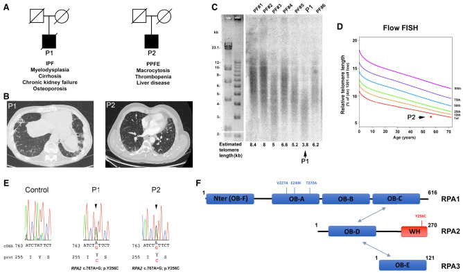
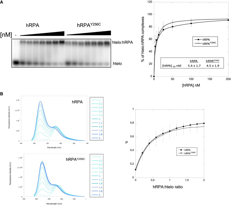
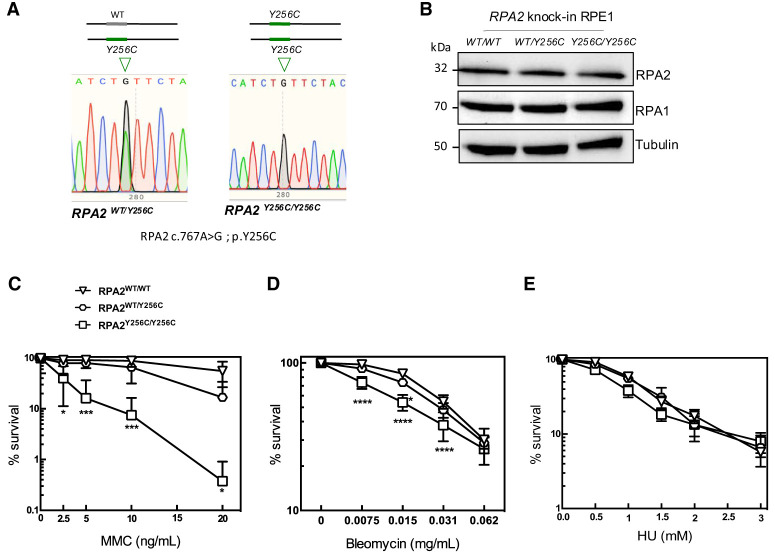
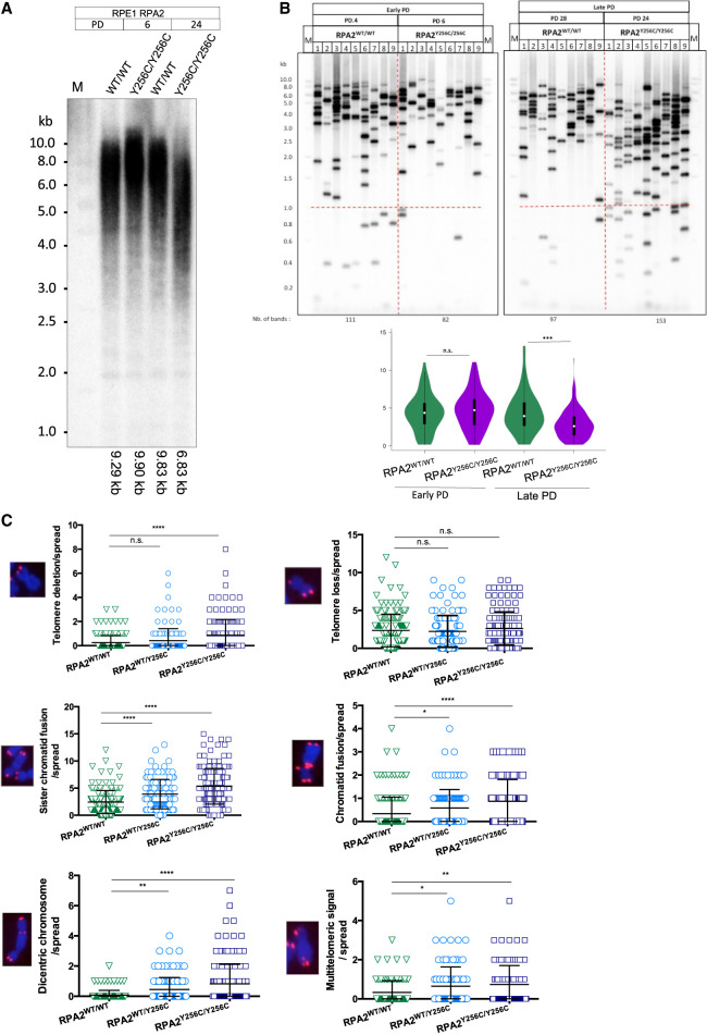
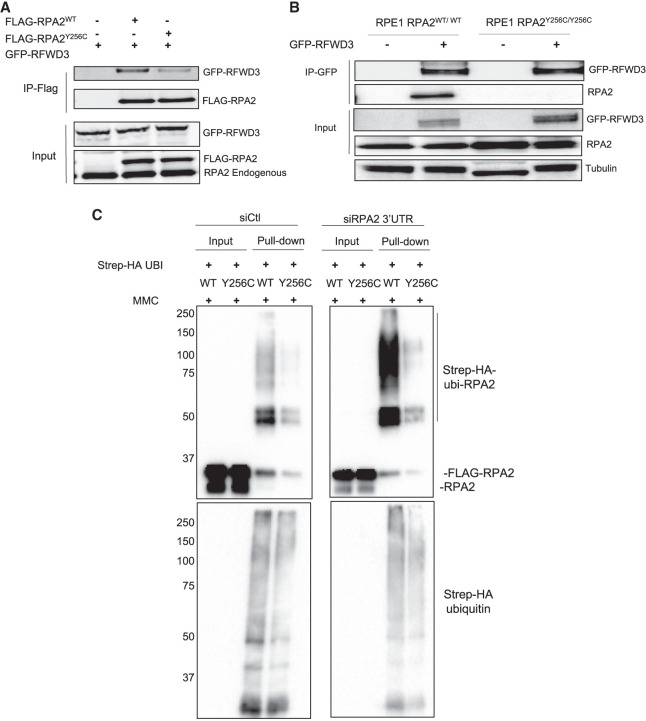
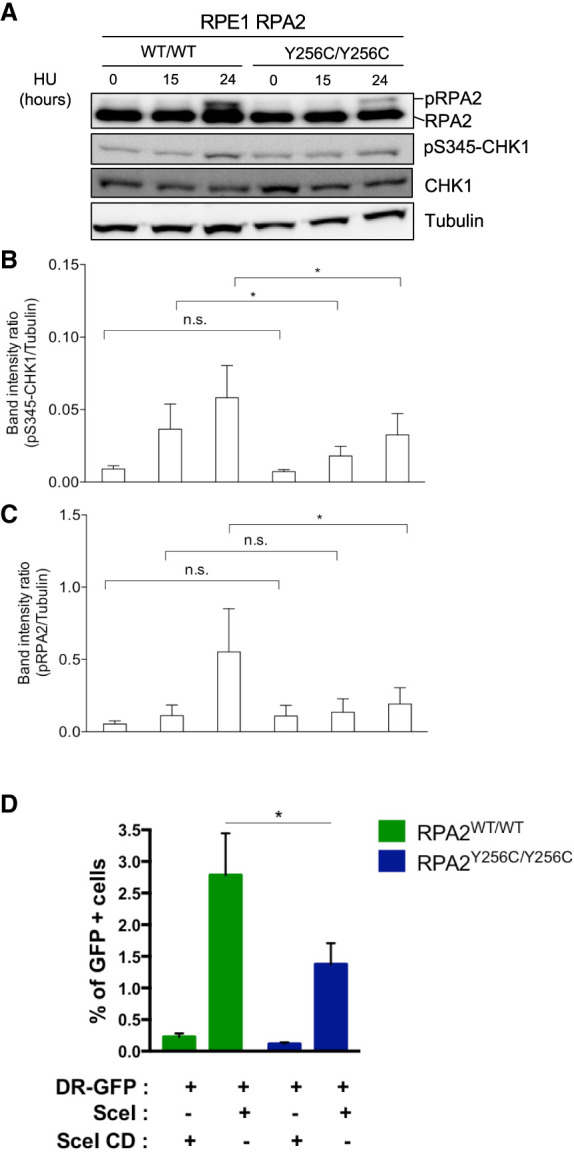
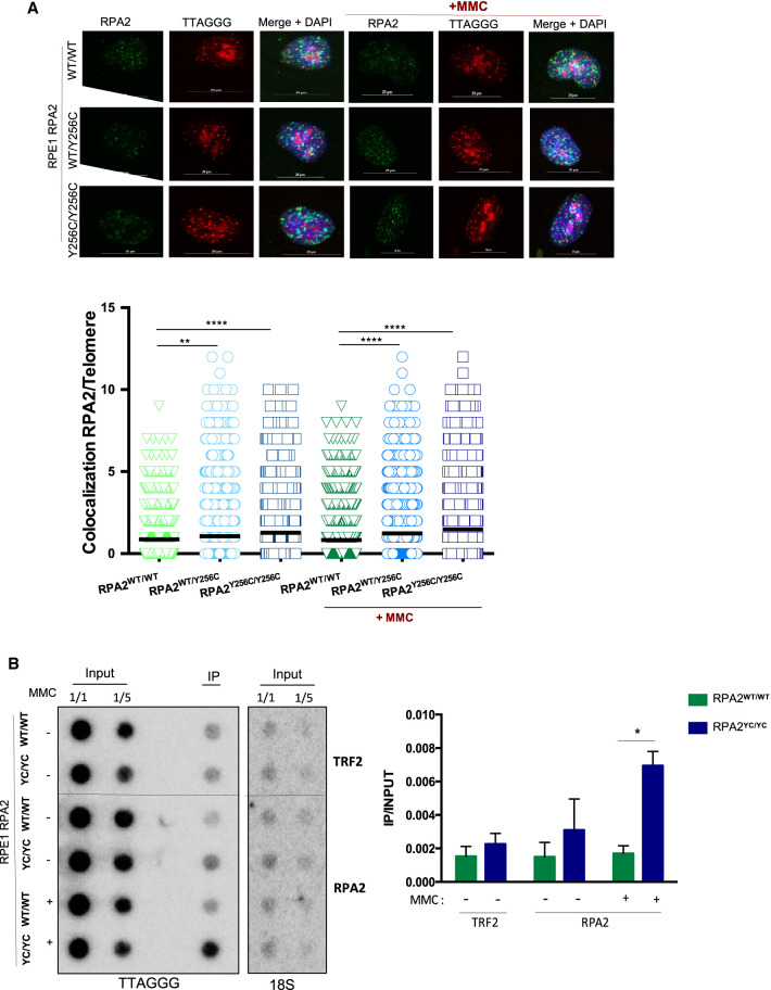
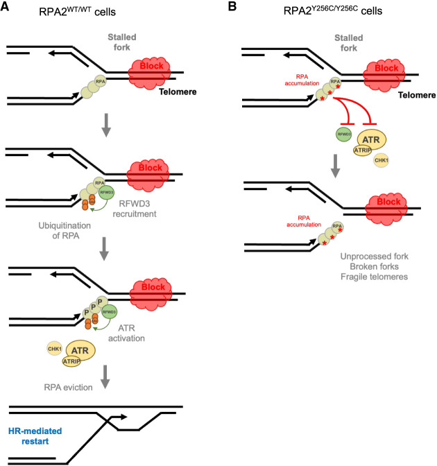
Premature telomere shortening or telomere instability is associated with a group of rare and heterogeneous diseases collectively known as telomere biology disorders (TBDs). Here we identified two unrelated individuals with clinical manifestations of TBDs and short telomeres associated with the identical monoallelic variant c.767A>G; Y256C in RPA2 Although the replication protein A2 (RPA2) mutant did not affect ssDNA binding and G-quadruplex-unfolding properties of RPA, the mutation reduced the affinity of RPA2 with the ubiquitin ligase RFWD3 and reduced RPA ubiquitination. Using engineered knock-in cell lines, we found an accumulation of RPA at telomeres that did not trigger ATR activation but caused short and dysfunctional telomeres. Finally, both patients acquired, in a subset of blood cells, somatic genetic rescue events in either POT1 genes or TERT promoters known to counteract the accelerated telomere shortening. Collectively, our study indicates that variants in RPA2 represent a novel genetic cause of TBDs. Our results further support the fundamental role of the RPA complex in regulating telomere length and stability in humans.

Keywords: RFWD3; RPA; telomere; telomere disorders; telomere replication.


Figures

Figure 1.
Figure 1.
Identification of two individuals carrying the same RPA2 variant. (A) Pedigree of the two unrelated individuals presenting with clinical manifestations of telomere biology disorder. The main clinical features are noted. (PPFE) Pleuroparenchymal fibroelastosis, (IPF) interstitial pulmonary fibrosis. (B) High-resolution computed tomography scans revealed a pattern of pleuroparenchymal fibroelastosis for both P1 and P2, including pleural fibrotic thickening with subpleural consolidations, bronchiectasis, and honeycombing in the upper regions, in particular in the upper right lobe (P1); in the lower lobes, the pattern of interstitial lung disease (ILD) is indeterminate for usual interstitial pneumonia, with the presence of reticulations, ground glass opacities, and focal areas of honeycombing. P2 shows the presence of subpleural reticulations without bronchiectasis, suggestive of indeterminate ILD. (C,D) Telomere length measurement by telomere restriction fragment in P1 (C) and flow-FISH in P2 (D) showed short telomeres in both patients. (E) Genetic analysis identified the same RPA2 variant c.767A>G; p.Y256C in both patients as represented by Sanger sequences. (F) Schematic representation of the RPA complex. The OB folds A–F are shown as blue rectangles, and the winged helix (WH) domain of RPA2 is in red. OB folds A–D (DBDs A–D) support the ssDNA-binding activity of the RPA complex, while OB fold F and the WH domain of RPA2 support protein–protein interactions. Heterotrimerization of the RPA complex is mediated by OB folds C of RPA1, D of RPA2, and E of RPA3 (gray arrows). Mutations of RPA isolated from patients with telomere syndrome are shown.
Figure 2.
Figure 2.
The Y256C mutation does not affect the DNA binding activity of RPA. (A) Electrophoretic mobility shift assays (EMSAs). 32P-htelo (2 nM) was incubated with various amounts of RPAWT or RPAY256C complexes (from 3.125 to 200 nM) in the presence of 100 mM KCl solutions and separated on a native 1% agarose gel. Quantification of htelo:RPA complexes is presented. (B) Fluorimetric titration of 100 nM F-htelo-T by increasing amounts of RPAWT and RPAY256C complexes was performed in the presence of KCl. The spectra were recorded after 2 min of incubation. P (see the Materials and Methods) increased as a function of the RPA:F-htelo-T ratio r (from 0.5 to 2), showing the unfolding of the G-quadruplex by both RPA complexes. The results correspond to the mean ± SD of four independent determinations.
Figure 3.
Figure 3.
RPA2Y256C/Y256C knock-in cell lines are sensitive to genotoxic agents. (A) Schematic representation of insertion of the RPA2 Y256C mutation mediated by CRISPR/Cas9 in RPE1 cells. We generated RPE1 cellular clones carrying the Y256C RPA2 variant in the heterozygous and homozygous states (RPA2WT/Y256C and RPA2Y256C/Y256C, respectively). (B) Cellular extracts from RPA2WT/WT, RPA2WT/Y256C, and RPA2Y256C/Y256C RPE1 cells were subjected to Western blotting. Membrane was revealed using the indicated antibodies. (CE) Clonogenic survival analysis of RPA2WT/WT, RPA2WT/Y256C, and RPA2Y256C/Y256C RPE1 cells exposed to the indicated genotoxins. For each cell type, the viability of untreated cells is defined as 100%. Data are represented as mean ± SEM; n = 3. A two-way ANOVA test was used to determine P-values. (*) P < 0.05, (***) P < 0.001, (****) P < 0.0001.
Figure 4.
Figure 4.
RPA2Y256C mutation causes telomere defects. (A) Terminal restriction fragment (TRF) analysis of RPA2WT/WT and RPA2Y256C/Y256C RPE1 cells at early and late population doublings. Telomere lengths were estimated according to the peaks of the signal density using ImageQuant. (B) Detection of the individual PCR-amplified telomeres by TeSLA performed in RPA2WT/WT and RPA2Y256C/Y256C RPE1 cells at early (four to six) and late (24–28) population doublings. Violin plots represent the distributions of TeSLA band sizes. The statistical significance of the differences was tested using unpaired two-tailed Student's t-tests. (***) P < 0.001. (C) Analysis of telomeric aberrations detected by telomere FISH RPA2WT/WT, RPA2WT/Y256C, and RPA2Y256C/Y256C RPE1 cells at early population doubling. Three independent experiments were performed (counted metaphase spreads: RPA2WT/WT: n = 163; RPA2WT/Y256C: n = 125; RPA2Y256C/Y256C: n = 163). Both ordinary one-way ANOVA and Mann–Whitney statistical tests were applied to determine P-values. (*) P < 0.05, (**) P < 0.01, (****) P < 0.0001.
Figure 5.
Figure 5.
The Y256C mutation impairs ubiquitination of RPA2 by RFWD3. (A) HEK293T cells were cotransfected with plasmids expressing GFP-RFWD3 (catalytically inactive C315A mutant) and FLAG-RPA2WT or FLAG-RPA2Y256C. Anti-FLAG immunoprecipitates (IP) were probed with anti-GFP and anti-RPA2 antibodies by immunoblotting. Picture is representative of three independent experiments. (B) RPA2WT/WT and RPA2Y256C/Y256C RPE1 cells were transfected with GFP-RFWD3 (catalytically inactive C315A mutant). Anti-GFP immunoprecipitates were analyzed by immunoblotting using anti-GFP and anti-RPA2 antibodies. Picture is representative of three independent experiments. (C) HEK293T cells were transfected with FLAG-RPA2WT and FLAG-RPA2Y256C, with control or RPA2 targeting siRNAs. Twenty-four hours later, cells were transfected with a Strep-HA ubiquitin construct for 24 h and treated or not with 100 ng/mL MMC for 24 h. Ubiquitylated proteins were collected by denaturing Strep-Tactin pull-down and blotted with anti-RPA2 (top panel) and anti-HA (bottom panel) antibodies.
Figure 6.
Figure 6.
ATR signaling and HR are defective in the RPA2Y256C/Y256C cell line. (A) Western blot analysis of phospho-RPA2 (S4/S8), RPA2, phospho-CHK1 (S345), CHK1 in RPA2WT/WT, and RPA2Y256C/Y256C RPE1 cells treated with 2 mM HU for 15 and 24 h. Tubulin levels were determined to control for protein loading. (B,C) Quantification of phospho-RPA2 and phospho-CHK1 was normalized to tubulin levels. (*) P < 0.05 by t-test (n = 3). (HU) Hydroxyurea. (D) HR-mediated repair assay. RPA2WT/WT and RPA2Y256C/Y256C RPE1 cells were cotransfected with pDR-GFP and pISceI-RFP or pISceI-catalytic-dead-RFP (Sce1 CD) plasmids. Cells were treated 24 h after transfection and analyzed by flow cytometry to determine the percentage of GFP-positive (GFP+) cells, indicative of HR efficiency. Results from three independent experiments. Error bars represent standard deviation from the mean. Two-way ANOVA statistical test was applied to determine P-values. (*) P < 0.05.
Figure 7.
Figure 7.
Y256C mutation leads to accumulation of RPA at telomeres. (A) Immunofluorescence FISH in RPA2WT/WT, RPA2WT/Y256C, and RPA2Y256C/Y256C RPE1 cells treated or not with 40 ng/mL MMC for 24 h. Fixed cells were labeled with telomeric FISH probe (red) and immunostained for RPA2 (green) and DAPI (blue) as indicated. Scale bar, 20 μm. Quantification of RPA2 colocalizations at telomeres is from at least 400 nuclei per replicate. Data are representative of three independent experiments and are presented as mean. The statistical significance of the differences was tested using ordinary one-way ANOVA and Mann–Whitney test. (**) P < 0.01, (****) P < 0.001. (MMC) Mytomycin-C. (B) Chromatin immunoprecipitation (ChIP) assays were performed in RPA2WT/WT and RPA2Y256C/Y256C RPE1 cells treated or not with 40 ng/mL MMC for 24 h and immunoprecipitated with anti-RPA2 and anti-TRF2 (as a control). Dot blots were hybridized with a telomeric probe or 18S probe. Quantification of ChIP dot blot expressed as IP/input of three biological replicates. (*) P < 0.05 by t-test and two-way ANOVA test.
Figure 8.
Figure 8.
Model of replication fork processing at telomeres involving RPA. (A) Telomeric sequences are hard to replicate regions because of the diverse sources of endogenous blocks that impede the progression of the fork. RPA is a first sensor and responder to replication stress and binds to ssDNA that may accumulate at stalled forks. This is the first step in the processing of stalled replication forks. In this model, RPA2–RFWD3 interaction promotes ubiquitination of chromatin-bound RPA. Ubiquitination by RFWD3 also contributes to phosphorylation of RPA, which activates the ATR pathway. ATR controls specific aspects of DNA damage signaling and restart of the fork, especially by HR. Ubiquitination of RPA by RFWD3 is also necessary to promote its eviction to facilitate HR-dependent repair. (B) In the context of Y256C RPA2 mutation, several aspects of mechanisms of fork protection are altered. Improper interaction with RFWD3 would lead to defects of ATR activation and would prevent efficient RPA eviction. This would cause accumulation of RPA at telomeres. We hypothesize that these dysfunctions, caused by Y256C mutation, could then lead to catastrophic events that may result in a deadlock in the resolution of blocked forks and cause the loss of telomeric sequences and telomere instability.

Similar articles

Cited by

References

    1. Audry J, Maestroni L, Delagoutte E, Gauthier T, Nakamura TM, Gachet Y, Saintomé C, Géli V, Coulon S. 2015. RPA prevents G-rich structure formation at lagging-strand telomeres to allow maintenance of chromosome ends. EMBO J 34: 1942–1958. 10.15252/embj.201490773 - DOI - PMC - PubMed
    1. Bansbach CE, Bétous R, Lovejoy CA, Glick GG, Cortez D. 2009. The annealing helicase SMARCAL1 maintains genome integrity at stalled replication forks. Genes Dev 23: 2405–2414. 10.1101/gad.1839909 - DOI - PMC - PubMed
    1. Bertrand A, Ba I, Kermasson L, Pirabakaran V, Chable N, Lainey E, Ménard C, Kallel F, Picard C, Hadiji S, et al. 2024. Characterization of novel mutations in the TEL-patch domain of the telomeric factor TPP1 associated with telomere biology disorders. Hum Mol Genet 33: 612–623. 10.1093/hmg/ddad210 - DOI - PubMed
    1. Bétous R, Mason AC, Rambo RP, Bansbach CE, Badu-Nkansah A, Sirbu BM, Eichman BF, Cortez D. 2012. SMARCAL1 catalyzes fork regression and Holliday junction migration to maintain genome stability during DNA replication. Genes Dev 26: 151–162. 10.1101/gad.178459.111 - DOI - PMC - PubMed
    1. Bhat KP, Cortez D. 2018. RPA and RAD51: fork reversal, fork protection, and genome stability. Nat Struct Mol Biol 25: 446–453. 10.1038/s41594-018-0075-z - DOI - PMC - PubMed

Publication types

LinkOut - more resources