. 2024 Dec 12;64(6):2301983.
doi: 10.1183/13993003.01983-2023. Print 2024 Dec.

Microenvironmental acidification by pneumococcal sugar consumption fosters barrier disruption and immune suppression in the human alveolus

Affiliations

Microenvironmental acidification by pneumococcal sugar consumption fosters barrier disruption and immune suppression in the human alveolus

Diana Fatykhova et al. Eur Respir J. .

Abstract

Streptococcus pneumoniae is the most common causative agent of community-acquired pneumonia worldwide. A key pathogenic mechanism that exacerbates severity of disease is the disruption of the alveolar-capillary barrier. However, the specific virulence mechanisms responsible for this in the human lung are not yet fully understood. In this study, we infected living human lung tissue with Strep. pneumoniae and observed a significant degradation of the central junctional proteins occludin and vascular endothelial cadherin, indicating barrier disruption. Surprisingly, neither pneumolysin, bacterial hydrogen peroxide nor pro-inflammatory activation were sufficient to cause this junctional degradation. Instead, pneumococcal infection led to a significant decrease of pH (∼6), resulting in the acidification of the alveolar microenvironment, which was linked to junctional degradation. Stabilising the pH at physiological levels during infection reversed this effect, even in a therapeutic-like approach. Further analysis of bacterial metabolites and RNA sequencing revealed that sugar consumption and subsequent lactate production were the major factors contributing to bacterially induced alveolar acidification, which also hindered the release of critical immune factors. Our findings highlight bacterial metabolite-induced acidification as an independent virulence mechanism for barrier disruption and inflammatory dysregulation in pneumonia. Thus, our data suggest that strictly monitoring and buffering alveolar pH during infections caused by fermentative bacteria could serve as an adjunctive therapeutic strategy for sustaining barrier integrity and immune response.


Conflict of interest statement

Conflict of interest: D. Beule reports grants from DFG and BMBF; two patent applications not related to the manuscript or project; and a leadership role as AEBC2 (Core Unit Network, unpaid); outside the submitted work. S. Hammerschmidt reports support for the present manuscript from Federal Excellence Initiative of Mecklenburg Western Pomerania and European Social Fund Grant KoInfekt (ESF_14-BM-A55-0001_16). A.D. Gruber reports support for the present manuscript from German Research Council (Grant No SFB-TR84 Z01b). A.C. Hocke reports support for the present manuscript from DFG, SFB-TR84, projects B6 and Z1a, Einstein Foundation Berlin, Einstein Center 3R. Outside the submitted work, A.C. Hocke reports grants from Charité – Zeiss Development Center for Multidimensional Microscopy. All other authors have nothing to disclose.

Figures

None
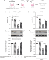
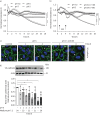
Microenvironmental acidification by pneumococcal sugar consumption fosters barrier disruption and immune suppression in the human alveolus. AM: alveolar macrophage; ATI/II: alveolar type I/II cells; EC: endothelial cells; ZO: zonula occludens protein; VE: vascular endothelial; Strep. pneumoniae: Streptococcus pneumoniae; IL: interleukin; H+: hydrogen; GM-CSF: granulocyte–macrophage colony-stimulating factor; COX: cyclo-oxygenase.
FIGURE 1
FIGURE 1
Degradation of junctional proteins occludin and vascular endothelial (VE)-cadherin during pneumococcal infection in human lungs is independent of pneumolysin (PLY), hydrogen peroxide (H2O2) or interleukin (IL)-1β. Lung explants were infected with a) 106 CFU·mL−1 Streptococcus pneumoniae wild-type (Strep. pneumoniae wt) and mutants Δply, ΔspxB or stimulated with b) 1 and 5 µg·mL−1 purified PLY, 1 and 10 mM·mL−1 H2O2 or c) 100 ng·mL−1 IL-1β for 24 h. d) Lung tissue was challenged with Strep. pneumoniae wt or Strep. pneumoniae Δply alone or treated with 50 µg·mL−1 pan-caspase inhibitor zVAD for 24 h. Total tissue lysates were analysed by Western blot and quantified by densitometry. Representative gels for occludin, VE-cadherin and cyclo-oxygenase (COX)-2 are shown. Values represent respective protein expression level relative to control and normalised to β-actin. Data are presented as mean±sd of at least three donors within independent experiments. ns: nonsignificant. *: p<0.05, **: p<0.01, ***: p<0.001.
FIGURE 2
FIGURE 2
Acidification during pneumococcal infection in human lungs leads to pH-dependent reduction of junctional proteins occludin and vascular endothelial (VE)-cadherin, which is prevented by HEPES buffering. Tissue was infected with a) and b) 106 CFU·mL−1 Streptococcus pneumoniae wild-type (Strep. pneumoniae wt) and a) mutants Δply, ΔspxB alone, or the medium was supplemented (volume-controlled) with 25 mmol·mL−1 HEPES. a) After 24 h, supernatants were collected and pH was measured. c) Lung explants were exposed to pH 7, 6.5 and 6 for 24 h. b) and c) Total tissue lysates were evaluated by Western blot and quantified by densitometry, representative gels for occludin and VE-cadherin are shown. Values represent respective protein expression level relative to control and normalised to β-actin. d) Spectral confocal microscopy illustrates VE-cadherin expression (green) in alveolar capillaries after 24 h under control (panel I), HEPES buffering (panel II), Strep. pneumoniae infection without HEPES buffering (panels III and IV) as well as pH 6 stimulation (panel V). Related inserts are indicated by white boxes showing high magnification. White arrows indicate intact VE-cadherin expression; black arrows show degraded VE-cadherin. Black asterisks demonstrate Strep. pneumoniae distribution in the alveoli. Cell nuclei are visualised by 4′,6-diamidino-2-phenylindole (DAPI) stain (blue). DIC: differential interference contrast. Scale bars=20 µm (panels I and III), 10 µm (panels II and IV). Data are presented as mean±sd of at least four donors within independent experiments. ns: nonsignificant; *: p<0.05, **: p<0.01, ***: p<0.001.
FIGURE 3
FIGURE 3
pH- and time-dependent degradation of vascular endothelial (VE)-cadherin in human lungs is reverted by pH normalisation. a) The experimental setting. Lung explants were challenged with b) pH 6 or c) pH 5 for 4 and 8 h, followed by medium exchange (ME; pH 7.2) for 16 h; or 16 h, followed by ME; pH 7.2 for 8 h. b) and c) Total tissue lysates were evaluated by Western blot and quantified by densitometry; representative gels for VE-cadherin are shown. Values represent VE-cadherin expression level relative to control and normalised to β-actin. Data are presented as mean±sd of at least seven donors within independent experiments. ns: nonsignificant. *: p<0.05, **: p<0.01, ***: p<0.001.
FIGURE 4
FIGURE 4
Reduction of vascular endothelial (VE)-cadherin expression during pneumococcal infection ex vivo and in vitro is reverted by HEPES buffering. a) The experimental setting. b) and c) Lung explants were infected with 106 CFU·mL−1 Streptococcus pneumoniae wild-type (Strep. pneumoniae wt) alone or the medium was supplemented (volume-controlled) with 25 mmol·mL−1 HEPES for 24 h. Afterwards supernatants of human lung tissue (SN) were collected for pH measurement. Next, human umbilical vein endothelial cells (HUVEC), grown to confluent monolayer, were incubated with filtered SN for 24 h. d) and e) HUVEC, grown to confluent monolayer, were infected with Strep. pneumoniae ΔcpsΔply (1 multiplicity of infection; 24 h) and the medium was supplemented (volume-controlled) with 10 U·mL−1 catalase in the presence or absence of HEPES. b) and d) pH measurements of HUVEC SN after 24 h. c) and e) Total tissue lysates were evaluated by Western blot and quantified by densitometry; representative gels for VE-cadherin are shown. Values represent VE-cadherin expression level relative to control and normalised to β-actin. Data are presented as mean±sd of at least four independent experiments. ns: nonsignificant. *: p<0.05, **: p<0.01, ***: p<0.001.
FIGURE 5
FIGURE 5
Acidic extracellular pH impairs endothelial barrier integrity, vascular endothelial (VE)-cadherin expression and distribution in human umbilical vein endothelial cells (HUVEC), which is reverted by pH normalisation. HUVEC, grown to confluent monolayer, were incubated in culture medium at pH 7, 6 and 5 for 18 h, or challenged with pH 5 for 1 or 4 h, after respective time points followed by medium exchange (ME; pH 7.2) to the end-point of 24 h. a) and b) Electric cell-substrate impedance sensing analysis of pH 7-, 6- and 5-treated HUVEC with or without ME for 18 h. c) Representative immunofluorescence confocal images of HUVEC stained for VE-cadherin (green) and 4′,6-diamidino-2-phenylindole (DAPI; blue). White arrowheads indicate regular structure of VE-cadherin in control (panel I) or after ME (panels IV and V) and linearisation of VE-cadherin in pH 5-treated HUVEC (panels II and III). Open arrowheads point to interruptions in VE-cadherin structure, indicating gap formation (asterisks). Representative figures of three independent experiments are shown. Scale bar=10 µm. d) Total tissue lysates were evaluated by Western blot and quantified by densitometry; representative gel for VE-cadherin is shown. Values represent VE-cadherin expression level relative to control and normalised to β-actin. Data are presented as mean±sd of at least four independent experiments. ns: nonsignificant. *: p<0.05, ***: p<0.001.
FIGURE 6
FIGURE 6
Lactate as main fermentation end-product of pneumococcal glucose catabolism leads to acidification in human lungs. a) Lung explants were infected with 106 CFU·mL−1 Streptococcus pneumoniae wild-type (Strep. pneumoniae wt) and Δldh mutant for 24 h. After the indicated time points, supernatants were collected, filtered and snap-frozen in liquid nitrogen. Extracellular metabolites were analysed using hydrogen-1 nuclear magnetic resonance. b–d) Lung explants were infected with 106 CFU·mL−1 Strep. pneumoniae wt in the absence or presence of 2 mM 2-deoxy-d-glucose (2-DG) inhibitor. After 24 h supernatants were collected and b) concentration of glucose, lactate and c) pH was measured using a blood gas analyser. d) Total tissue lysates were evaluated by Western blot and quantified by densitometry, representative gel for vascular endothelial (VE)-cadherin is shown. Values represent VE-cadherin expression level relative to control and normalised to β-actin. Data are presented as mean±sd of at least three donors within independent experiments. ns: nonsignificant. *: p<0.05; **: p<0.01; ***: p<0.001.
FIGURE 7
FIGURE 7
Human-selective lactate dehydrogenase (LDH) inhibitor oxamate does not affect lactate production in Streptococcus pneumoniae, but reduces lactate production in human lung tissue. a–c) Lung explants were infected with 106 CFU·mL−1 Strep. pneumoniae wild-type (wt) or d) and e) 102 CFU·mL−1 Strep. pneumoniae wt was grown in medium (RPMI+10% fetal calf serum) in the absence or presence of 0.1, 1 and 10 mM oxamate for 24 h. After respective time point supernatants were collected and (a) and (e) concentration of glucose and lactate and (b) pH was measured using a blood gas analyser. c) Total tissue lysates were evaluated by Western blot and quantified by densitometry; representative gel for vascular endothelial (VE)-cadherin is shown. Values represent VE-cadherin expression level relative to control and normalised to β-actin. d) Bacterial growth was assessed in colony-forming units after 0 and 24 h. Data are presented as mean±sd of at least two donors within independent experiments. ns: nonsignificant. *: p<0.05, **: p<0.01.

Comment in

References

    1. European Centre for Disease Prevention and Control . Invasive Pneumococcal Disease – Annual Epidemiological Report for 2017. www.ecdc.europa.eu/en/publications-data/invasive-pneumococcal-disease-an.... Date last updated: 14 May 2019. Date last accessed: 26 October 2023.
    1. Birukov KG, Zebda N, Birukova AA. Barrier enhancing signals in pulmonary edema. Compr Physiol 2013; 3: 429–484. doi:10.1002/cphy.c100066 - DOI - PubMed
    1. Berkowitz DM, Danai PA, Eaton S, et al. . Accurate characterization of extravascular lung water in acute respiratory distress syndrome. Crit Care Med 2008; 36: 1803–1809. doi:10.1097/CCM.0b013e3181743eeb - DOI - PMC - PubMed
    1. Flemming S, Burkard N, Renschler M, et al. . Soluble VE-cadherin is involved in endothelial barrier breakdown in systemic inflammation and sepsis. Cardiovasc Res 2015; 107: 32–44. doi:10.1093/cvr/cvv144 - DOI - PubMed
    1. McAuley DF, Laffey JG, O'Kane CM, et al. . Simvastatin in the acute respiratory distress syndrome. N Engl J Med 2014; 371: 1695–1703. doi:10.1056/NEJMoa1403285 - DOI - PubMed

MeSH terms

LinkOut - more resources