. 2024 Sep 4;12(1):50.
doi: 10.1038/s41413-024-00357-1.

Targeting Fascin1 maintains chondrocytes phenotype and attenuates osteoarthritis development

Affiliations

Targeting Fascin1 maintains chondrocytes phenotype and attenuates osteoarthritis development

Panpan Yang et al. Bone Res. .

Abstract

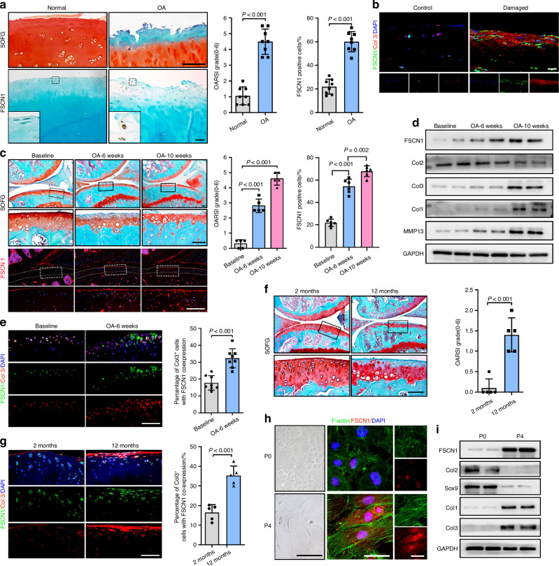
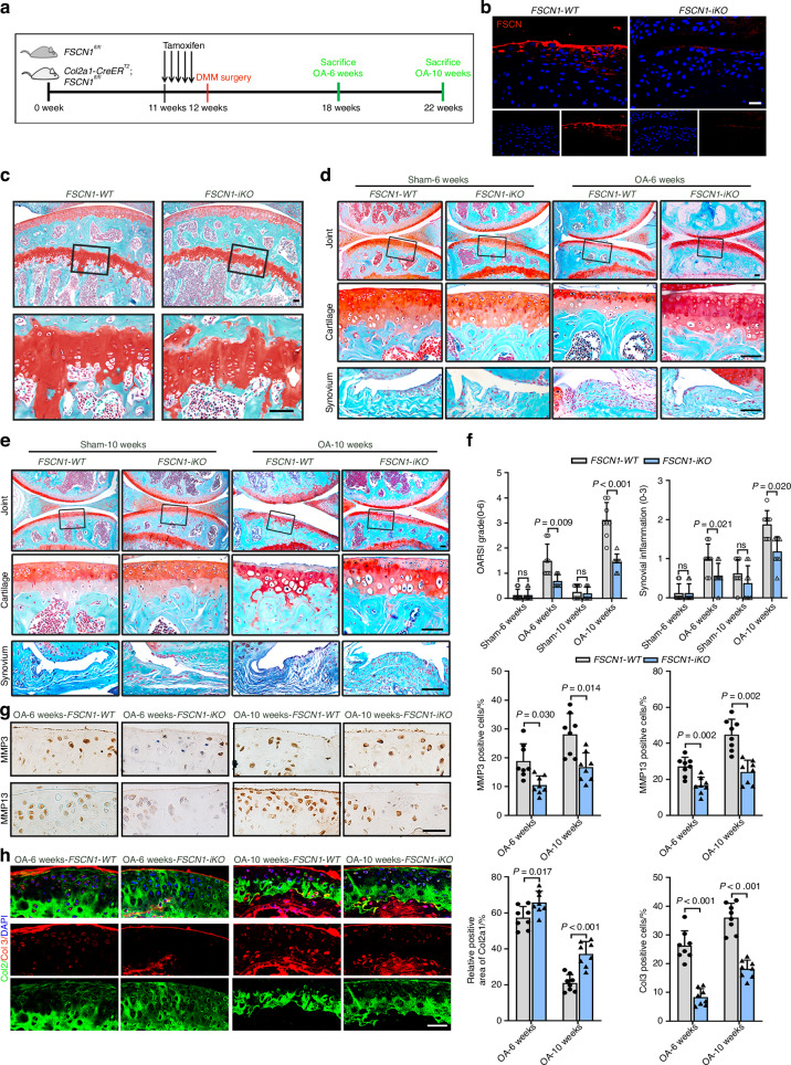
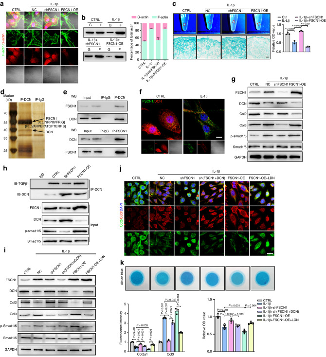
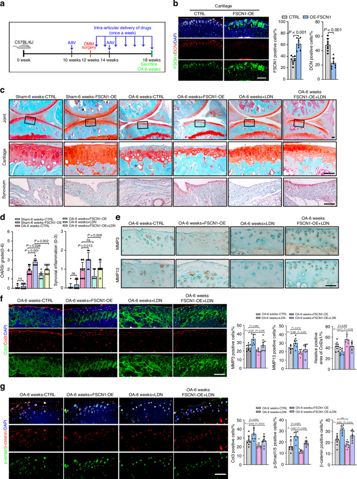
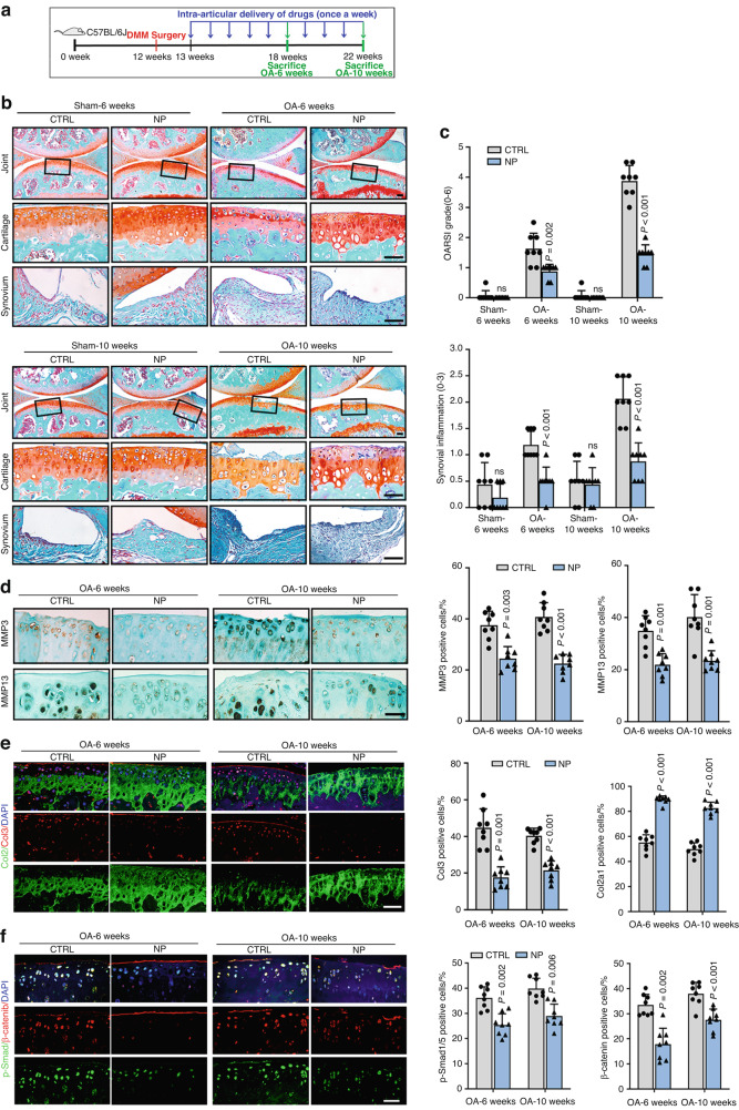
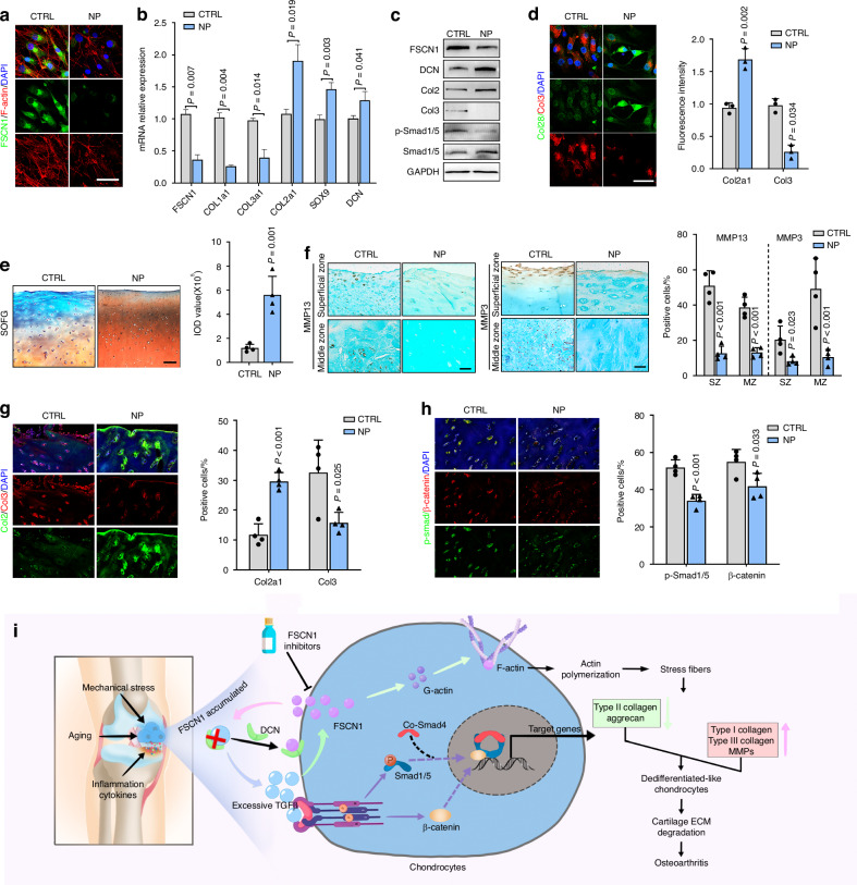
Osteoarthritis (OA) is the most common form of arthritic disease, and phenotypic modification of chondrocytes is an important mechanism that contributes to the loss of cartilage homeostasis. This study identified that Fascin actin-bundling protein 1 (FSCN1) plays a pivotal role in regulating chondrocytes phenotype and maintaining cartilage homeostasis. Proteome-wide screening revealed markedly upregulated FSCN1 protein expression in human OA cartilage. FSCN1 accumulation was confirmed in the superficial layer of OA cartilage from humans and mice, primarily in dedifferentiated-like chondrocytes, associated with enhanced actin stress fiber formation and upregulated type I and III collagens. FSCN1-inducible knockout mice exhibited delayed cartilage degeneration following experimental OA surgery. Mechanistically, FSCN1 promoted actin polymerization and disrupted the inhibition of Decorin on TGF-β1, leading to excessive TGF-β1 production and ALK1/Smad1/5 signaling activation, thus, accelerated chondrocyte dedifferentiation. Intra-articular injection of FSCN1-overexpressing adeno-associated virus exacerbated OA progression in mice, which was mitigated by an ALK1 inhibitor. Moreover, FSCN1 inhibitor NP-G2-044 effectively reduced extracellular matrix degradation in OA mice, cultured human OA chondrocytes, and cartilage explants by suppressing ALK1/Smad1/5 signaling. These findings suggest that targeting FSCN1 represents a promising therapeutic approach for OA.


Conflict of interest statement

The authors declare no competing interests.

Figures

Fig. 1
Fig. 1
FSCN1 expression is upregulated along with chondrocyte dedifferentiation and OA pathogenesis. a, b SOFG staining and IHC of FSCN1, Osteoarthritis Research Society International (OARSI) grades and quantification of FSCN1-positive cells (a), IF staining of FSCN1 (green) and Collagen type III (red) (b) in cartilage from intact (control) and damaged articular cartilage sections collected from OA patients (n = 8). c, d Safranin O/Fast Green (SOFG) staining, immunofluorescence (IF) staining of FSCN1, OARSI grades (c), western blot analysis of FSCN1, Collagen type II, MMP13, Collagen type I and III protein levels (d) in articular cartilage from mice with baseline or DMM-induced OA at 6- or 10-weeks post-surgery. The inset in each image is shown as a magnified image in the bottom row (n = 5). e IF staining and quantification of FSCN1 (green) and Collagen type III (red) in articular cartilage from mice with sham or DMM-induced OA at 6 weeks post-surgery (n = 5). SOFG staining and OARSI grades (f), IF staining of FSCN1 (green) and Collagen type III (red) (g) in cartilage from young (2-month-old) and aged (12-month-old) mice (n = 5). h-i. Optical microscopy, IF staining of FSCN1 (red) and phalloidin staining of F-actin structures (green) (h), western blot analysis of the protein level of FSCN1, Collagen type II, Sox9, Collagen type I and III protein levels (i) in mouse primary chondrocytes at passage 0 (P0) and passage 4 (P4) (n = 3). Scale bars, 50 μm. All data are presented as means ± SEM
Fig. 2
Fig. 2
Targeted deletion of FSCN1 in chondrocytes prevents OA development in mice. a Twelve-week-old FSCN1flox/flox (FSCN1-WT) and FSCN1-iKO mice underwent DMM or sham surgery 1 week after tamoxifen injection. The knees were harvested at 6 or 10 weeks postoperatively for histological analysis (n = 8). b IF staining of FSCN1 in articular cartilage from FSCN1-WT and FSCN1-iKO mice at 6-weeks post-surgery. c SOFG staining of joints from FSCN1-WT and FSCN1-iKO mice in sham group displayed with growth plate. d, e SOFG staining of joints from FSCN1-WT and FSCN1-iKO mice with sham or DMM-induced OA at 6- or 10-weeks post-surgery. The inset in each image is shown as a magnified image in the bottom row. f Cartilage destruction (OARSI grades) and synovial inflammation were determined by SOFG staining and scored (n = 8). g IHC staining of MMP3 and MMP13 in articular cartilage from FSCN1-WT and FSCN1-iKO mice at 6 or 10 weeks post-surgery. h IF staining of Collagen type II (green) and III (red) in articular cartilage from FSCN1-WT and FSCN1-iKO mice at 6- or 10-weeks post-surgery. Quantified results of each set of data are shown below (n = 8). Scale bars, 50 μm. All data are presented as means ± SEM
Fig. 3
Fig. 3
FSCN1 promotes chondrocyte shift towards dedifferentiation phenotype via inducing actin polymerization and activating ALK1/Smad1/5 signaling. a Representative images of phalloidin staining of F-actin structures (green) and IF staining for globular (G) actin (red) in chondrocytes with control, IL-1β (10 ng/mL), IL-1β plus FSCN1 knockdown or IL-1β plus FSCN1 overexpression treatment for 24 h. b G-/F-actin ratio quantified by western blotting analysis in all groups (n = 3). c Representative images, alcian blue staining and absorbance quantification of high-density pellet cultured chondrocytes with control, IL-1β (10 ng/mL), IL-1β plus FSCN1 knockdown or IL-1β plus FSCN1 overexpression treatment for 10 days. d Cell extracts were subjected to immunoprecipitation with an anti-DCN antibody. The immunoprecipitate–protein complex was separated with SDS–PAGE and the gel was then stained with silver stain. The identified peptide sequences of FSCN1 by LC–MS/MS analysis are shown. e FSCN1 or DCN was immunoprecipitated from chondrocytes with an anti-FSCN1 or anti-DCN antibody. The presence of FSCN1 and DCN in these immunoprecipitates was evaluated by immunoblotting. f Co-localization of FSCN1 (green) and DCN (red) in chondrocytes with control (saline) or IL-1β (10 ng/mL) treatment for 24 h was analyzed by confocal microscopy. g Western blot analysis of FSCN1, DCN, Collagen type II and III, p-Smad1/5 and Smad1/5 in chondrocytes treated with control, IL-1β (10 ng/mL), IL-1β plus FSCN1 knockdown and with IL-1β plus FSCN1 overexpression for 24 h (n = 3). h Chondrocytes with control, FSCN1 knockdown, or FSCN1 overexpression were subjected to immunoprecipitation with an anti-DCN antibody. The presence of TGF-β1 and DCN in immunoprecipitates was evaluated by immunoblotting. Western blot analysis of FSCN1, DCN, Collagen type II and III, p-Smad1/5 and Smad1/5 (i), IF staining and quantification of Collagen type II (green) and III (red) (j), alcian blue staining and absorbance quantification (k) in chondrocytes with control, IL-1β (10 ng/mL), IL-1β plus FSCN1 knockdown or IL-1β plus FSCN1 and DCN knockdown, IL-1β plus FSCN1 overexpression, IL-1β plus FSCN1 overexpression and LDN-193719 (5 μg/mL) treatment for 48 h (n = 3). Scale bars, 50 μm. All data are presented as means  ± SEM
Fig. 4
Fig. 4
Local injection of FSCN1 overexpressing adeno-associated virus exacerbates OA progression through activation of the ALK1/Smad1/5 signaling in vivo. a Twelve-week-old C57BL6/J mice underwent DMM or sham surgery, AAV-FSCN1 or AAV-control was injected into the knee joints at ten and fourteen week age, and saline (control), or LDN-193719 (20 mmol/L) was injected into the knee joints every week after surgery. The knees were harvested at 6 weeks postoperatively for histological analysis (n = 8). b IF staining and quantification of FSCN1 (red) and DCN (green) in articular cartilage from control and FSCN1-OE mice at 6 weeks post-surgery (n = 8). c SOFG staining of joints from mice with DMM-induced OA in control, FSCN1-OE, LDN-193719 and FSCN1-OE with LDN-193719 group at 6 weeks post-surgery. The insets in the images are shown as magnified images in the bottom row (n = 8). d Cartilage destruction (OARSI grades) and synovial inflammation were determined by SOFG staining and scored (n = 8). e IHC staining of MMP3 and MMP13 in articular cartilage from mice with DMM-induced OA in all groups at 6 weeks post-surgery. f,g IF staining of Collagen type II (green) and III (red) (f), p-Smad1/5 (green) and β-catenin (red) (g) in articular cartilage from mice with DMM-induced OA in all groups at 6 weeks post-surgery. Quantified results of each data set are shown below (n = 8). Scale bars, 50 μm. All data are presented as means ± SEM
Fig. 5
Fig. 5
FSCN1 inhibitor NP-G2-044 protects chondrocytes against dedifferentiation in vitro via suppressing ALK1/Smad1/5 signaling. a IF staining of FSCN1 (red) and phalloidin (green) staining for F-actin structures in chondrocytes treated with control, IL-1β (10 ng/mL), or IL-1β plus NP-G2-044 (10 μmol/L) for 24 h. Heat map (b) and volcano plot (c) showing differentially-expressed genes (DEGs) (fold change > 2 or <0.5) from RNA-sequencing of chondrocytes treated with IL-1β or IL-1β plus NP-G2-044; n = 3 per condition and seven independent experiments were analyzed. KEGG pathway (d) and GSEA (e) analysis for DRGs demonstrating TGF-β and Wnt signaling pathway enrichment in chondrocytes treated with IL-1β or IL-1β plus NP-G2-044. f Images of optical microscopy, phalloidin staining for F-actin structures (green) and IF staining for globular actin (red) in chondrocytes treated with control, IL-1β (10 ng/mL), or IL-1β plus NP-G2-044 (10 μmol/L) for 24 h. g G-/F-actin ratio quantified by western blotting analysis; n = 3 independent experiments. Alcian blue staining and absorbance quantification (h), IF staining and quantification of Collagen II (green) and III (red) (i), IF staining and quantification of p-Smad1/5 (green) and β-catenin (red) (j) in chondrocytes treated with control, IL-1β, or IL-1β plus NP for 24 h (n = 3). Relative mRNA expression of chondrogenic and dedifferentiation genes (k), Western blot analysis of FSCN1, DCN, Collagen type II and III, p-Smad1/5 and Smad1/5 (l) in chondrocytes treated with control, IL-1β, or IL-1β plus NP-G2-044 for 24 h (n = 3). Scale bars, 50 μm. All data are presented as means ± SEM
Fig. 6
Fig. 6
FSCN1 inhibitor NP-G2-044 ameliorates OA progression at early and middle stages in mice. a Twelve-week-old C57BL6/J mice underwent DMM or sham surgery, and saline (control), or NP-G2-044 (10 mmol/L) was injected into the knee joints every week after surgery. The knees were harvested at 6 or 10 weeks postoperatively for histological analysis (n = 8). b SOFG staining of joints from mice with DMM-induced OA and treated with control or with NP-G2-044 at 6 or 10 weeks post-surgery. The insets in the images are shown as magnified images in the bottom row. c cartilage destruction (OARSI grades) and synovial inflammation were determined by SOFG staining and scored (n = 8). IHC staining of MMP3 and MMP13 (d), IF staining of Collagen type II (green) and III (red) (e) and p-Smad1/5 (green) and β-catenin (red) (f) in articular cartilage from control mice and those treated with NP-G2-044 at 6 or 10 weeks post-surgery (n = 8). Scale bars, 50 μm. All data are presented as means ± SEM
Fig. 7
Fig. 7
FSCN1 serves as a potential therapeutic target for human OA. IF staining of FSCN1 (green) and phalloidin (red) staining for F-actin structures (a), relative mRNA expression of chondrogenic and dedifferentiation genes (b), western blot analysis of FSCN1, DCN, Collagen type II and III, p-Smad1/5 and Smad1/5 (c), IF staining and quantification of Collagen II (green) and III (red) (d) in human chondrocytes treated with control or NP-G2-044 (10 μmol/L) for 24 h (n = 3). e SOFG staining and absorbance quantification in explant-cultured cartilage tissue from OA donors treated with control or NP-G2-044 (10 μmol/L) for 7 days (n = 4). f-h. IHC staining and quantification of MMP13 and MMP3 (f), IF staining and quantification of Collagen type II (green) and III (red) (g), p-Smad1/5 (green) and β-catenin (red) (h) in explant-cultured cartilage tissue from OA doners treated with control or NP-G2-044 (10 μmol/L) for 7 days (n = 4). Scale bars, 100 μm. All data are presented as means ± SEM. i Schematic diagram showing the role and mechanism of FSCN1 in the regulation of chondrocyte dedifferentiation and the pathogenesis of OA

References

    1. Hunter, D. J. & Bierma-Zeinstra, S. Osteoarthritis. Lancet393, 1745–1759 (2019). 10.1016/S0140-6736(19)30417-9 - DOI - PubMed
    1. Loeser, R. F., Goldring, S. R., Scanzello, C. R. & Goldring, M. B. Osteoarthritis: a disease of the joint as an organ. Arthritis Rheum.64, 1697–1707 (2012). 10.1002/art.34453 - DOI - PMC - PubMed
    1. Guilak, F., Nims, R. J., Dicks, A., Wu, C. L. & Meulenbelt, I. Osteoarthritis as a disease of the cartilage pericellular matrix. Matrix Biol.: J. Int. Soc. Matrix Biol.71-72, 40–50 (2018). 10.1016/j.matbio.2018.05.008 - DOI - PMC - PubMed
    1. Jiang, Y. & Tuan, R. S. Origin and function of cartilage stem/progenitor cells in osteoarthritis. Nat. Rev. Rheumatol.11, 206–212 (2015). 10.1038/nrrheum.2014.200 - DOI - PMC - PubMed
    1. Aigner, T., Bertling, W., Stoss, H., Weseloh, G. & von der Mark, K. Independent expression of fibril-forming collagens I, II, and III in chondrocytes of human osteoarthritic cartilage. J. Clin. Investig.91, 829–837 (1993). 10.1172/JCI116303 - DOI - PMC - PubMed

MeSH terms