. 2024 Sep 4;15(9):654.
doi: 10.1038/s41419-024-07028-3.

B4GALT1-dependent galectin-8 binding with TGF-β receptor suppresses colorectal cancer progression and metastasis

Affiliations

B4GALT1-dependent galectin-8 binding with TGF-β receptor suppresses colorectal cancer progression and metastasis

Tzu-Hui Hsu et al. Cell Death Dis. .

Abstract

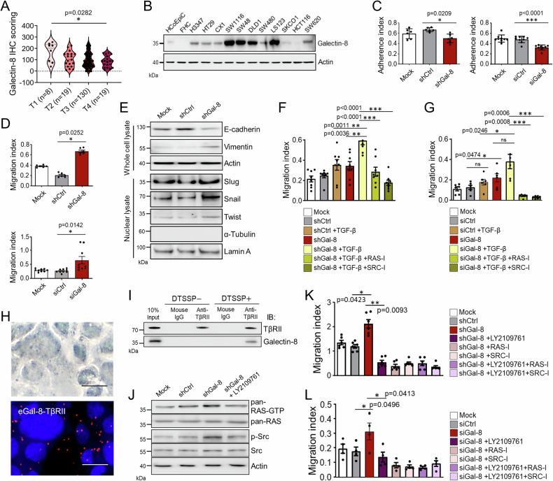
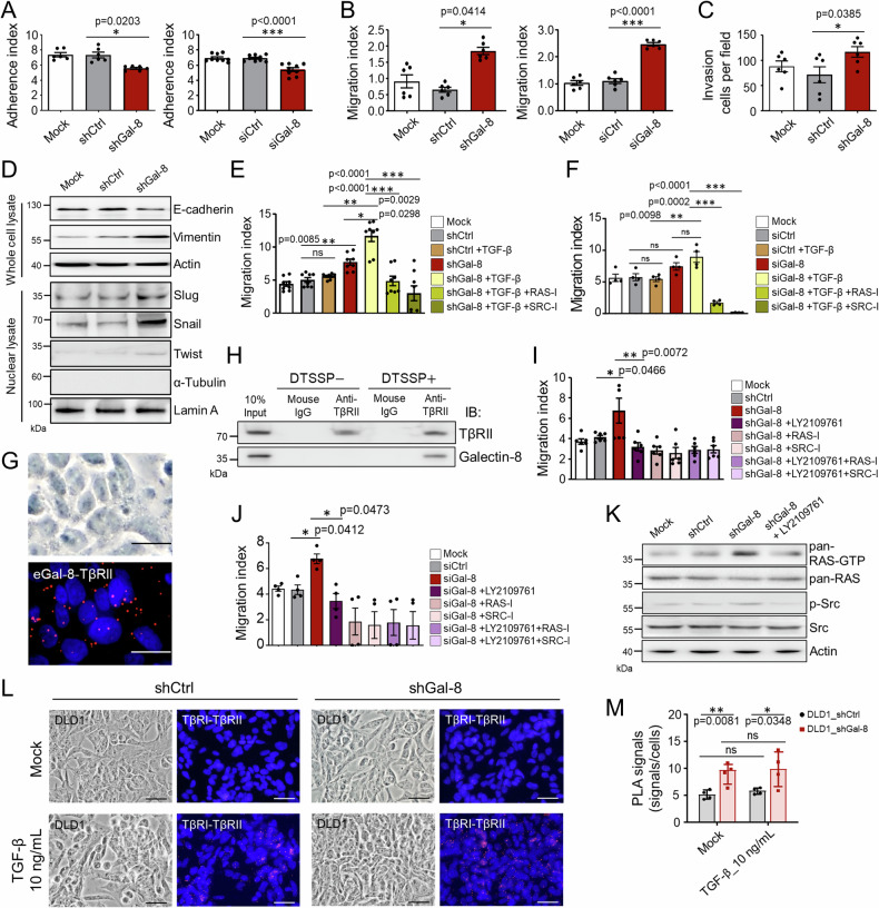
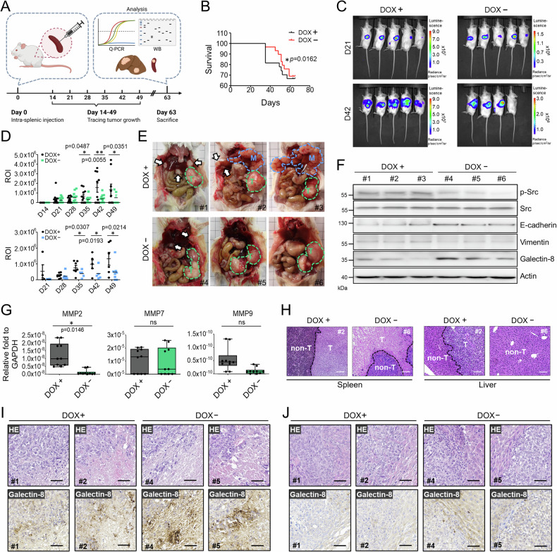
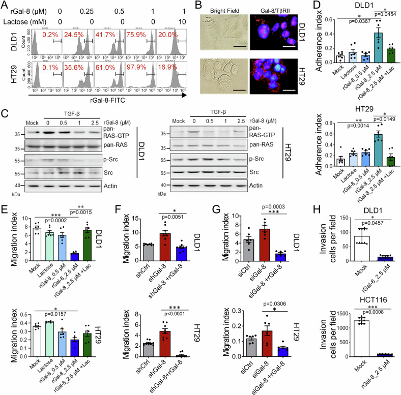
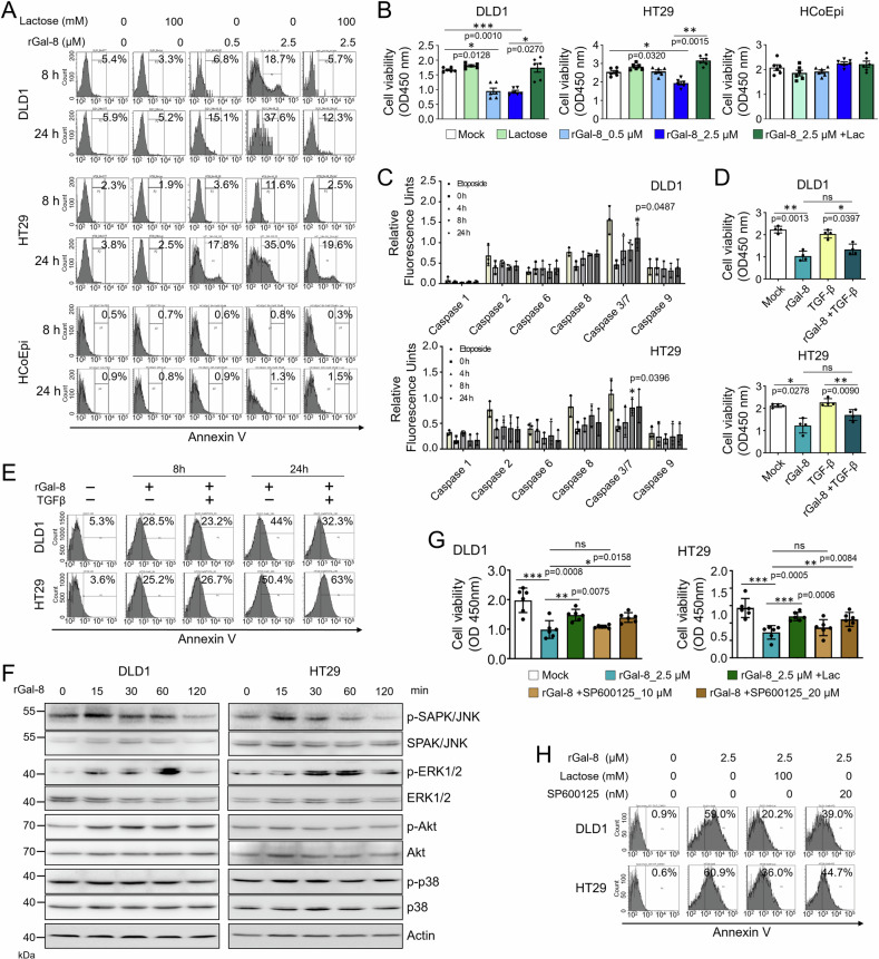
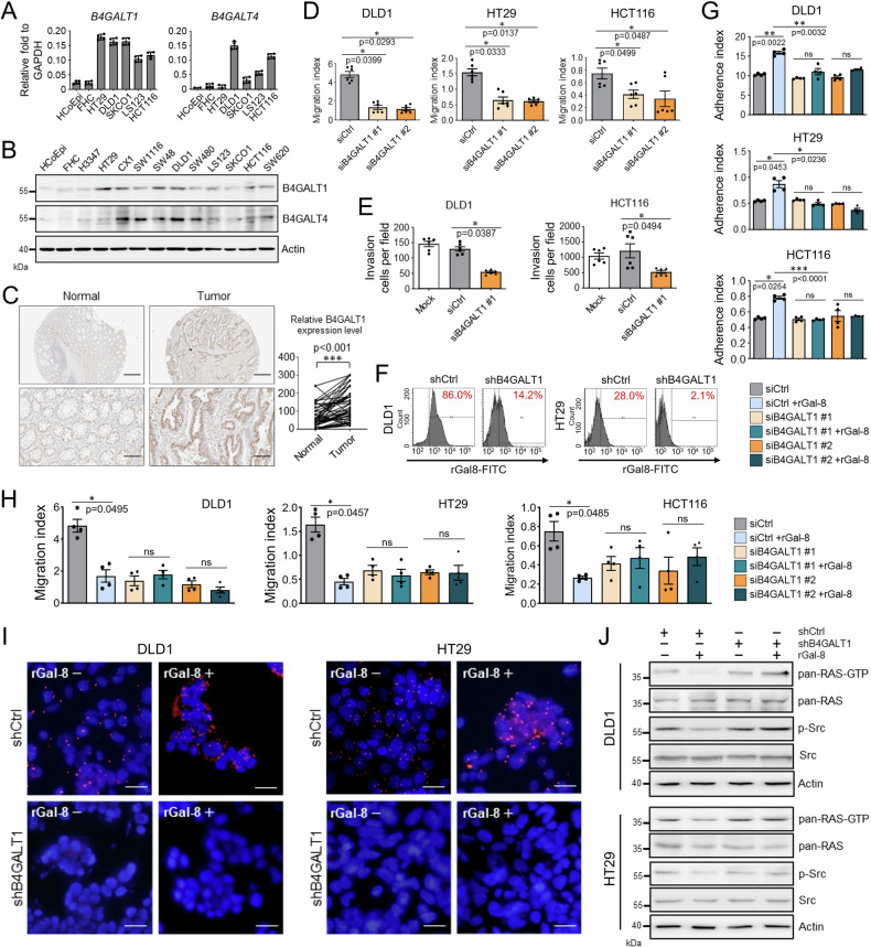
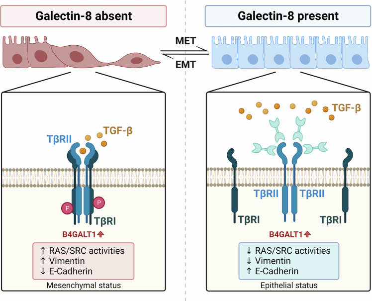
Transforming growth factor (TGF)-β signaling is critical for epithelial-mesenchymal transition (EMT) and colorectal cancer (CRC) metastasis. Disruption of Smad-depednent TGF-β signaling has been shown in CRC cells. However, TGF-β receptor remains expressed on CRC cells. Here, we investigated whether the cooperation between tumor-associated N-glycosylation and a glycan-binding protein modulated the TGF-β-driven signaling and metastasis of CRC. We showed that galectin-8, a galactose-binding lectin, hampered TGF-β-induced EMT by interacting with the type II TGF-β receptor and competing with TGF-β binding. Depletion of galectin-8 promoted the migration of CRC cells by increasing TGF-β-receptor-mediated RAS and Src signaling, which was attenuated after recombinant galectin-8 treatment. Treatment with recombinant galectin-8 also induces JNK-dependent apoptosis in CRC cells. The anti-migratory effect of galectin-8 depended on β4-galactosyltransferase-I (B4GALT1), an enzyme involved in N-glycan synthesis. Increased B4GALT1 expression was observed in clinical CRC samples. Depletion of B4GALT1 reduced the metastatic potential of CRC cells. Furthermore, inducible expression of galectin-8 attenuated tumor development and metastasis of CRC cells in an intra-splenic injection model. Our results thus demonstrate that galectin-8 alters non-canonical TGF-β response in CRC cells and suppresses CRC progression.


Conflict of interest statement

The authors declare no competing interests.

Figures

Fig. 1
Fig. 1. Knockdown of LGALS8 potentiates the migratory characteristics of TGF-β responsive HT29 cells by increasing RAS and Src activities.
A Scoring results from IHC staining of galectin-8 in tumor tissues from CRC patients classified by T-classification. B Western blotting shows the expression of galectin-8 protein in various CRC cells, FHC and normal human intestinal epithelial cells (HcoEpi). Cell adhesion activities measured 6 h after seeding (C) and migration activities measured 24 h after seeding (D) of HT29 cells expressing shCtrl or shGal-8 (top) or transfected with a mixture of three different siRNAs targeting galectin-8 (siGal-8) or a scrambled control siRNA (siCtrl) (bottom) were analyzed by xCELLigence RTCA. E Western blotting shows the expression of EMT markers in the whole cell lysate and nuclear lysate isolated from the mock-treated, shCtrl-expressing, and shGal-8-expressing HT29 cells. Actin and Lamin A serves as the whole cell lysate and nuclear lysate loading control, respectively. α-tubulin serves as the negative control of nuclear lysate preparation. Cell migration activities of shCtrl- and shGal-8-expressing HT29 cells (F) or siCtrl- and siGal-8-transfected HT29 cells (G) in the presence or absence of TGF-β (10 ng/mL), the pan-RAS inhibitor lonafarnib (RAS-I, 100 μM), and the Src inhibitor dasatinib (SRC-I, 30 μM), assessed by xCELLigence RTCA at 48 h after seeding. H Proximity ligation assay (PLA) to determine the interaction of endogenous galectin-8 with TβRII in HT29 cells. Scale bar = 10 μm. I HT29 cells were treated with or without DTSSP crosslinker before cell lysis. Co-immunoprecipitation (Co-IP) was used to examine the interaction of galectin-8 and TβRII by IP of lysates with anti-TβRII antibody or isotype control mouse IgG, followed by immunoblotting (IB) with the indicated antibodies. J Western blotting shows the expression of activated, phosphorylated, and total proteins of RAS or Src in mock-treated, shCtrl, shGal-8 alone or with LY2109761 treated HT29 cells. Actin serves as the loading control. Migration activities of HT29 cells expressing the indicated shRNA (K) or siCtrl- and siGal-8-transfected HT29 cells (L) and/or treated with TGF-β signaling antagonist LY2109761 (10 µM), RAS-I (100 μM), and SRC-I (30 μM) for 48 h. Results in C, D, F, G, K, L are shown as mean ± SD (3 biological replicates with technical duplicates in C (left), D (top), G and K; 3 biological replicates with technical tripilicates in C (right) and D (bottom); 3 biological replicates with technical quintuplicates in F; 2 biological replicates with technical duplicates in L). Statistical tests were calculated by one-way ANOVA for A and t-test for C, D, F, G, K, L. ns not significant.
Fig. 2
Fig. 2. Knockdown of LGALS8 promotes migratory characteristics and ameliorates the resposnes to TGF-β in DLD1 cells.
A, B Cell adhesion activities measured at 6 h after seeding (A) and migration activities measured at 48 h after seeding of DLD1 cells expressing shCtrl or shGal-8 (left) or transfected with siGal-8 or siCtrl (right) were analyzed by xCELLigence RTCA. C Cell invasion activities of DLD1 cells expressing shCtrl or shGal-8 were analyzed 24 h after seeding by xCELLigence RTCA. D Western blotting shows the expression of EMT markers in mock-treated, shCtrl-expressing, and shGal-8-expressing DLD1 cells. Actin and Lamin A serves as the whole cell lysate and nuclear lysate loading control, respectively. α-tubulin serves as the negative control of nuclear lysate preparation. Cell migration activities of shCtrl- and shGal-8-expressing DLD1 cells (E) or siCtrl- and siGal-8-transfected DLD1 cells (F) in the presence or absence of TGF-β (10 ng/mL), RAS-I, (100 μM), and SRC-I (30 μM) evaluated by xCELLigence RTCA at 48 h. G PLA determining the interaction of endogenous galectin-8 with TβRII in DLD1 cells. Scale bar = 10 μm. H DLD1 cells were treated with or without DTSSP crosslinker before cell lysis. Co-immunoprecipitation (Co-IP) showing the interaction of galectin-8 and TβRII by IP of lysates with anti-TβRII antibody and isotype control mouse IgG, followed by immunoblotting (IB) with the indicated antibodies. Migration activities of DLD1 cells expressing indicated shRNA (I) or transfected with siRNAs (J) and/or with LY2109761 (10 µM), RAS-I, (100 μM), and SRC-I (30 μM) for 48 h. K Western blotting shows the expression of activated, phosphorylated, and total proteins of RAS or Src in mock-treated, shCtrl, shGal-8 alone or with LY2109761 treatment in DLD1 cells. Actin serves as the loading control. PLA images (L) and statistical analysis of PLA signal quantification (M) show the effect of endogenous galectin-8 on the interaction of TβRI with TβRII. DAPI staining was used to label cell nuclei. Scale bar = 20 µm. Results in A, B, C, E, F, I, J, M are shown as mean ± SD (3, 3, 3, 4, 2, 3, and 2 biological replicates with technical duplicates in A (left), B, C, E, F, I, J, respectively; 3 biological replicates with technical triplicates in A (right); 4 biological replicates for M). Statistical tests were calculated by t-test (AC, E, F, I, J, M). ns not significant.
Fig. 3
Fig. 3. Induction of galectin-8 suppresses liver metastases from intra-splenically implanted CRC cells.
A Scheme of intra-splenic implantation of galectin-8 Tet-off DLD1-Luc cells and subsequent examination of metastasis. Implanted NOD-SCID mice were treated with or without doxycycline (DOX, 200 µM in drinking water), monitored for tumor burden, or sacrificed for various analyses at indicated days. B Kaplan–Meier survival curves of NOD-SCID mice implanted intra-splenically with galectin-8 Tet-off DLD1-Luc cells with or without DOX treatment (200 µM). n = 10 per group. Statistical significance was determined using the log-rank test. C Representative bioluminescence images of NOD-SCID mice treated with or without DOX at 21 days and 42 days after intra-splenic implantation of galectin-8 Tet-off DLD1-Luc cells. D The bioluminescence intensity of the regions of interest (ROI) detected from primary tumors (upper panel) and metastatic nodules (lower panel) at indicated days after implantation. E Gross images of galectin-8 Tet-off DLD1-Luc tumor-bearing mice treated with or without DOX show the primary tumors (green dotted circles, marked as T) and metastatic nodules (arrows and blue dotted circles, marked as M) at day 63 after implantation. F Western blot analysis of the expression of phosphorylated and total Src, E-cadherin, Vimentin, and galectin-8 in the primary tumor mass of spleen from galectin-8 Tet-off DLD1-Luc tumor-bearing mice with or without DOX treatment. Actin was used as the loading control. G RT-qPCR analysis of mRNA levels of MMP-2, MMP-7, and MMP9 in tumor tissues from the spleen in (E). H Hematoxylin-eosin (HE) staining showing the boundary between tumorous (T) and non-tumorous (non-T) tissues in the liver and spleen of galectin-8 Tet-off DLD1-Luc tumor-bearing mice. Scale bar = 100 μm. (I and J) HE staining and IHC staining of galectin-8 expression with sections derived from the primary lesions in spleen (I) and metastatic lesions in liver (J) in (E). Scale bar = 50 μm. Results in D and G are shown as mean ± SD (3 independent experiments in the upper panel of D; 3 independent experiments in the lower panel of D; 5 biological replicates with technical duplicate for G). Statistical tests in D and G were calculated by t-test. ns not significant.
Fig. 4
Fig. 4. rGal-8 suppresses migration and invasion of CRC cells by targeting TGF-β signaling.
A FACS shows binding of the indicated amounts of FITC-conjugated rGal-8 with DLD1 and HT29 cells. Lactose (10 mM) blocks the binding of rGal-8 with CRC cells. B PLA shows the interaction of galectin-8 (Gal-8) and TβRII on CRC cells. Scale bar = 10 μm. C Western blotting shows that rGal-8 affects TGF-β-mediated RAS and Src activities in CRC cells. Cell lysates from untreated DLD1 and HT29 cells (mock) or from cells treated with TGF-β (100 ng/mL) and various amounts of rGal-8 were subjected to immunoblotting with the indicated antibodies. Actin serves as the loading control. Cell adhesion (D) and migration (E) of DLD1 and HT29 cells treated with the indicated doses of rGal-8 and/or lactose (Lac, 100 mM), analyzed by xCELLigence RTCA. Bar graphs show the average cell indexes at 6 h (D) and 48 h (E). F Cell migration of DLD1 and HT29 cells expressing shCtrl or shGal-8 in the absence or presence of rGal-8 (2.5 µM) analyzed by xCELLigence RTCA at 48 h. G Cell migration of DLD1 and HT29 cells treated with siCtrl or siGal-8 in the absence or presence of rGal-8 (2.5 µM) analyzed by xCELLigence RTCA at 48 h. H Invasive activities of DLD1 and HCT116 cells treated with rGal-8 for 24 h analyzed by xCELLigence RTCA. Results in D, E, F, G, H are shown as mean ± SD (3, 3, 4 and 3 biological replicates with technical duplicates for each in D, E, F, G, respectively; 2 biological replicates with technical quintuplicates in H). Statistical tests were calculated by t-test.
Fig. 5
Fig. 5. rGal-8 triggers apoptosis in CRC cells by inducing transient JNK activation.
The level of cell apoptosis, determined by Annexin V staining (A), and cell viability, determined by MTT assay (B), of DLD1, HT29, and HCoEpi cells treated with various doses of rGal-8 in the presence or absence of lactose (Lac, 100 mM). C The activities of caspases in DLD1 and HT29 cells treated with rGal-8 (2.5 µM) or etoposide (10 µM, positive control of caspase activation) for the indicated time points were analyzed based on the cleavage of each substrate. D Cell viability of DLD1 and HT29 cells treated with rGal-8 (2.5 µM) or/and TGF-β (100 ng/mL) for 72 h determined by MTT assay. E FACS shows the percentage of Annexin V+ apoptotic DLD1 or HT29 cells treated with rGal-8 (2.5 µM) and/or TGF-β (100 ng/mL) for the indicated time points. F Western blotting shows the expression level of phosphorylated and total JNK, ERK1/2, AKT and p38 in DLD1 and HT29 cells treated with rGal-8 (2.5 µM) for the indicated time points. Cell viability determined by MTT assay (G), and apoptosis determined by Annexin V staining (H) of DLD1 and HT29 cells treated with rGal-8 (2.5 µM) alone or together with JNK inhibitor (SP600125) at the indicated doses for 72 h (G) and 24 h (H). Lactose (Lac, 100 mM) was used to block the effect of rGal-8 in some groups. Results in B, C, D, G are shown as mean ± SD (3 biological replicates with technical duplicates for each in B, C, G; two biological replicates with technical duplicates for D). Statistical tests were calculated by t-test.
Fig. 6
Fig. 6. B4GALT1 supports the migratory activity of CRC cells and is required for the anti-metastatic effect of galectin-8.
A RT-qPCR shows the relative B4GALT1 and B4GALT4 mRNA levels normalized by GAPDH mRNA levels in non-tumorous cells (HCoEpi and FHC) and various CRC lines. B Western blotting shows protein expression of B4GALT1 and B4GALT4 in non-tumorous cells (HCoEpi and FHC) and in different CRC lines. Actin was used as a protein loading control. C Immunohistochemical staining of B4GALT1 in paired non-tumor and tumor tissues from CRC patients (left). The right panel shows the distribution of immunoactivity scores of B4GALT1 expression in normal and primary CRC tissues (n = 57). Scores were determined as the product of staining intensity and percentage of positive cells. Scale bar = 200 μm (top); 50 μm (bottom). Cell migration (D) and invasive activities (E) of the indicated CRC cells treated with siCtrl or siB4GALT1 (#1 and #2 in D, #1 in E) were analyzed at endpoint (D, 48 h; E, 24 h) using xCELLigence RTCA. F FACS shows the percentage of cells binding with FITC-conjugated rGal-8 among DLD1 and HT29 cells expressing shCtrl or shB4GALT1. Cell adhesion (G) and migration (H) activities of DLD1, HT29, and (Fig. 5D, E) cells treated with siCtrl or siB4GALT1 (#1 or #2) (25 pmol/106 cells) in the presence or absence of rGal-8 (2.5 µM). Results were analyzed by xCELLigence RTCA at 6 h (E) and 48 h (F) after treatment. I PLA shows the effects of knockdown of B4GALT1 on the interaction between TβRII and galectin-8 in DLD1 and HT29 cells with (rGal-8+) or without (rGal-8−) rGal-8 treatment. DAPI staining was used to label cell nuclei. Scale bar = 10 µm. J Western blotting shows the expression of activated, phosphorylated, and total proteins of RAS or Src in DLD1 and HT29 cells treated as indicated. rGal-8 at 2.5 µM was used. Actin was used as the loading control. Results in A, D, E, G, H are shown as mean ± SD (2 biological replicates with technical duplicates for each in A, G, H; 3 biological replicates with technical duplicates for D, E). Statistical tests were calculated by t-test. ns not significant.
Fig. 7
Fig. 7. Proposed model of the action of galectin-8 in CRC.
CRC cells showed increased levels of B4GALT1. In the absence of galectin-8 (left), TGF-β binds to TβRII, thereby promoting EMT. In the presence of galectin-8 (right), galectin-8 binds to TβRII through B4GALT1-mediated galactosylation of N-glycans, resulting in a decrease of TGF-β signaling-mediated EMT.

References

    1. Bray F, Ferlay J, Soerjomataram I, Siegel RL, Torre LA, Jemal A. Global cancer statistics 2018: GLOBOCAN estimates of incidence and mortality worldwide for 36 cancers in 185 countries. CA Cancer J Clin. 2018;68:394–424. 10.3322/caac.21492 - DOI - PubMed
    1. Kinzler KW, Vogelstein B. Lessons from hereditary colorectal cancer. Cell. 1996;87:159–70. 10.1016/S0092-8674(00)81333-1 - DOI - PubMed
    1. Konishi M, Kikuchi-Yanoshita R, Tanaka K, Muraoka M, Onda A, Okumura Y, et al. Molecular nature of colon tumors in hereditary nonpolyposis colon cancer, familial polyposis, and sporadic colon cancer. Gastroenterology. 1996;111:307–17. 10.1053/gast.1996.v111.pm8690195 - DOI - PubMed
    1. Massagué J. TGFbeta in cancer. Cell. 2008;134:215–30. 10.1016/j.cell.2008.07.001 - DOI - PMC - PubMed
    1. Massague J, Blain SW, Lo RS. TGFbeta signaling in growth control, cancer, and heritable disorders. Cell. 2000;103:295–309. 10.1016/S0092-8674(00)00121-5 - DOI - PubMed

MeSH terms

Substances