. 2024 Sep 4;15(9):649.
doi: 10.1038/s41419-024-06796-2.

L3MBTL1, a polycomb protein, promotes Osimertinib acquired resistance through epigenetic regulation of DNA damage response in lung adenocarcinoma

Affiliations

L3MBTL1, a polycomb protein, promotes Osimertinib acquired resistance through epigenetic regulation of DNA damage response in lung adenocarcinoma

Zihe Zhang et al. Cell Death Dis. .

Abstract

Osimertinib is a third-generation epidermal growth factor receptor (EGFR) tyrosine kinase inhibitor (EGFR-TKI) approved for patients with EGFR T790M resistance mutations as first- or second-line treatment of EGFR-positive patients. Resistance to Osimertinib will inevitably develop, and the underlying mechanisms are largely unknown. In this study, we discovered that acquired resistance to Osimertinib is associated with abnormal DNA damage response (DDR) in lung adenocarcinoma cells. We discovered that the polycomb protein Lethal(3) Malignant Brain Tumor-Like Protein 1 (L3MBTL1) regulates chromatin structure, thereby contributing to DDR and Osimertinib resistance. EGFR oncogene inhibition reduced L3MBTL1 ubiquitination while stabilizing its expression in Osimertinib-resistant cells. L3MBTL1 reduction and treatment with Osimertinib significantly inhibited DDR and proliferation of Osimertinib-resistant lung cancer cells in vitro and in vivo. L3MBTL1 binds throughout the genome and plays an important role in EGFR-TKI resistance. It also competes with 53BP1 for H4K20Me2 and inhibits the development of drug resistance in Osimertinib-resistant lung cancer cells in vitro and in vivo. Our findings suggest that L3MBTL1 inhibition is a novel approach to overcoming EGFR-TKI-acquired resistance.


Conflict of interest statement

The authors declare no competing interests.

Figures
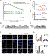
Fig. 1
Fig. 1. Comparison of DNA Damage Response Variations in Relapse Resistant and Sensitive Tissues and Cells.
A DNA damage response pathway differential gene enrichment is observed in 85 lung adenocarcinoma patients screened with dataset GSE30219 using KEGG analysis. B, C CCK-8 detection of H1975 and H1975/AR, PC9, and PC9/OR cells viability after treatment with various doses of Osimertinib for 48 h. The experiments included three replicates, and the results are presented as mean ± SEM. D Immunoblotting was used to detect the effect of Osimertinib treatment in H1975 and H1975/AR, PC9, and PC9/OR on the expression of γ-H2AX over time. E Immunoblotting was used to detect the effect of Etoposide treatment in H1975 and H1975/AR, PC9, and PC9/OR on the expression of γ-H2AX over time. F, G Immunofluorescence staining of H1975 and H1975/AR cells with γ-H2AX antibody after treatment with Osimertinib over time. Count and analyze cells with γ-H2AX focis>5. Scale bar: 10 µm. The experiments included three replicates, and the results are presented as mean ± SEM. H H1975 and H1975/AR were treated with Osimertinib, and the level of ROS was measured using DCFH-DA staining. (mean ± SD, n = 3, *P < 0.05, **P < 0.01, ***P < 0.001).
Fig. 2
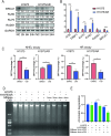
Fig. 2. A comparison of the differences between acquired resistant and sensitive cells’ chromatin structures and DNA repair pathways.
A Immunoblotting was used to determine the effect of Osimertinib treatment in H1975 and H1975/AR on the expression of key NHEJ and HR proteins over time. B qRT-PCR measured mRNA levels of ATM, 53BP1, KU70, KU80, RAD51 and BRCA1 between H1975 and H1975/AR (mean ± SD, n = 3, *P < 0.05, **P < 0.01, ***P < 0.001). C NHEJ efficiency and HR efficiency were determined by quantifying GFP expression (by flow cytometry) in H1975 and H1975/AR cells harboring pDR-GFP reporter or pimEJ5GFP after transfection with a pCBASceI plasmid with or without treated Osimertinib for 48 h. (mean ± SD, n = 3, *P < 0.05, **P < 0.01, ***P < 0.001). D, E Micrococcal Nuclease digestion assay determined the extent of MNase sensitivity in H1975 and H1975/AR cells with Osimertinib treated.
Fig. 3
Fig. 3. Osimertinib-resistant cells showed more stable L3MBTL1 expression at the protein level.
A Immunoblotting was used to measure the protein level of L3MBTL1 in H1975 and H1975/AR, as well as PC9 and PC9/OR. B, C H1975 and H1975/AR were treated with Osimertinib and cycloheximide (CHX) alone or in combination over time. L3MBTL1 protein expression was detected by immunoblotting and relative density was assessed using Image J (mean ± SD, n = 3, *P < 0.05, **P < 0.01, ***P < 0.001). D H1975 and H1975/AR cells were treated with Osimertinib and MG132 separately or together over time. L3MBTL1 protein expression was detected by immunoblotting, and relative density was measured using Image J (mean ± SD, n = 3, *P < 0.05, **P < 0.01, ***P < 0.001). E, F H1975/AR has a lower ubiquitin level than the corresponding H1975, MG132 combination for rescue. After 48 h of treatment with 2 μM Osimertinib, cell lysates were immunoprecipitated with rabbit anti-L3MBTL1 antibody, followed by anti-ubiquitin antibody for immunoblotting, and relative density was measured using Image J (mean ± SD, n = 3, *P < 0.05, **P < 0.01, ***P < 0.001). G Immunoblotting was used to assess the effect of Osimertinib treatment in H1975 and H1975/AR on the expression of RNF8. H The co-immunoprecipitate assay was used to determine the binding of EGFR to RNF8 in H1975 and H1975/AR.
Fig. 4
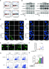
Fig. 4. L3MBTL1 compacts the chromatin structure, reducing cellular DNA damage.
A The enrichment of signaling pathways among differentially expressed genes was investigated using gene set enrichment analysis (GSEA). DEGs were discovered to be enriched in DDR-related pathways such as apoptosis, homologous recombination, mismatch repair, and nucleotide excision repair. B H1975 and H1975/AR were transfected into OE-L3MBTL1 and sh-L3MBTL1, respectively, and treated with or without Osimertinib. 53BP1, L3MBTL1, γ-H2AX, and H4K20Me2 protein levels were evaluated by immunoblotting. C H1975/AR sh-NC and H1975/AR sh-L3MBTL1 were treated with Osimertinib or DMSO, γ-H2AX and 53BP1 focis can be observed by immunofluorescence staining. Scale bar: 2 µm. D H1975 OE-NC and H1975 OE-L3MBTL1 were treated with Osimertinib or DMSO, γ-H2AX and 53BP1 focis can be observed by immunofluorescence staining. Scale bar: 2 µm. E, F H1975/AR si-NC and H1975/AR si-L3MBTL1-3 were treated with DMSO or Osimertinib, DNA damage was measured using the Comet assay, and the tail olive moment was analyzed using Cometscore (mean ± SD, n = 3, *P < 0.05, **P < 0.01, ***P < 0.001). G, H After H1975/AR was transfected into si-NC, si-L3MBTL1-1 and si-L3MBTL1-3 for 48 h, DMSO or Osimertinib were treated, respectively, and the apoptosis level was detected by flow cytometry (mean ± SD, n = 3, *P < 0.05, **P < 0.01, ***P < 0.001).
Fig. 5
Fig. 5. L3MBTL1 plays an important role in EGFR-TKI resistance.
A Distribution of L3MBTL1 Peaks in Chromatin. B, C The distribution of peaks in the functional regions of the genome accounted for 4.8% of the peaks in Promoter-TSS. D Distribution and heatmap of Peaks above and below TSS. E A GO analysis of genes related to L3MBTL1 promoter peaks revealed enrichment in cellular processes such as cell cycle, cell metabolism, and cellular stress response. F A KEGG analysis revealed that L3MBTL1 promoter peaks-related genes were highly enriched in EGFR and its downstream pathways, as well as some EGFR-TKI-related bypass pathways.
Fig. 6
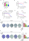
Fig. 6. L3MBTL1 inhibition enhances the antitumor effect of EGFR-TKIs in vitro.
After cells transfected with si-NC, si-L3MBTL1-1, or si-L3MBTL1-3 48 h as indicated, with treated for DMSO or Osimertinib, IC50 were determined using the CCK-8 assay (A) in H1975/AR. (mean ± SD, n = 3, *P < 0.05, **P < 0.01, ***P < 0.001). B After transfecting H1975/AR into mock lentiviral or sh-L3MBTL1, CCK-8 was used to detect cell viability after various doses of Osimertinib were treated for 48 h. The experiments included three replicates, and the results are presented as mean ± SEM. C Cell viability of H1975/AR was assessed using the CCK-8 assay when treated with Osimertinib and UNC669 alone and in combination. The experiments included three replicates, and the results are presented as mean ± SEM. D After transfecting H1975 into mock lentiviral or OE-L3MBTL1, CCK-8 was used to detect cell viability after various doses of Osimertinib were treated for 48 h. The experiments included three replicates, and the results are presented as mean ± SEM. EH Cell proliferation was assessed using clonogenic assays in H1975/AR and PC9/OR cells after transfection with empty vector or L3MBTL1 knockdown for the indicated number of hours and treated with DMSO or Osimertinib, Etoposide for positive control (mean ± SD, n = 3, *P < 0.05, **P < 0.01, ***P < 0.001).
Fig. 7
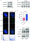
Fig. 7. L3MBTL1 primarily competes with 53BP1 for binding to H4K20Me2 in H1975/AR cells.
Immunoblotting revealed protein levels of H4K20Me1 and H4K20Me2 in nuclear proteins of H1975 and H1975/AR (A), as well as changes over time in H1975/AR (B). C, D After treating H1975/AR sh-NC and H1975/AR sh-L3MBTL1 with Osimertinib, immunofluorescence staining revealed γ-H2AX and 53BP1 foci. Scale bar: 2 µm. Pearson’s coefficient was assessed by FluoView10-ASW 4.2. (mean ± SD, n = 3, *P < 0.05, **P < 0.01, ***P < 0.001). E H1975 and H1975/AR were transfected into si-NC or si-53BP1, 53BP1, L3MBTL1, and H4K20Me2, and nuclear protein levels were determined using immunoblotting.
Fig. 8
Fig. 8. L3MBTL1 inhibition enhances the antitumor effect and DNA damage of EGFR-TKIs in vivo.
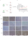
A Schematic diagram depicting tumor formation and drug treatment time in nude mice. The design was created by Figdraw. B Tumor volume was measured following 2 weeks of Osimertinib treatment. Results are presented as mean ± standard deviation. C Immunohistochemistry was used to identify L3MBTL1, Ki67, 53BP1, and γ-H2AX in tumor sections of nude mice.
Fig. 9
Fig. 9. A working model for the role of L3MBTL1 acting on DNA damage response in EGFR-TKI acquired-resistant cells.
In Osimertinib-resistant NSCLC, the inhibition of Osimertinib leads to a decrease in the level of L3MBTL1 ubiquitination, thereby making the chromatin more condensed, resulting in a decrease in ROS and DNA damage levels, and an increase in DNA damage repair, which increases the tumor's drug resistance. The working model is created with BioRender.com.

Similar articles

References

    1. Siegel RL, Miller KD, Fuchs HE, Jemal A. Cancer statistics, 2022. CA Cancer J Clin. 2022;72:7–33. 10.3322/caac.21708 - DOI - PubMed
    1. Sun JM, Lee SH, Ahn JS, Park K, Ahn MJ. Osimertinib for the treatment of non-small cell lung cancer. Expert Opin Pharmacother. 2017;18:225–31. 10.1080/14656566.2017.1285283 - DOI - PubMed
    1. Roper N, Brown AL, Wei JS, Pack S, Trindade C, Kim C, et al. Clonal evolution and heterogeneity of osimertinib acquired resistance mechanisms in EGFR mutant lung cancer. Cell Rep Med. 2020;1:100007. 10.1016/j.xcrm.2020.100007 - DOI - PMC - PubMed
    1. Polo SE, Almouzni G. Chromatin assembly: a basic recipe with various flavours. Curr Opin Genet Dev. 2006;16:104–11. 10.1016/j.gde.2006.02.011 - DOI - PubMed
    1. Lukas J, Lukas C, Bartek J. More than just a focus: the chromatin response to DNA damage and its role in genome integrity maintenance. Nat Cell Biol. 2011;13:1161–9. 10.1038/ncb2344 - DOI - PubMed

MeSH terms