. 2024 Sep 4;15(1):7711.
doi: 10.1038/s41467-024-52193-w.

EXTL3 and NPC1 are mammalian host factors for Autographa californica multiple nucleopolyhedrovirus infection

Affiliations

EXTL3 and NPC1 are mammalian host factors for Autographa californica multiple nucleopolyhedrovirus infection

Yuege Huang et al. Nat Commun. .

Abstract

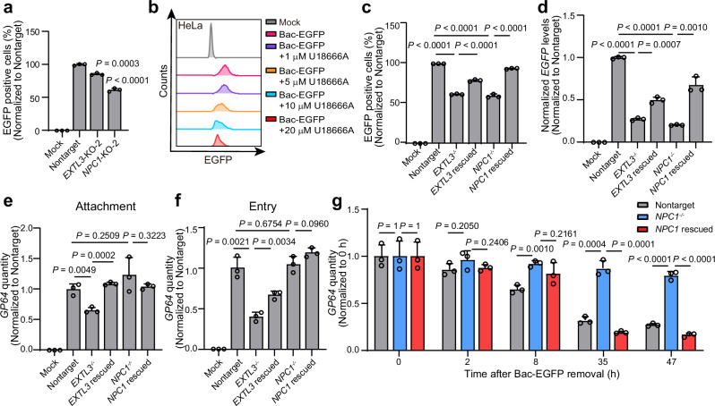
Baculovirus is an obligate parasitic virus of the phylum Arthropoda. Baculovirus including Autographa californica multiple nucleopolyhedrovirus (AcMNPV) has been widely used in the laboratory and industrial preparation of proteins or protein complexes. Due to its large packaging capacity and non-replicative and non-integrative natures in mammals, baculovirus has been proposed as a gene therapy vector for transgene delivery. However, the mechanism of baculovirus transduction in mammalian cells has not been fully illustrated. Here, we employed a cell surface protein-focused CRISPR screen to identify host dependency factors for baculovirus transduction in mammalian cells. The screening experiment uncovered a series of baculovirus host factors in human cells, including exostosin-like glycosyltransferase 3 (EXTL3) and NPC intracellular cholesterol transporter 1 (NPC1). Further investigation illustrated that EXTL3 affected baculovirus attachment and entry by participating in heparan sulfate biosynthesis. In addition, NPC1 promoted baculovirus transduction by mediating membrane fusion and endosomal escape. Moreover, in vivo, baculovirus transduction in Npc1-/+ mice showed that disruption of Npc1 gene significantly reduced baculovirus transduction in mouse liver. In summary, our study revealed the functions of EXTL3 and NPC1 in baculovirus attachment, entry, and endosomal escape in mammalian cells, which is useful for understanding baculovirus transduction in human cells.


Conflict of interest statement

J. Liu is the founder and shareholder of Shanghai AsiFlyer Biotechnology. The remaining authors declare no competing interests.

Figures

Fig. 1
Fig. 1. Identification of NPC1 and EXTL3 as baculovirus host factors in HEK-293T.
a Flowchart showing the negative selection approach for baculovirus host factor identification using surfaceome CRISPR screen. Candidate gene hits identified from the first (b) and second (c) rounds of selection. d The effects of knockout of the seven candidate genes on Bac-EGFP transduction, as determined by flow cytometry analyses. The p-value between Nontarget and EXTL3-KO-1, EXTL3-KO-2, NPC1-KO-1, NPC1-KO-2, MBTPS2-KO-1 and MBTPS2-KO-2 groups are 1e-5, 1e-5, 6e-6, 1e-5, 4e-5 and 4e-5, respectively. e Fluorescence imaging of Bac-EGFP infected cells. The experiment is repeated three times independently with similar results. f RT-qPCR quantification of EGFP mRNA in the lysate of EXTL3 and NPC1 knockout cells at 48 h post-Bac-EGFP transduction. β-actin is used as an internal control. g Evaluation of the effects of EXTL3 and NPC1 knockdown on Bac-EGFP transduction. The p-value between SiRNA-nontarget and SiRNA-NPC1-2 groups is 1e-5. h Evaluation of the effects of NPC1 inhibitor U18666A on Bac-EGFP transduction. For (d, f, and g), Data are presented as mean ± SD (n = 3) from three biological replicates. The significant difference is analyzed by two-tailed unpaired Student’s t-test. Mock group is the PBS treatment without baculovirus. Source data are provided as a Source Data file.
Fig. 2
Fig. 2. Determination of the functions of EXTL3 and NPC1 in HeLa cells.
a Evaluation of the effects of EXTL3 and NPC1 knockouts on Bac-EGFP transduction, as determined by flow cytometry analyses. The p-value between Nontarget and NPC1-KO-2 groups is 2e-5. b Evaluation of the effects of NPC1 inhibitor U18666A on Bac-EGFP transduction. Evaluation of the effects of EXTL3 and NPC1 knockouts and overexpression rescue on Bac-EGFP transduction, as determined by flow cytometry (EGFP protein) (c) and RT-qPCR (EGFP mRNA) (d). β-actin is used as an internal control for RT-qPCR. For (c), the p-values between Nontarget and EXTL3−/− and NPC1−/− groups are 5e-8 and 5e-6, respectively. The p-value between EXTL3−/− and EXTL3 rescued groups is 2e-5. The p-value between NPC1−/− and NPC1 rescued groups is 1e-5. For (d), the p-value between Nontarget and EXTL3−/ and NPC1−/− groups are 4e-7 and 7e-8, respectively. Evaluation of the effects of EXTL3 or NPC1 knockout and overexpression rescue on Bac-EGFP attachment (e) and entry (f). g qPCR quantification of the intracellular baculovirus genome GP64 gene after Bac-EGFP transduction in non-targeting sgRNA, NPC1−/− and NPC1 rescued HeLa cells. Bac-EGFP is incubated with cells at an MOI of 2 for 1 h and then removed from the medium. The total GP64 in each well is quantified. The p-values between NPC1−/− and Nontarget and NPC1 rescued group at 47 h are 5e-5 and 3e-5, respectively. For (a), (cg), Data are presented as mean ± SD (n = 3) from three biological replicates. The significant difference is analyzed by two-tailed unpaired Student’s t-test. Mock group is the PBS treatment without baculovirus. Source data are provided as a Source Data file.
Fig. 3
Fig. 3. Investigation of the functions of EXTL3-mediated heparin biosynthesis in baculovirus transduction in HEK-293T cells.
a Structural organization of EXTL3 protein. b Evaluation of baculovirus attachment and entry in EXTL3−/− cells. GP64 gene was quantified by qPCR. The p-value between Nontarget and EXTL3−/− in entry groups is 4e-5. c Western blotting detection of heparan sulfate proteoglycan 2 (HSPG2) in wild-type and EXTL3−/− cells. The experiment is repeated three times independently with similar results. d Analysis of the effects of heparin sodium on Bac-EGFP transduction, as determined by flow cytometry quantification of EGFP-positive cells. e Analysis of the effects of heparin sodium on Bac-EGFP attachment and entry, as determined by qPCR quantification of attached or internalized baculovirus GP64 gene. f Evaluation of the effects of HPSE on baculovirus transduction. The p-value between Nontarget and Nontarget+HPSE groups is 2e-5. g Schematic presentation of HSPG biosynthesis. h Evaluation of the effects of EXT2 knockout on baculovirus transduction. The p-value between Nontarget and EXT2-KO groups is 7e-6. i Investigation of the relationship between EXTL3 and NPC1 functions in baculovirus transduction. The p-value between No heparin sodium and With heparin sodium in NPC1−/− groups is 1e-6. The p-value between No heparin sodium and With heparin sodium in NPC1 rescued groups is 2e-6. For (b, e, f, h, and i), Data are presented as mean ± SD (n = 3) from three biological replicates. The significant difference is analyzed by two-tailed unpaired Student’s t-test. Mock group is the PBS treatment without baculovirus. Source data are provided as a Source Data file.
Fig. 4
Fig. 4. Dissection of the function of NPC1 in baculovirus endosomal escape.
DiOC18-staining experiment for illustration of NPC1 function in baculovirus membrane fusion, as analyzed by confocal microscopy (a) or flow cytometry analyses (b). The p-value between DiOC18 only and Nontarget groups is 1e-5. The p-value between NPC1−/− and NPC1 rescued groups is 4e-6. c The structural organization of NPC1. d Design of NPC1 constructs. Analyses of the interactions between HA-tagged GP64 and Myc-tagged full-length NPC1, NPC1-A, NPC1-C, NPC1-I (e), NPC1-ΔA, NPC1-ΔC or NPC1-ΔI (f). g Investigation of the effects of overexpression rescue with full-length or truncation constructs of NPC1 on baculovirus transduction, as determined by the EGFP-positive cells via flow cytometry. Mock, PBS treatment without baculovirus. The p-values between NPC1−/− and Nontarget and NPC1 rescued groups are 8e-8 and 1e-7, respectively. h Neutralization of Bac-EGFP transduction in HEK-293T cells by recombinant protein of NPC1 C domain, as determined by flow cytometry analyses. i ESI-MS confirmation of purified GST-I protein (n = 1). j Far-western analysis of the interaction between GP64 and GST-I. For (b, g, and h), Data are presented as mean ± SD (n = 3) from three biological replicates. The significant difference is analyzed by two-tailed unpaired Student’s t-test. For (e, f, and j), The experiment is repeated three times independently with similar results. Source data are provided as a Source Data file.
Fig. 5
Fig. 5. Baculovirus transduction in mouse liver is dependent on NPC1.
a Investigation of the tissue tropism of Bac-EGFP in wild-type mice at 72 h post intravenous injection, as determined by EGFP expression at protein (n = 3). The fluorescence images are acquired using confocal microscopy on flash-frozen tissues that are fixed with optimal cutting temperature compound (OCT). b RT-qPCR quantification of NPC1 mRNA in the lysate of liver, kidney, lung, brain, and spleen in wild-type and Npc1−/+ mice. The p-value between WT and Npc1−/+ in spleen groups is 1e-8. RT-qPCR quantification of EGFP mRNA in the lysate of liver (c) and other tissues including kidney, lung, brain, and spleen (d) in wild-type and heterozygous Npc1−/+ knockout mice at 72 h post-Bac-EGFP injection. The EGFP mRNA in the lysates of kidney, lung, brain, and spleen (d) in heterozygous Npc1−/+ knockout mice is below the limit of detection. For (bd), β-actin is used as an internal control. The data in this figure are from independent biological replicates. The significant difference is analyzed by two-tailed unpaired Student’s t-test. For (b and d), Data are presented as mean ± SD of biological replicates (n = 5 for (b) and = 3 for (d)). For c, Data are collected from nine biological replicates (n = 9). The thick horizontal dashed line represents the median and thin horizontal dashed line indicates interquartile range between 25th and 75th percentile. Source data are provided as a Source Data file.

Similar articles

References

    1. Ihalainen, T. O. et al. Morphological characterization of baculovirus Autographa californica multiple nucleopolyhedrovirus. Virus Res.148, 71–74 (2010). 10.1016/j.virusres.2009.11.017 - DOI - PubMed
    1. Ayres, M. D., Howard, S. C., Kuzio, J., Lopez-Ferber, M. & Possee, R. D. The complete DNA sequence of Autographa californica nuclear polyhedrosis virus. Virology202, 586–605 (1994). 10.1006/viro.1994.1380 - DOI - PubMed
    1. Rohrmann, G. F. Baculovirus structural proteins. J. Gen. Virol.73, 749–761 (1992). 10.1099/0022-1317-73-4-749 - DOI - PubMed
    1. Summers, M. D. & Volkman, L. E. Comparison of biophysical and morphological properties of occluded and extracellular nonoccluded baculovirus from in vivo and in vitro host systems. J. Virol.17, 962–972 (1976). 10.1128/jvi.17.3.962-972.1976 - DOI - PMC - PubMed
    1. Blissard, G. W. & Rohrmann, G. F. Location, sequence, transcriptional mapping, and temporal expression of the gp64 envelope glycoprotein gene of the Orgyia pseudotsugata multicapsid nuclear polyhedrosis virus. Virology170, 537–555 (1989). 10.1016/0042-6822(89)90445-5 - DOI - PubMed

Publication types

Supplementary concepts

Associated data

LinkOut - more resources