. 2024 Sep 4;15(1):7715.
doi: 10.1038/s41467-024-51973-8.

Structural basis for the evolution and antibody evasion of SARS-CoV-2 BA.2.86 and JN.1 subvariants

Affiliations

Structural basis for the evolution and antibody evasion of SARS-CoV-2 BA.2.86 and JN.1 subvariants

Haonan Yang et al. Nat Commun. .

Abstract

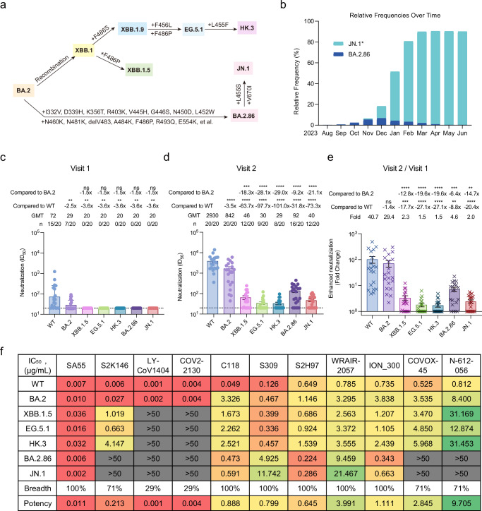
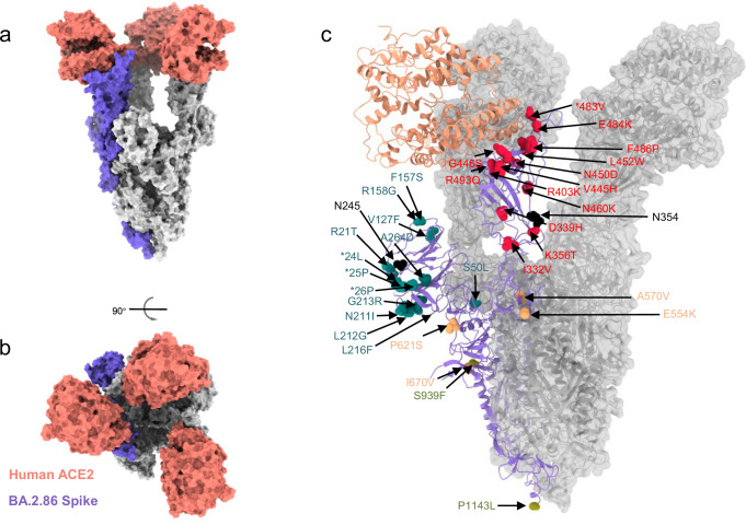
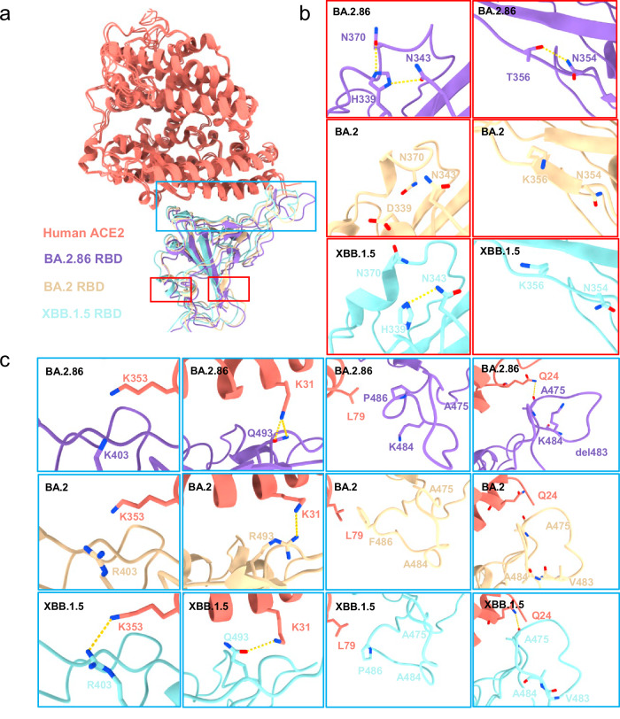
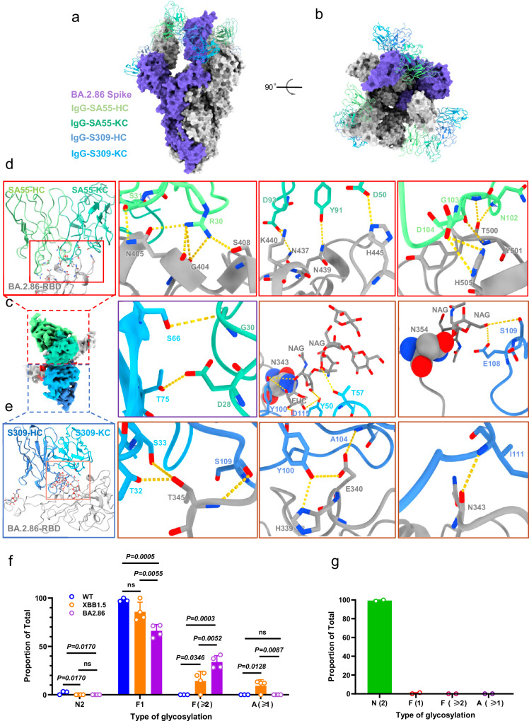
The Omicron subvariants of SARS-CoV-2, especially for BA.2.86 and JN.1, have rapidly spread across multiple countries, posing a significant threat in the ongoing COVID-19 pandemic. Distinguished by 34 additional mutations on the Spike (S) protein compared to its BA.2 predecessor, the implications of BA.2.86 and its evolved descendant, JN.1 with additional L455S mutation in receptor-binding domains (RBDs), are of paramount concern. In this work, we systematically examine the neutralization susceptibilities of SARS-CoV-2 Omicron subvariants and reveal the enhanced antibody evasion of BA.2.86 and JN.1. We also determine the cryo-EM structures of the trimeric S proteins from BA.2.86 and JN.1 in complex with the host receptor ACE2, respectively. The mutations within the RBDs of BA.2.86 and JN.1 induce a remodeling of the interaction network between the RBD and ACE2. The L455S mutation of JN.1 further induces a notable shift of the RBD-ACE2 interface, suggesting the notably reduced binding affinity of JN.1 than BA.2.86. An analysis of the broadly neutralizing antibodies possessing core neutralizing epitopes reveals the antibody evasion mechanism underlying the evolution of Omicron BA.2.86 subvariant. In general, we construct a landscape of evolution in virus-receptor of the circulating Omicron subvariants.


Conflict of interest statement

The authors declare no competing interests.

Figures

Fig. 1
Fig. 1. Overall mutations in the spike, prevalence, and antibody evasion of SARS-CoV-2 BA.2.86 variant.
a The schematic diagram of several SARS-CoV-2 BA.2-related subvariants evolution. Some additional mutations in the spike acquired by XBB.1.5, EG.5.1, HK.3, BA.2.86, and JN.1 were displayed. b The relative frequencies of BA.2.86 and JN.1* over time. The data to produce the chart were collected from the GISAID database and updated on 30 June 2024. Of which, JN.1* combine the JN.1 and its main sublineages including JN.1.1, JN.1.4, JN.1.4.5, JN.1.7, JN.1.11, JN.1.16, JN.1.16.1, KP.1.1, KP.2, KP.2.3, KP.3, KP.3.1.1, KP.3.2, and KP.3.3 variants. The neutralization of 20 BA.4 or BA.5 breakthrough infected human plasma samples collected in the early stage of breakthrough infection (c: Visit 1) and in another follow-up (d: Visit 2, an interval of 7–15 days) against WT, BA.2, XBB.1.5, EG.5.1, HK.3, BA.2.86, and JN.1, respectively. The 50% inhibitory dilution (ID50) values are means of two independent experiments. Data are presented as geometric mean values ± standard deviation (SD). The number, GMT, fold change, and significance of difference are labeled on the top. “-” represents decreased value. e Fold change in the enhanced neutralization of WT, BA.2, XBB.1.5, EG.5.1, HK.3, BA.2.86, and JN.1 by the BA.4 or BA.5 breakthrough infection. The fold change was obtained through the calculation of ID50 in Visit 2 divided by ID50 in Visit 1. Data are presented as geometric mean values ± SD. The statistical significance in (ce) was performed using two-tailed Kruskal–Wallis test with paired Wilcoxon’s multiple-comparison test. ns: P > 0.05, **P < 0.01, ***P < 0.001; ****P < 0.0001. f The neutralization of mAbs against WT, BA.2, XBB.1.5, EG.5.1, HK.3, BA.2.86, and JN.1 pseudoviruses. The 50% inhibitory concentration (IC50) values are means of two independent experiments. The neutralization potency is marked in the different color. Red: high, yellow: moderate, green: weak, gray: non-neutralization (IC50 > 50 μg/mL). The neutralization breadth is defined as the percentage of pseudoviruses neutralized by each mAb. The neutralization potency is calculated by the geometric mean of neutralizing values <50 μg/mL. Source data are provided as a Source Data file.
Fig. 2
Fig. 2. Structural and biological analysis of Omicron BA.2.86 subvariant.
a, b Surface presentation of domain-colored cryo-EM structures of extracellular domain of S protein (S-ECD) from Omicron BA.2.86, respectively, in complex with the PD of human ACE2, which show top and side view. Three RBDs are extended upward. ACE2 colored salmon. c Location of mutations detected in BA.2.86 spike, relative to its ancestral BA.2 (PDB: 7XO7). The red, cyan, orange, and laurel green mutations are in RBD, NTD, SD, and S2, respectively. The additional N-glycosylation sites colored in black. *deletion.
Fig. 3
Fig. 3. Interactions between SARS-CoV-2 BA.2.86-RBD and ACE2.
a Cryo-EM density of the BA.2.86-RBD–ACE2 interface. The “up” RBD is shown in medium purple. The ACE2 is shown in salmon. This is an overall cryo-EM density of the RBD–ACE2 region. b The cryo-EM density map between the RBD and ACE2 interface. Residues are shown in sticks, with the corresponding cryo-EM density represented in mesh. c Overall structural model of Omicron RBD–ACE2-bound region. The regions enclosed by the red, green, and blue solid lines are illustrated in detail in (d). d Detailed analysis of the interface between RBD and ACE2. Polar interactions are indicated by yellow dotted lines.
Fig. 4
Fig. 4. Interface comparison between BA.2.86-RBD, BA.2-RBD, and XBB.1.5 with ACE2.
a Structural alignment of the BA.2.86-RBD (this paper), BA.2-RBD (PDB:7XO9), and XBB.1.5-RBD (PDB:8WRL). The regions enclosed by the red and blue dashed lines are illustrated in detail in (b) to (c), respectively. BA.2.86-RBD and ACE2 in our cryo-EM structure are colored medium purple and salmon, respectively; BA.2-RBD and XBB.1.5-RBD are colored light yellow and light blue, respectively. b, c Variation of the mutation residues between BA.2.86-RBD (labeled in medium purple), BA.2-RBD (labeled in light yellow), and XBB.1.5-RBD (labeled in light blue).
Fig. 5
Fig. 5. Overall cryo-EM structure of S-ECD (BA.2.86) in complex with S309 and SA55.
a, b Overall structural model of RBD-SA55-S309–bound region. The SA55 and S309 were shown as cartoons and the spike of BA.2.86 was shown with surface map. c Cryo-EM density of the BA.2.86-RBD-S309-SA55 interface. BA.2.86-RBD are colored dark gray. d The regions enclosed by the red solid lines are illustrated in detail in (d). Residues are shown in sticks. The heavy and light chains of SA55 are colored pale green and medium aquamarine, respectively. e The regions enclosed by the pink solid lines are illustrated in detail in (e). The heavy and light chains of S309 are colored cornflower blue and deep sky blue, respectively. The ball indicates the N-glycosylation sites in N343 and N354. The purple solid lines box shows the interaction SA55 with S309. f Comparison of N-linked glycosylation profiles of the site Asn343 derived from S proteins of WT, XBB.1.5, and BA.2.86. Data are presented as mean values ± SD. Each experiment was repeated three times. g N-linked glycosylation profile of the site Asn354 that was identified from S protein of SARS-CoV-2 BA.2.86. N-linked glycans were divided into four basic classifications, including high mannose glycosylation (N2), mono-fucosylation (F1), multi-fucosylation (with ≥2 fucose residues, F ≥ 2), and sialylation (A ≥ 1). H represents hexose, N represents N-acetylglucosamine, F represents fucose, A represents sialic acid. Data are presented as mean values ± SD. Each experiment was repeated four times. In two of these sets, the site was not detected. Source data are provided as a Source Data file.
Fig. 6
Fig. 6. The comparison of neutralizing epitopes among various antibodies.
a, b The comparison of overall structural model of RBD-antibody-bound region. S309 (this paper) is colored medium blue. SA55 (this paper) is colored lime green. S2K146 (PDB:7TAT) are colored light yellow. N-612-056 (PDB:7S0B) are colored light cyan. COVOX-45 (PDB:7PRY) is colored orange. WT (PDB:6WPS) are colored plum. XBB.1.5 (PDB:8WRL) are colored wheat. BA.2 (PDB:7XO9) is colored sky blue. BA.2.86 (this paper) is colored medium purple. c, d Compared to WT, XBB.1.5, and BA.2, the specific details of the antigenic epitopes on BA.2.86 for the five antibodies S309, SA55, S2K146, N-612-056, and COVOX-45 are mapped.
Fig. 7
Fig. 7. Interactions and interface comparison between SARS-CoV-2 BA.2.86-RBD and JN.1-RBD with ACE2.
a Cryo-EM density of the JN.1-RBD–ACE2 interface. The JN.1 RBD is shown in cyan. The ACE2 is shown in salmon. This is an overall cryo-EM density of the RBD–ACE2 region. b The cryo-EM structural alignment of JN.1 and BA.2.86 between the RBD and ACE2 interface. Residues are shown in sticks, with the corresponding cryo-EM density represented in mesh. The BA.2.86 RBD is shown in medium purple. c Detailed analysis of 455 site in JN.1 or BA.2.86 RBD–ACE2-bound region. The region illustrates the disappearance of hydrophobic interactions at 455 site. L455S generates the hydrogen bond interaction with Gln493 in intra-RBD. Polar interactions are indicated by yellow dotted lines. d Detailed analysis of the same interacting residues between RBD and ACE2. The region displays the variation in interaction bond lengths between JN.1 and BA.2.86 at the same interacting residue positions. Polar interactions are indicated by yellow dotted lines. e Detailed analysis of the different interacting residues between RBD and ACE2. The region shows the interacting residues altered in JN.1 compared to BA.2.86. Polar interactions are indicated by yellow dotted lines.

Similar articles

Cited by

References

    1. Iketani, S. et al. Antibody evasion properties of SARS-CoV-2 Omicron sublineages. Nature604, 553–556 (2022). 10.1038/s41586-022-04594-4 - DOI - PMC - PubMed
    1. Miller, J. et al. Substantial neutralization escape by SARS-CoV-2 Omicron variants BQ.1.1 and XBB.1. N. Engl. J. Med.388, 662–664 (2023). 10.1056/NEJMc2214314 - DOI - PMC - PubMed
    1. Yisimayi, A. et al. Repeated Omicron exposures override ancestral SARS-CoV-2 immune imprinting. Nature625, 148–156 (2024). - PMC - PubMed
    1. Chen, X. et al. Protective effect of plasma neutralization from prior SARS-CoV-2 Omicron infection against BA.5 subvariant symptomatic reinfection. Lancet Reg. Health West. Pac.33, 100758 (2023). - PMC - PubMed
    1. Nehlmeier, I. et al. Host cell entry and neutralisation sensitivity of the SARS-CoV-2 XBB.1.16 lineage. Cell. Mol. Immunol.20, 969–971 (2023). 10.1038/s41423-023-01030-z - DOI - PMC - PubMed

Publication types

MeSH terms

Supplementary concepts