. 2024 Sep 4;14(1):20591.
doi: 10.1038/s41598-024-70892-8.

Enabling SENSE accelerated 2D CSI for hyperpolarized carbon-13 imaging

Affiliations

Enabling SENSE accelerated 2D CSI for hyperpolarized carbon-13 imaging

Ayaka Shinozaki et al. Sci Rep. .

Abstract

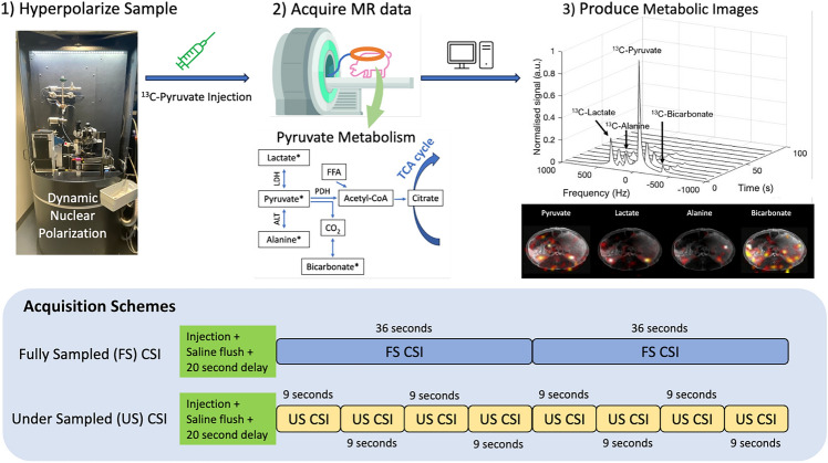
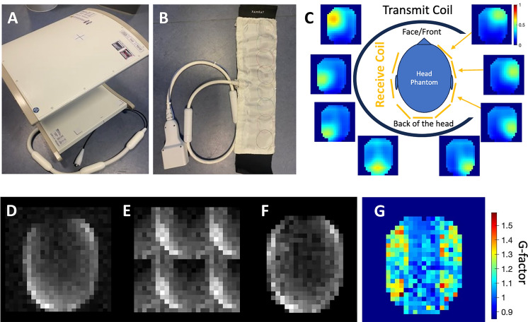
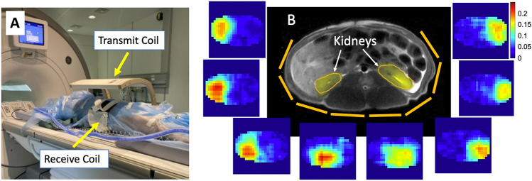
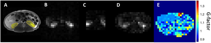
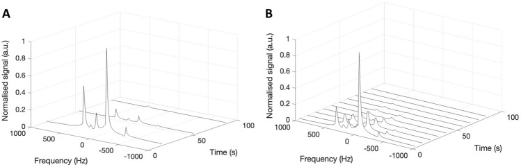
As hyperpolarized (HP) carbon-13 (13C) metabolic imaging is clinically translated, there is a need for easy-to-implement, fast, and robust imaging techniques. However, achieving high temporal resolution without decreasing spatial and/or spectral resolution, whilst maintaining the usability of the imaging sequence is challenging. Therefore, this study looked to accelerate HP 13C MRI by combining a well-established and robust sequence called two-dimensional Chemical Shift Imaging (2D CSI) with prospective under sampling and SENSitivity Encoding (SENSE) reconstruction. Due to the low natural abundance of 13C, the sensitivity maps cannot be pre-acquired for the reconstruction. As such, the implementation of sodium (23Na) sensitivity maps for SENSE reconstructed 13C CSI was demonstrated in a phantom and in vivo in the pig kidney. Results showed that SENSE reconstruction using 23Na sensitivity maps corrected aliased images with a four-fold acceleration. With high temporal resolution, the kidney spectra produced a detailed metabolic arrival and decay curve, useful for further metabolite kinetic modelling or denoising. Metabolic ratio maps were produced in three pigs demonstrating the technique's ability for repeat metabolic measurements. In cases with unknown metabolite spectra or limited HP MRI specialist knowledge, this robust acceleration method ensures comprehensive capture of metabolic signals, mitigating the risk of missing spectral data.


Conflict of interest statement

An author declares the following competing interests: Rolf F. Schulte is an employee of GE Healthcare. All other authors declare no competing interests.

Figures

Fig. 1
Fig. 1
Overview of the HP 13C experimental work flow (steps 1–3) carried out in this study. The illustrations of the two CSI acquisition schemes are both FID CSIs taken consecutively following an injection, saline flush, and a 20 second delay. The fully sampled (FS) CSI (36 seconds per acquisition) and the under sampled (US) CSI (9 s per acquisition) are collected after separate HP 13C injections about 90min apart. To visualize the four-fold acceleration, the schemes are illustrated directly on top of each other, where four consecutive US CSIs are taken within a single FS CSI.
Fig. 2
Fig. 2
(A) 13C clamshell transmit coil (Rapid Biomedical, Germany) (B) an eight-channel array, flexible 23Na/13C receive coil (fabricated by Juan D. Sanchez-Heredia) (C) a phantom experiment set up is illustrated, where the head phantom sits inside the clamshell transmit coil and the head is wrapped with the receive coil. The location of the receive elements are depicted as yellow lines. Coil-wise sodium sensitivity images surround the illustration. (D) The fully sampled 13C phantom image. (E) The retrospectively under sampled 13C phantom image. (F) The under sampled SENSE reconstructed image. (G) The g-factor map of R = 4, where acquisition is accelerated by R = 2 in both the frequency and phase directions.
Fig. 3
Fig. 3
(A) A porcine experiment set up with a supine pig inside the clamshell transmit coil and the torso wrapped with the flexible receive coil. (B) Axial proton image of the porcine torso shows the location of the kidneys. The yellow lines visualize the flexible receive coil element positions and coil-wise sodium sensitivity images surround the proton image.
Fig. 4
Fig. 4
(A) Axial T2 proton image of the pig anatomy with the kidneys highlighted in yellow. (B) [13C -1] pyruvate signal from fully sampled CSI taken over 36 seconds. (C) [13C -1] Pyruvate signal from under sampled CSI taken over 9 seconds. (D) [13C -1] pyruvate SENSE Reconstructed CSI based on the under sampled CSI and the sodium sensitivity image. (E) The g-factor map of R = 4, where acquisition is accelerated by R = 2 in both the frequency and phase directions.
Fig. 5
Fig. 5
(A) Dynamic 13C spectra of the fully sampled CSI in the kidneys of a pig. (B) Dynamic 13C spectra of the under sampled CSI prior to the SENSE reconstruction in the kidneys of the same pig.
Fig. 6
Fig. 6
Ratio maps of lactate-to-pyruvate, alanine-to-pyruvate, and bicarbonate-to-pyruvate were calculated to show metabolism in the kidneys in the single scan fully sampled image, the under sampled image, and the SENSE reconstructed under sampled image of a healthy representative porcine subject. The axial T2 images have also been shown to indicate the kidney locations in yellow ROIs.

References

    1. Kurhanewicz, J. et al. Hyperpolarized 13c MRI: Path to clinical translation in oncology. Neoplasia21, 1–16. 10.1016/J.NEO.2018.09.006 (2019). 10.1016/J.NEO.2018.09.006 - DOI - PMC - PubMed
    1. Wolber, J. et al. Generating highly polarized nuclear spins in solution using dynamic nuclear polarization. Nucl. Instrum. Methods Phys. Res., Sect. A526, 173–181. 10.1016/J.NIMA.2004.03.171 (2004).10.1016/J.NIMA.2004.03.171 - DOI
    1. Ardenkjaer-Larsen, J. H. et al. Increase in signal-to-noise ratio of> 10,000 times in liquid-state nmr. Proc. Natl. Acad. Sci.100, 10158–10163. 10.1073/PNAS.1733835100 (2003). 10.1073/PNAS.1733835100 - DOI - PMC - PubMed
    1. Hurd, R. E., Yen, Y. F., Chen, A. & Ardenkjaer-Larsen, J. H. Hyperpolarized 13c metabolic imaging using dissolution dynamic nuclear polarization. J. Magn. Reson. Imaging36, 1314–1328. 10.1002/JMRI.23753 (2012). 10.1002/JMRI.23753 - DOI - PubMed
    1. Ardenkjaer-Larsen, J. H. et al. Cryogen-free dissolution dynamic nuclear polarization polarizer operating at 3.35 t, 6.70 t, and 10.1 t. Magn. Reson. Med.81, 2184–2194. 10.1002/MRM.27537 (2019). 10.1002/MRM.27537 - DOI - PubMed