. 2024 Oct;9(10):2488-2505.
doi: 10.1038/s41564-024-01796-6. Epub 2024 Sep 4.

Diverse RNA viruses of parasitic nematodes can elicit antibody responses in vertebrate hosts

Affiliations

Diverse RNA viruses of parasitic nematodes can elicit antibody responses in vertebrate hosts

Shannon Quek et al. Nat Microbiol. 2024 Oct.

Abstract

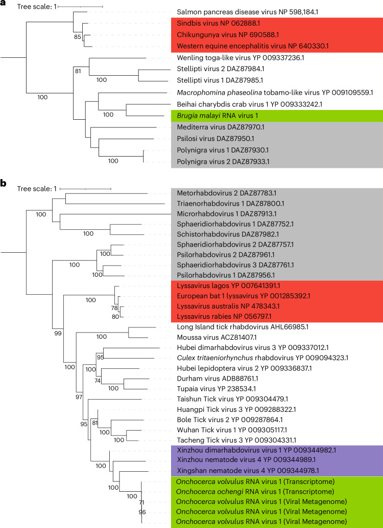
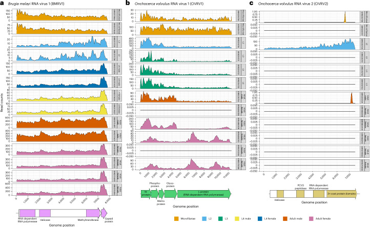
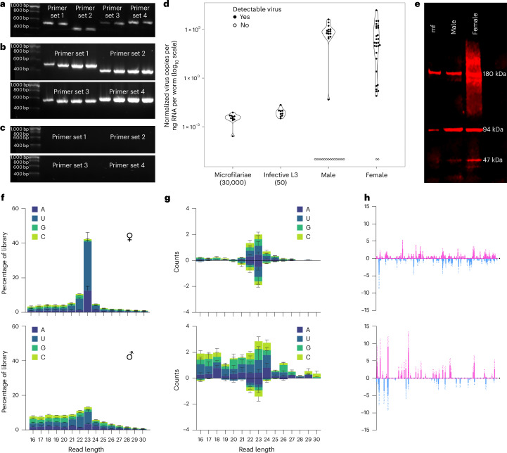
Parasitic nematodes have an intimate, chronic and lifelong exposure to vertebrate tissues. Here we mined 41 published parasitic nematode transcriptomes from vertebrate hosts and identified 91 RNA viruses across 13 virus orders from 24 families in ~70% (28 out of 41) of parasitic nematode species, which include only 5 previously reported viruses. We observe widespread distribution of virus-nematode associations across multiple continents, suggesting an ancestral acquisition event and host-virus co-evolution. Characterization of viruses of Brugia malayi (BMRV1) and Onchocerca volvulus (OVRV1) shows that these viruses are abundant in reproductive tissues of adult parasites. Importantly, the presence of BMRV1 RNA in B. malayi parasites mounts an RNA interference response against BMRV1 suggesting active viral replication. Finally, BMRV1 and OVRV1 were found to elicit antibody responses in serum samples from infected jirds and infected or exposed humans, indicating direct exposure to the immune system.


Conflict of interest statement

The authors declare no competing interests.

Figures

Fig. 1
Fig. 1. Phylogenetic analysis of BMRV1 and OVRV1 based on alignment of their respective RdRP sequences.
a, BMRV1. b, OVRV1 or the variant found in O. ochengi (Onchocerca ochengi RNA Virus 1). Green highlight, viruses found in this study; red highlight, human pathogenic viruses; grey highlight, viruses previously found in flatworms; purple highlight, viruses previously found in other nematodes. Trees were constructed with 1,000 non-parametric bootstrap replicates, and only values above 70% are shown in the figures.
Fig. 2
Fig. 2. Transcriptome read alignment depths.
a, BMRV1. b, OVRV1. c, OVRV2. For each graph, the x-axis represents positions in the respective virus genomes, and the y-axis represents the number of reads from the transcriptome dataset for different life cycle stages (defined by the grey columns on the right side of each graph and the colour code on the bottom right of the figure), which aligned to the virus genome. An annotation of the virus genomes is at the bottom of each graph, with identified domains and proteins labelled. Source data
Fig. 3
Fig. 3. Molecular and sequencing evidence for BMRV1, including RT-PCR, qPCR, western blot and small RNA sequencing results.
ac, Reverse-transcription PCR experiments show that BMRV1 can be found in LSTM-reared (a) and FR3-reared (b) B. malayi and that BMRV1 can be amplified only from reverse-transcribed RNA and not DNA (c). More details on the primers used and their target in the BMRV1 genome are given in Supplementary Table 5. The individual lanes shown correspond to different biological replicates of 50,000 pooled B. malayi microfilariae (for tests on RNA and cDNA, n = 6 total; for gDNA, n = 4). d, The qPCR results reflect the differences in viral abundance across the life cycle. Note the difference in the number of adult males and females that appear positive for the virus (48% (15 out of 39) and 93% (28 out of 30) of surveyed individuals, respectively). All data points are shown in the graph (pools of mf, n = 10; pools of L3, n = 9; adult male, n = 39; adult female, n = 30). e, Western blots with antibodies raised against the BMRV1 capsid protein show multimerization and different signal intensities between microfilariae, males and females (n = 3 per life cycle stage). f, Histogram of total small RNA sequencing of six pools of adult male and female B. malayi (n = 3 per gender) showing an active siRNA machinery with a length bias at 23 nt that is most visible in samples derived from female nematodes. The histogram shows the mean values of three independent biological replicates (pools) of each gender (n = 3) ± s.d. g, The size distribution of small RNAs mapping to the BMRV1 genome shows no discernible strand bias, with 23 nt vsiRNAs predominant (especially in females), indicating active processing of BMRV1-derived dsRNA by a Dicer protein. The histogram shows the mean values of three independent biological replicates (pools) of each gender (n = 3) ± s.d. h, The 23 nt vsiRNA mapping against the BMRV1 genome (pink) and antigenome (blue) (x-axis) shows roughly equal coverage against both viral RNA strands, indicating active viral replication. The graph shows the mean values of three independent biological replicates (pools) of each gender (n = 3), with error bars indicating ±s.e.m. Source data
Fig. 4
Fig. 4. Representative FISH microscopy images of B. malayi showing localization of virus RNA within nematode tissues, alongside the Wolbachia endosymbiont as a technical control.
Virus RNA stained red; Wolbachia stained green; DAPI nuclear stain blue. ae, Note the different levels of viral infection in microfilariae (a), localization of the viral stain in male testes (b) and the hypodermal cells near the male spicule (c). Virus signal within adult female reproductive tracts appears between developing eggs within the paired uteri of adult females, with early embrys in the left uteri and ‘pretzel-stage’ microfilariae in the right (d), with the developing eggs casting a ‘shadow’ in between virus staining, visible in 3D images of female uteri (e). fj, In older adults (>12 months), we observed ‘epicuticular inflations’ often with an intense viral signal (f), typically occurring near the head (g) or tail regions of the nematodes. They can appear as single separate inflations at different nematode orientations, either next to internal organs (h) or the hypodermal chords (i), or as a continuous inflation along the nematode flank (j). Scale bars measure 20 µm (a,b,h,i) or 50 µm (d,g). Gridlines in three-dimensional z-stack figures (c,e,f,j) measure 40 µm by 40 µm. A total of 15 adult male and female parasites were processed in separate experiments. Parasites with epicuticular inflations were typically between 12 and 19 months at the time of sampling, with jird animal hosts being 15–22 months of age, respectively. Parasites without were typically 3–6 months of age, with the jird animal hosts being 6–9 months of age.
Fig. 5
Fig. 5. Validation of OVRV1 using RT-PCR, western blot and representative IFA staining of O. volvulus nodules with anti-OVRV1 glycoprotein antibodies.
Anti-OVRV1 glycoprotein antibodies stained green; DAPI nuclear stain blue. a, RT-PCR experiments show that OVRV1 can be amplified only from reverse-transcribed RNA, from both O. volvulus (lane 1, n = 1) and O. ochengi (lanes 2–4, n = 3). b, Western blots against the OVRV1 glycoprotein show different molecular weight bands occurring depending on the life cycle stage of O. volvulus (n = 3). All IFA images include the DAPI nuclear stain (blue). c,d, Images of the paired uteri from adult O. volvulus females show virus stains surrounding and entering developing embryos within the uteri (solid arrow), while surrounding but not within the early embryos (hollow arrow). Developing embryos can show either complete infection rates (c) or a much smaller proportion (d). e, Mature microfilariae released from the female, located within surrounding nodule tissues, stain heavily for OVRV1 glycoprotein. f,g, Intense antibody staining is observed surrounding the nematode rachis, where eggs are first formed (solid arrows). The heavily stained rachis is either surrounded by early-stage eggs with green staining surrounding them (f) or without surrounding eggs (g). h,i, Cellular inflations containing intense antibody staining are observed on the external face of the adult female uterine walls (solid arrows). j,k, Male O. volvulus are frequently observed to be infected, with viral stains occurring in different tissues (j), as well as surrounding and entering the male testes (k). Parasites were obtained from sections of fixed O. volvulus nodules from human patients (n = 8 nodules). Source data
Fig. 6
Fig. 6. ELISA distribution graphs to illustrate host antibody responses against BMRV1 and OVRV1.
Each data point represents the results from one infected individual. a, Statistically significant difference in the antibody serology of jirds infected with B. malayi against BMRV1 capsid protein when compared with uninfected jirds (P = 0.0027). b, Statistically significant difference in the antibody serology of jirds infected with O. ochengi against OVRV1 glycoprotein when compared with uninfected jirds (P = 0.0053). c, Serology of O. volvulus-infected individuals (‘active infection’ or from the CDC serum bank, dark blue or lavender violin respectively) and individuals without detectable active infection, but resident in the community for at least 20 years (‘putatively immune’, orange violin) against OVRV1 glycoprotein. This shows that all individuals in Africa infected or exposed to O. volvulus transmission are seropositive, with 4 samples from Uganda and an additional 26 samples from Ecuador appearing seronegative, when compared with 3× the mean of UK uninfected controls (the dotted line is 3× the mean antibody concentration of UK negative controls). Serum was taken from individuals from Uganda (n = 58, P = 7.88 × 10−11) Cameroon (n = 200, P = 6.56 × 10−13), Nigeria (n = 54, P = 8.58 × 10−11), Togo (n = 67, P = 3.09 × 10−11), Ecuador (n = 54, P = 5.97 × 10−9) and uninfected UK controls (‘uninfected control’, yellow violin). Significance is defined by number of asterisks (**P < 0.01, ***P < 0.001). Significance for all panels was determined via a two-sided Wilcoxon rank-sum test with continuity correction, after identifying the distribution of points as non-normal via a Shapiro–Wilk normality test. Panel c uses Benjamini–Hochberg’s P-adjustment method for multiple testing correction. ELISA optical density results from human samples were converted into a concentration of IgG antibodies against the OVRV1 glycoprotein using a standard curve of human total IgG.

References

    1. Donohue, R. E., Cross, Z. K. & Michael, E. The extent, nature, and pathogenic consequences of helminth polyparasitism in humans: a meta-analysis. PLoS Negl. Trop. Dis.13, 1–41 (2019). - PMC - PubMed
    1. Hotez, P. J. & Kamath, A. Neglected tropical diseases in sub-Saharan Africa: review of their prevalence, distribution, and disease burden. PLoS Negl. Trop. Dis.3, 2–11 (2009). - PMC - PubMed
    1. Jex, A. R., Gasser, R. B. & Schwarz, E. M. Transcriptomic resources for parasitic nematodes of veterinary importance. Trends Parasitol.35, 72–84 (2019). - PubMed
    1. Else, K. J. et al. Whipworm and roundworm infections. Nat. Rev. Dis. Primers6, 1–23 (2020). - PubMed
    1. Abbafati, C. et al. Global burden of 369 diseases and injuries in 204 countries and territories, 1990–2019: a systematic analysis for the Global Burden of Disease Study 2019. Lancet396, 1204–1222 (2020). - PMC - PubMed

LinkOut - more resources