. 2024 Nov 7;31(11):1612-1629.e8.
doi: 10.1016/j.stem.2024.08.007. Epub 2024 Sep 3.

Human vascularized macrophage-islet organoids to model immune-mediated pancreatic β cell pyroptosis upon viral infection

Affiliations

Human vascularized macrophage-islet organoids to model immune-mediated pancreatic β cell pyroptosis upon viral infection

Liuliu Yang et al. Cell Stem Cell. .

Abstract

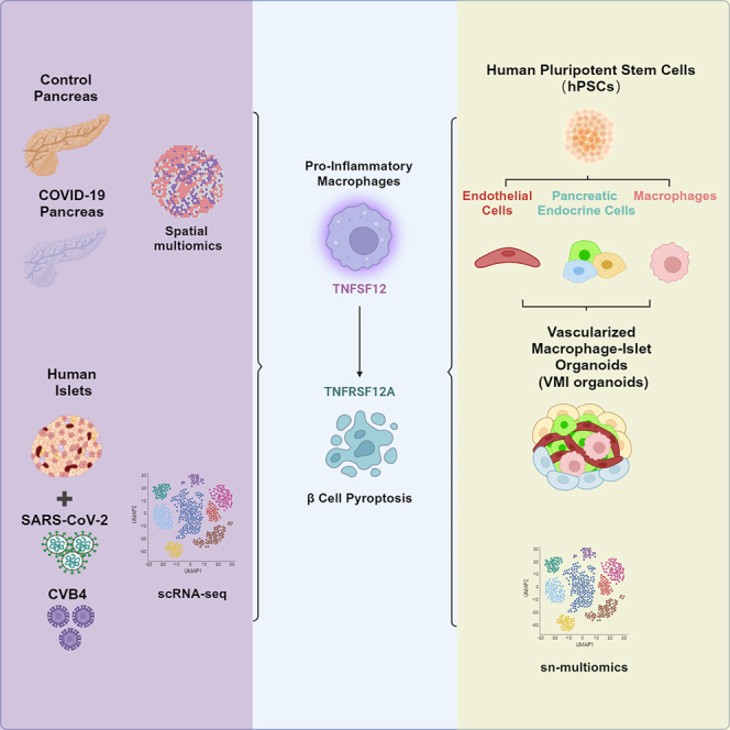
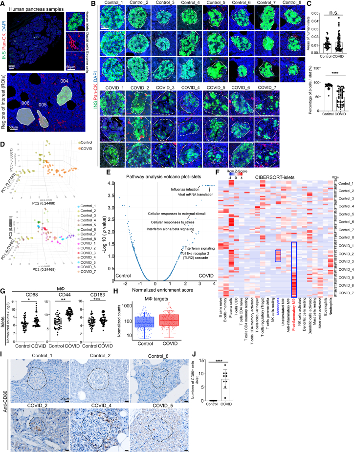
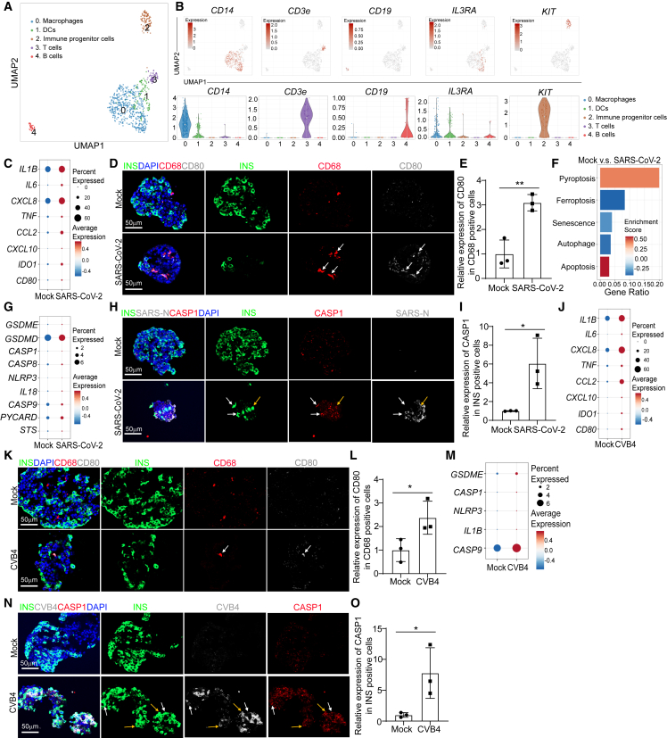
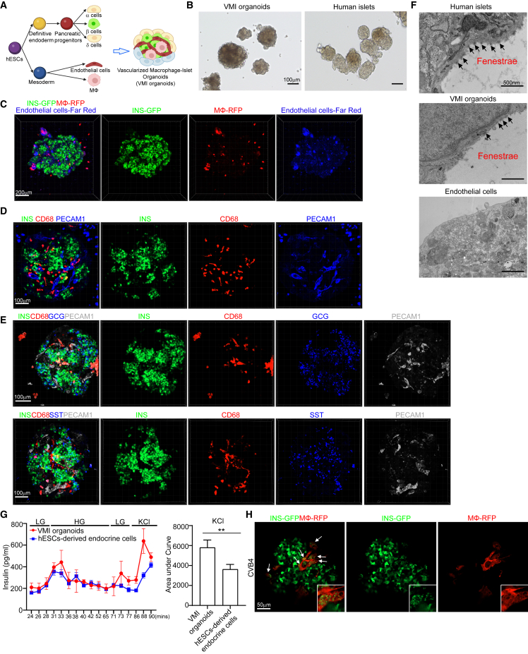
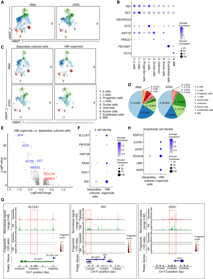
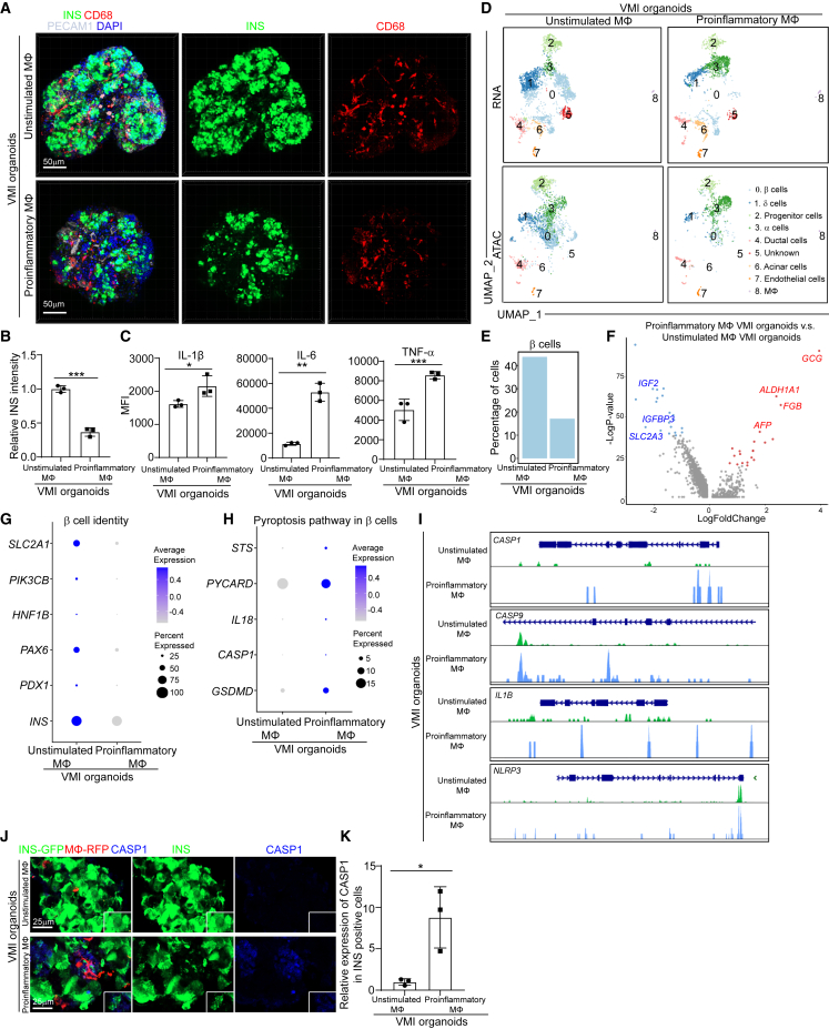
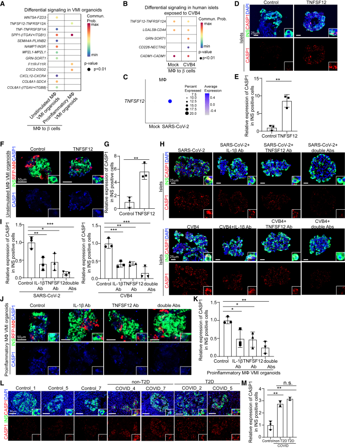
There is a paucity of human models to study immune-mediated host damage. Here, we utilized the GeoMx spatial multi-omics platform to analyze immune cell changes in COVID-19 pancreatic autopsy samples, revealing an accumulation of proinflammatory macrophages. Single-cell RNA sequencing (scRNA-seq) analysis of human islets exposed to severe acute respiratory syndrome coronavirus 2 (SARS-CoV-2) or coxsackievirus B4 (CVB4) viruses identified activation of proinflammatory macrophages and β cell pyroptosis. To distinguish viral versus proinflammatory-macrophage-mediated β cell pyroptosis, we developed human pluripotent stem cell (hPSC)-derived vascularized macrophage-islet (VMI) organoids. VMI organoids exhibited enhanced marker expression and function in both β cells and endothelial cells compared with separately cultured cells. Notably, proinflammatory macrophages within VMI organoids induced β cell pyroptosis. Mechanistic investigations highlighted TNFSF12-TNFRSF12A involvement in proinflammatory-macrophage-mediated β cell pyroptosis. This study established hPSC-derived VMI organoids as a valuable tool for studying immune-cell-mediated host damage and uncovered the mechanism of β cell damage during viral exposure.

Keywords: SARS-CoV-2; coxsackievirus B4; diabetes; endothelial cells; human pluripotent stem cells; organoids; pancreatic endocrine cells; proinflammatory macrophages; pyroptosis.


Conflict of interest statement

Declaration of interests R.E.S. is on the scientific advisory board of Miromatrix Inc. and Lime Therapeutics and is a consultant and speaker for Alnylam Inc. S.C. and T.E are the co-founders of OncoBeat, LLC. S.C. is a consultant of Vesalius Therapeutics and co-founder of iOrganBio.

Figures

None
Graphical abstract
Figure 1
Figure 1
Macrophage accumulation in islets of COVID-19 pancreatic autopsy samples (A) Representative images illustrating morphology marker and selection of ROIs using GeoMx platform. 004, islet area; 005, ductal area; 006, exocrine area. Scale bars, 1 mm or 50 μm. (B) Representative images illustrating the insulin (INS) staining in COVID-19 (N = 7) and control (N = 8) pancreatic autopsy samples. Dotted lines encircle the islet regions. Scale bars, 75 μm. (C) Quantification of areas of islets and percentages of INS+ β cells per islet in COVID-19 (N = 7) and control (N = 8) pancreatic autopsy samples. (D) 3D PCA plot of data from human islet areas of COVID-19 (N = 7) and control (N = 8) pancreatic autopsy samples. (E) Volcano plot of transcriptome sequencing data highlighting the pathways enriched in human islet areas of COVID-19 (N = 7) versus control (N = 8) pancreatic autopsy samples. (F) Heatmap of the CIBERSORT analysis of immune cells (LM22) using the GeoMx whole transcriptome sequencing data of human islet areas of COVID-19 (N = 7) and control (N = 8) pancreatic autopsy samples. (G) Normalized counts (log2) of marker proteins associated with macrophages from human islet areas of COVID-19 (N = 7) and control (N = 8) pancreatic autopsy samples. Each dot represents one count in each ROI. (H) Boxplot of normalized counts of macrophage-associated targets in human islet areas of COVID-19 (N = 7) and control (N = 8) pancreatic autopsy samples. Each dot represents one count in each ROI. (I and J) Immunohistochemistry staining (I) and quantification (J) of CD80 in COVID-19 (N = 3) and control (N = 3) pancreatic autopsy samples. Dotted lines encircle the regions of the islets. Scale bars, 20 μm. p values were calculated by unpaired two-tailed Student’s t test. n.s., no significance; p < 0.05, ∗∗p < 0.01, ∗∗∗p < 0.001. See also Figure S1.
Figure 2
Figure 2
Single-cell RNA-seq analysis of human islets upon SARS-CoV-2 or CVB4 exposure (A) UMAP of immune cell populations in human islets exposed to mock, SARS-CoV-2 (MOI = 1), or CVB4 (2 × 106 PFU/mL). (B) UMAP and violin plots of immune cell markers. (C) Dot plot analysis of proinflammatory-macrophage-associated genes in macrophages of human islets exposed to mock or SARS-CoV-2 (MOI = 1). (D and E) Confocal images (D) and quantification (E) of the relative expression of CD80 in CD68+ cells in human islets exposed to mock or SARS-CoV-2 (MOI = 0.5). The white arrows highlight the CD68+CD80+ cells. Scale bars, 50 μm. (F) Pathway enrichment analysis of cell death pathways in β cell cluster of human islets exposed to mock or SARS-CoV-2 (MOI = 1). (G) Dot plot analysis of pyroptosis-associated genes in β cell cluster of human islets exposed to mock or SARS-CoV-2 (MOI = 1). (H and I) Confocal images (H) and quantification (I) of the relative expression of CAPS1 in human islets exposed to mock or SARS-CoV-2 (MOI = 0.5). The yellow arrows highlight the expression of CASP1 in SARS-N+INS+ cells while the white arrows highlight the expression of CASP1 in SARS-NINS+ cells. Scale bars, 50 μm. (J) Dot plot analysis of proinflammatory-macrophage-associated genes in macrophage cluster of human islets exposed to mock or CVB4 (2 × 106 PFU/mL). (K and L) Confocal images (K) and quantification (L) of the relative expression of CD80 in CD68+ cells in human islets exposed to mock or CVB4 (2 × 106 PFU/mL). The white arrows highlight the CD68+CD80+ cells. Scale bars, 50 μm. (M) Dot plot analysis of pyroptosis-pathway-associated genes in β cell cluster of human islets exposed to mock or CVB4 (2 × 106 PFU/mL). (N and O) Fluorescent images (N) and quantification (O) of the relative expression of CAPS1 in human islets exposed to mock or CVB4 (2 × 106 PFU/mL). The yellow arrows highlight the expression of CASP1 in SARS-N+INS+ cells while the white arrows highlight the expression of CASP1 in SARS-N-INS+ cells. Scale bars, 50 μm. n = 3 independent biological replicates. Data were presented as mean ± STDEV. p values were calculated by unpaired two-tailed Student’s t test. p < 0.05, ∗∗p < 0.01. See also Figures S2 and S3.
Figure 3
Figure 3
Construction of hPSC-derived VMI organoids (A) Schematic representation of VMI organoids construction. (B) Phase contract images of VMI organoids at day 14 after reaggregation and human islets. Scale bars, 200 μm. (C) Composite z stack confocal images of live VMI organoids at day 14 after reaggregation. β cells, INS-GFP; macrophages, RFP; and endothelial cells, far red. Scale bars, 200 μm. (D) Composite z stack confocal images of VMI organoids at day 14 after reaggregation stained with antibodies against INS, CD68, and PECAM1 (CD31). Scale bars, 100 μm. (E) Composite z stack confocal images of VMI organoids at day 14 after reaggregation stained with antibodies against INS, CD68, GCG, SST, and PECAM1 (CD31). Scale bars, 100 μm. (F) Transmission electron microscope (TEM) images of human islets, VMI organoids at day 14 after reaggregation, and endothelial cells without reaggregation. Arrows indicate fenestrae. Scale bars, 500 nm. (G) Dynamic glucose stimulated insulin secretion of VMI organoids at day 14 after reaggregation and hPSC-derived endocrine cells. Low glucose (LG), 2 mM D-glucose; high glucose (HG), 20 mM D-glucose; KCl, 30 mM KCl. Quantification was performed using the areas under curve of KCl stimulation from 86 to 90 min. (H) Composite z stack confocal images of VMI organoids at day 7 after reaggregation upon CVB4 infection (2 × 106 PFU/mL). β cells, INS-GFP; macrophages, RFP. Scale bars, 50 μm. Arrows highlight RFP+ macrophages that have phagocytosed damaged INS-GFP+ β cells. n = 3 independent biological replicates. Data were presented as mean ± STDEV. p values were calculated by unpaired two-tailed Student’s t test. ∗∗p < 0.01. See also Figure S4.
Figure 4
Figure 4
Single-cell multi-omics analysis of VMI organoids (A) Integrative UMAP of scRNA-seq and snATAC-seq analysis of VMI organoids at day 7 after reaggregation and separately cultured cells. (B) Dot plot displaying cell markers of each cluster using scRNA-seq dataset. (C) Individual UMAP of scRNA-seq and snATAC-seq analysis of VMI organoids at day 7 after reaggregation and separately cultured cells. (D) Pie chart shows the relative percentages of each cell types in VMI organoids at day 7 after reaggregation. (E) Volcano plot of DE genes in β cell cluster of VMI organoids at day 7 after reaggregation versus separately cultured cells. (F) Dot plot analysis of β cell-associated genes in β cell cluster of VMI organoids at day 7 after reaggregation and separately cultured cells. (G) Chromatin accessibility signals of SLC2A1, INS, and PDX1 in β cell cluster of VMI organoids at day 7 after reaggregation and separately cultured cells. The normalized signal shows the averaged frequency of sequenced DNA fragments within a genomic region. The fragment shows the frequency of sequenced fragments within a genomic region for individual cells. (H) Dot plot analysis of endothelial-cell-associated genes in endothelial cell cluster of VMI organoids at day 7 after reaggregation and separately cultured cells. See also Figure S4.
Figure 5
Figure 5
Construction and multi-omics analysis of VMI organoids containing unstimulated and proinflammatory macrophages (A and B) Composite z stack confocal images (A) and quantification (B) of INS intensity in INS+ cells of VMI organoids at day 7 after reaggregation containing unstimulated or proinflammatory macrophages stained with the antibodies against INS, CD68, and PECAM1 (CD31). Scale bars, 50 μm. (C) Measurements of cytokine secretions in the supernatant of VMI organoids at day 5 after reaggregation containing unstimulated or proinflammatory macrophages. (D) Integrative UMAP of VMI organoids at day 7 after reaggregation containing unstimulated or proinflammatory macrophages. (E) Percentage of cells in β cell cluster in VMI organoids at day 7 after reaggregation containing unstimulated or proinflammatory macrophages. (F) Volcano plot of DE genes in β cell cluster of VMI organoids at day 7 after reaggregation containing proinflammatory versus unstimulated macrophages. (G) Dot plot analysis of β cell-identity-associated genes in β cell cluster of VMI organoids at day 7 after reaggregation containing unstimulated or proinflammatory macrophages. (H) Dot plot analysis of pyroptosis-pathway-associated genes in β cell cluster of VMI organoids at day 7 after reaggregation containing unstimulated or proinflammatory macrophages. (I) Chromatin accessibility signals of CASP1, CASP9, IL1B, and NLRP3 in β cell cluster of VMI organoids at day 7 after reaggregation containing unstimulated or proinflammatory macrophages. The normalized signal shows the averaged frequency of sequenced DNA fragments within a genomic region. The fragment shows the frequency of sequenced fragments within a genomic region for individual cells. (J and K) Immunostaining (J) and quantification (K) of CASP1 staining in INS+ cells of VMI organoids at day 7 after reaggregation containing unstimulated or proinflammatory macrophages. β cells, INS-GFP; macrophages, RFP; endothelial cells, far red. CASP1, gray. Scale bars, 25 μm. n = 3 independent biological replicates. Data were presented as mean ± STDEV. p values were calculated by unpaired two-tailed Student’s t test. p < 0.01, ∗∗p < 0.05, ∗∗∗p < 0.001. See also Figure S5.
Figure 6
Figure 6
TNFSF12-TNFRSF12A as a candidate pathway that contributes to proinflammatory-macrophage-mediated β cell pyroptosis (A) Dot plot showed the differential signaling from macrophages to β cells in VMI organoids containing unstimulated or proinflammatory macrophages at day 7 after reaggregation. (B) Dot plot showed the differential signaling from macrophages to β cells in human islets exposed to mock or CVB4 virus (2 × 106 PFU/mL). (C) Dot plot of the expression level of TNFSF12 in macrophage of human islets exposed to mock or SARS-CoV-2 virus (MOI = 1). (D and E) Confocal images (D) and quantification (E) of CASP1 expression in INS+ cells in control or 10 ng/mL TNFSF12-treated human islets. Scale bars, 25 μm. (F and G) Confocal images (F) and quantification (G) of CASP1 expression in INS+ cells in control or 10 ng/mL TNFSF12-treated VMI organoids at day 7 after reaggregation. Scale bars, 50 μm. (H and I) Confocal images (H) and quantification (I) of CASP1 expression in INS+ cells of SARS-CoV-2-exposed (MOI = 0.5) or CVB4-exposed (2 × 106 PFU/mL) human islets treated with control, 10 μg/mL TNFSF12 blocking antibody, 5 μg/mL IL-1β blocking antibody, or 10 μg/mL TNFSF12 + 5 μg/mL IL-1β blocking antibodies. Scale bars, 25 μm. (J and K) Confocal images (J) and quantification (K) of CASP1 expression in INS+ cells of VMI organoids containing proinflammatory macrophages at day 7 after reaggregation and treated with control, 10 μg/mL TNFSF12 blocking antibody, 5 μg/mL IL-1β blocking antibody, or 10 μg/mL TNFSF12 + 5 μg/mL IL-1β blocking antibodies. Scale bars, 50 μm. (L and M) Confocal images (L) and quantification (M) of the CASP1 expression in INS+ cells of pancreas autopsy samples from control (N = 3) and COVID-19 (N = 4) subjects. The insert shows a high magnification of cells. Scale bars, 50 μm. n = 3 independent biological replicates. Data were presented as mean ± STDEV. p values were calculated by unpaired two-tailed Student’s t test or one-way ANOVA with a common control. n.s., no significance; p < 0.05, ∗∗p < 0.01, ∗∗∗p < 0.001. See also Figure S6.

Update of

Similar articles

Cited by

References

    1. Hollstein T., Schulte D.M., Schulz J., Glück A., Ziegler A.G., Bonifacio E., Wendorff M., Franke A., Schreiber S., Bornstein S.R., Laudes M. Autoantibody-negative insulin-dependent diabetes mellitus after SARS-CoV-2 infection: a case report. Nat. Metab. 2020;2:1021–1024. doi: 10.1038/s42255-020-00281-8. - DOI - PubMed
    1. Soliman A.T., Al-Amri M., Alleethy K., Alaaraj N., Hamed N., De Sanctis V. Newly-onset type 1 diabetes mellitus precipitated by COVID-19 in an 8-month-old infant. Acta Biomed. 2020;91 doi: 10.23750/abm.v91i3.10074. ahead of print. - DOI - PMC - PubMed
    1. Heaney A.I., Griffin G.D., Simon E.L. Newly diagnosed diabetes and diabetic ketoacidosis precipitated by COVID-19 infection. Am. J. Emerg. Med. 2020;38:2491.e3–2491.e4. doi: 10.1016/j.ajem.2020.05.114. - DOI - PMC - PubMed
    1. Unsworth R., Wallace S., Oliver N.S., Yeung S., Kshirsagar A., Naidu H., Kwong R.M.W., Kumar P., Logan K.M. New-Onset Type 1 Diabetes in Children During COVID-19: Multicenter Regional Findings in the U.K. Diabetes Care. 2020;43:e170–e171. doi: 10.2337/dc20-1551. - DOI - PubMed
    1. Chee Y.J., Ng S.J.H., Yeoh E. Diabetic ketoacidosis precipitated by Covid-19 in a patient with newly diagnosed diabetes mellitus. Diabetes Res. Clin. Pract. 2020;164 doi: 10.1016/j.diabres.2020.108166. - DOI - PMC - PubMed

LinkOut - more resources