

Role of the GalNAc-galectin pathway in the healing of premature rupture of membranes
- PMID: 39232672
- PMCID: PMC11375961
- DOI: 10.1186/s10020-024-00908-6
Role of the GalNAc-galectin pathway in the healing of premature rupture of membranes
Abstract
Background: Premature rupture of the membranes (PROM) is a key cause of preterm birth and represents a major cause of neonatal mortality and morbidity. Natural products N-acetyl-d-galactosamine (GalNAc), which are basic building blocks of important polysaccharides in biological cells or tissues, such as chitin, glycoproteins, and glycolipids, may improve possible effects of wound healing.
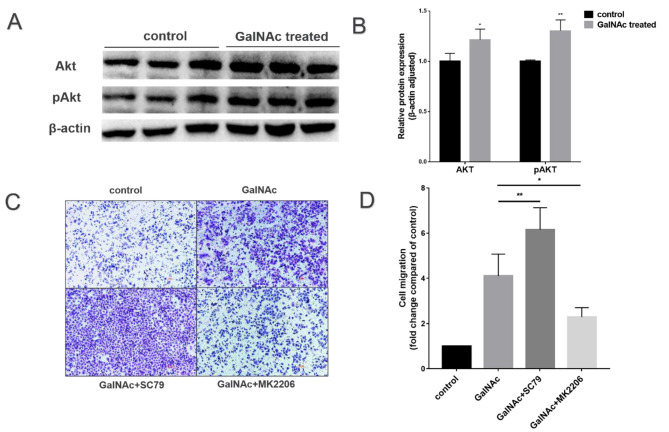
Methods: An in vitro inflammation and oxidative stress model was constructed using tumor necrosis-α (TNF-α) and lipopolysaccharide (LPS) action on WISH cells. Human amniotic epithelial cells (hAECs) were primarily cultured by digestion to construct a wound model. The effects of GalNAc on anti-inflammatory and anti-oxidative stress, migration and proliferation, epithelial-mesenchymal transition (EMT), glycosaminoglycan (GAG)/hyaluronic acid (HA) production, and protein kinase B (Akt) pathway in hAECs and WISH cells were analyzed using the DCFH-DA fluorescent probe, ELISA, CCK-8, scratch, transwell migration, and western blot to determine the mechanism by which GalNAc promotes amniotic wound healing.
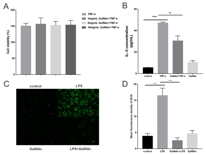
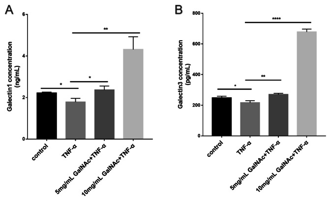
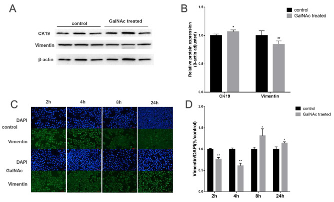
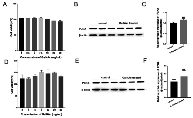
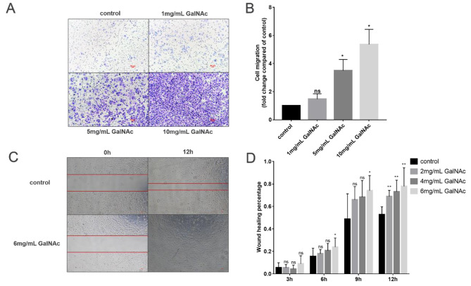
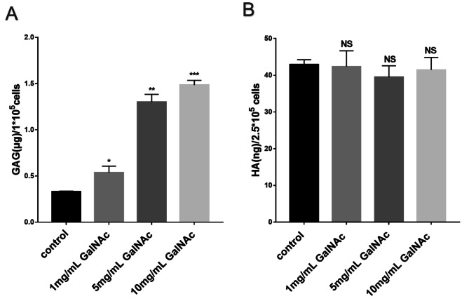
Results: GalNAc decreased IL-6 expression in TNF-α-stimulated WISH cells and ROS expression in LPS-stimulated WISH cells (P < 0.05). GalNAc promoted the expression of Gal-1 and Gal-3 with anti-inflammatory and anti-oxidative stress effects. GalNAc promoted the migration of hAECs (50% vs. 80%) and WISH cells through the Akt signaling pathway, EMT reached the point of promoting fetal membrane healing, and GalNAc did not affect the activity of hAECs and WISH cells (P > 0.05). GalNAc upregulated the expression of sGAG in WISH cells (P < 0.05) but did not affect HA levels (P > 0.05).
Conclusions: GalNAc might be a potential target for the prevention and treatment of PROM through the galectin pathway, including (i) inflammation; (ii) epithelial-mesenchymal transition; (iii) proliferation and migration; and (iv) regression, remodeling, and healing.
Keywords: Galectin; Healing; N-acetyl-d-galactosamine; PROM.
© 2024. The Author(s).
Conflict of interest statement
The authors declare that they have no competing interests.
Figures
Similar articles
-
The effect of lipopolysaccharide on anti-inflammatory and pro-inflammatory cytokines production of human amniotic epithelial cells.Reprod Biol. 2018 Dec;18(4):404-409. doi: 10.1016/j.repbio.2018.09.005. Epub 2018 Sep 13. Reprod Biol. 2018. PMID: 30220549
-
Healing of Preterm Ruptured Fetal Membranes.Sci Rep. 2017 Oct 13;7(1):13139. doi: 10.1038/s41598-017-13296-1. Sci Rep. 2017. PMID: 29030612 Free PMC article.
-
The regulatory role of the nuclear scaffold protein Emerin on the migration of amniotic epithelial cells and oxidative stress in a pressure environment.Placenta. 2025 May 23;165:102-113. doi: 10.1016/j.placenta.2025.04.010. Epub 2025 Apr 12. Placenta. 2025. PMID: 40245600
-
Possible important roles of galectins in the healing of human fetal membranes.Front Endocrinol (Lausanne). 2022 Aug 9;13:941029. doi: 10.3389/fendo.2022.941029. eCollection 2022. Front Endocrinol (Lausanne). 2022. PMID: 36017312 Free PMC article. Review.
-
Mini-review: Wound healing of amnion and macrophages.J Obstet Gynaecol Res. 2022 Mar;48(3):563-567. doi: 10.1111/jog.15161. Epub 2022 Jan 23. J Obstet Gynaecol Res. 2022. PMID: 35068017 Review.
References
MeSH terms
Substances
Grants and funding
LinkOut - more resources
Full Text Sources
Research Materials
