. 2024 Sep 4;22(1):823.
doi: 10.1186/s12967-024-05639-0.

RBM8A, a new target of TEAD4, promotes breast cancer progression by regulating IGF1R and IRS-2

Affiliations

RBM8A, a new target of TEAD4, promotes breast cancer progression by regulating IGF1R and IRS-2

Fang Li et al. J Transl Med. .

Abstract

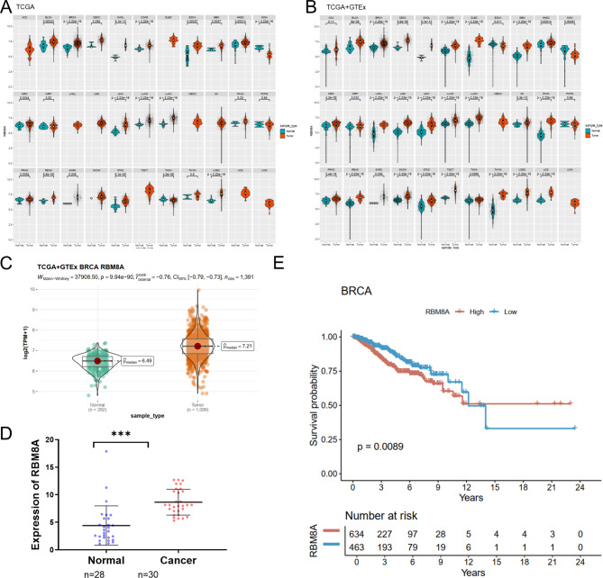
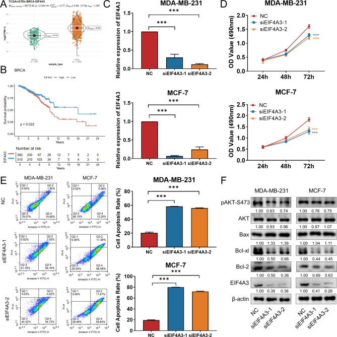
Background: Breast cancer (BC) is the most common malignant tumor in women worldwide, and further elucidation of the molecular mechanisms involved in BC pathogenesis is essential to improve the prognosis of BC patients. RNA Binding Motif Protein 8 A (RBM8A), with high affinity to a myriad of RNA transcripts, has been shown to play a crucial role in genesis and progression of multiple cancers. We attempted to explore its functional significance and molecular mechanisms in BC.

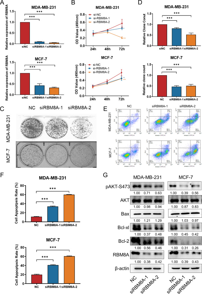
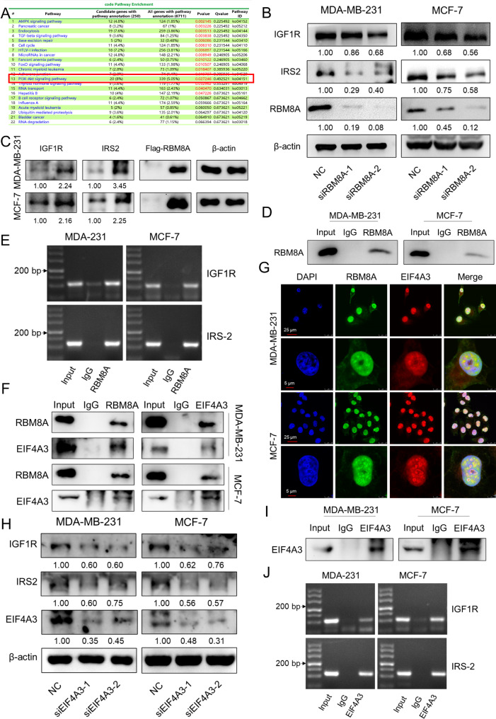
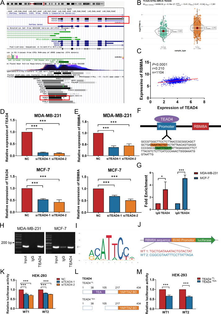
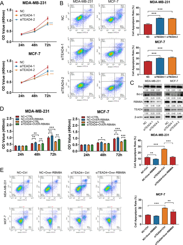
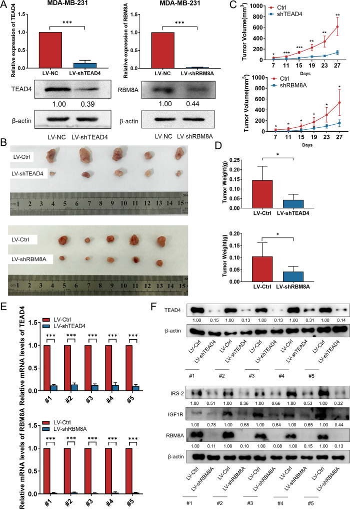
Methods: Bioinformatics analysis was performed on publicly available BC datasets. qRT-PCR was used to determine the expression of RBM8A in BC tissues. MTT assay, clone formation assay and flow cytometry were employed to examine BC cell proliferation and apoptosis in vitro. RNA immunoprecipitation (RIP) and RIP-seq were used to investigate the binding of RBM8A/EIF4A3 to the mRNA of IGF1R/IRS-2. RBM8A and EIF4A3 interactions were determined by co-immunoprecipitation (Co-IP) and immunofluorescence. Chromatin immunoprecipitation (Ch-IP) and dual-luciferase reporter assay were carried out to investigate the transcriptional regulation of RBM8A by TEAD4. Xenograft model was used to explore the effects of RBM8A and TEAD4 on BC cell growth in vivo.

Results: In this study, we showed that RBM8A is abnormally highly expressed in BC and knockdown of RBM8A inhibits BC cell proliferation and induces apoptosis in vitro. EIF4A3, which phenocopy RBM8A in BC, forms a complex with RBM8A in BC. Moreover, EIF4A3 and RBM8A complex regulate the expression of IGF1R and IRS-2 to activate the PI3K/AKT signaling pathway, thereby promoting BC progression. In addition, we identified TEAD4 as a transcriptional activator of RBM8A by Ch-IP, dual luciferase reporter gene and a series of functional rescue assays. Furthermore, we demonstrated the in vivo pro-carcinogenic effects of TEAD4 and RBM8A by xenograft tumor experiments in nude mice.

Conclusion: Collectively, these findings suggest that TEAD4 novel transcriptional target RBM8A interacts with EIF4A3 to increase IGF1R and IRS-2 expression and activate PI3K/AKT signaling pathway, thereby further promoting the malignant phenotype of BC cells.

Keywords: Breast cancer; EIF4A3; PI3K/AKT; RBM8A; TEAD4.


Conflict of interest statement

The authors declare no competing interests.

Figures

Fig. 1
Fig. 1
The expression of RBM8A is upregulated in breast cancer. (A) Comparison of RBM8A expression between tumor and normal tissues across different cancer types from TCGA database. (B) Comparison of RBM8A expression between tumor and normal tissues across different cancer types from TCGA and GTEx databases. (C) The expression of RBM8A in breast cancer tissues based on the TCGA and GTEx database. (D) qRT-PCR was used to detect RBM8A expression in breast cancer tissues and normal tissues. (E) Association between the RBM8A expression and the PFI of BC patients. ACC: Adrenocortical carcinoma; BLCA: Bladder Urothelial Carcinoma; BRCA: Breast invasive carcinoma; CESC: Cervical squamous cell carcinoma and endocervical adenocarcinoma; CHOL: Cholangiocarcinoma; COAD: Colon adenocarcinoma; DLBC: Lymphoid Neoplasm Diffuse Large B-cell Lymphoma; ESCA: Esophageal carcinoma; GBM: Glioblastoma multiforme; HNSC: Head and Neck squamous cell carcinoma; KICH: Kidney Chromophobe; KIRC: Kidney renal clear cell carcinoma; KIRP: Kidney renal papillary cell carcinoma; LAML: Acute Myeloid Leukemia; LGG: Brain Lower Grade Glioma; LIHC: Liver hepatocellular carcinoma; LUAD: Lung adenocarcinoma; LUSC: Lung squamous cell carcinoma; MESO: Mesothelioma; OV: Ovarian serous cystadenocarcinoma; PAAD: Pancreatic adenocarcinoma; PCPG: Pheochromocytoma and Paraganglioma; PRAD: Prostate adenocarcinoma; READ: Rectum adenocarcinoma; SARC: Sarcoma; SKCM: Skin Cutaneous Melanoma; STAD: Stomach adenocarcinoma; TGCT: Testicular Germ Cell Tumors; THCA: Thyroid carcinoma; THYM: Thymoma; UCEC: Uterine Corpus Endometrial Carcinoma; UCS: Uterine Carcinosarcoma; UVM: Uveal Melanoma.*p < 0.05, **p < 0.01, ***p < 0.001
Fig. 2
Fig. 2
Knockdown of RBM8A inhibits BC cell proliferation. (A) The mRNA expression of RBM8A was detected by qRT-PCR to confirm the knockdown efficiency of two siRNAs in MDA-MB-231 and MCF-7 cells. (B) MTT assay was used to detect the effect of RBM8A knockdown on the proliferation of BC cells. (C) Colony formation assay was used to detect the effect of RBM8A knockdown on the colony formation ability of BC cells. (D) Quantification of colony formation assay in BC cells transfected with RBM8A siRNAs. (E) Flow cytometry assay for apoptosis was used to detect the effect of knockdown of RBM8A on BC cell apoptosis. (F) Quantification of cell apoptosis assay in BC cells transfected with RBM8A siRNAs. (G) Western blotting was used to detect the effect of RBM8A knockdown on the expression of cell apoptosis related molecules at protein level in BC cells. *p < 0.05, **p < 0.01, ***p < 0.001
Fig. 3
Fig. 3
RBM8A/EIF4A3 complex promotes translation of IGF1R and IRS-2 (A) Pathway analysis of genes enriched by RIP-seq. (B) Western blotting was used to detect protein levels of IGF1R and IRS-2 in BC cells after knockdown of RBM8A. (C) Western blotting was used to detect protein levels of IGF1R and IRS-2 in BC cells after overexpression of RBM8A. (D) Western blotting was used to detect whether the target antibody pulls down the corresponding protein in RIP experiments. (E) After RIP experiments using RBM8A antibody, PCR and agarose gel electrophoresis were performed with primers for IGF1R and IRS-2. (F) MDA-MB-231 and MCF-7 cell lysates were subjected to Co-IP to detect RBM8A and EIF4A3 interaction. (G) Immunofluorescence assay was used to detect the co-localization of RBM8A and EIF4A3 in BC cells. (H) Western blotting was used to detect protein levels of IGF1R and IRS-2 in BC cells after knockdown of EIF4A3. (I) Western blotting was used to detect whether the target antibody pulls down the corresponding protein in RIP experiments. (J) After RIP experiments using EIF4A3 antibody, PCR and agarose gel electrophoresis were performed with primers for IGF1R and IRS-2
Fig. 4
Fig. 4
EIF4A3 is upregulated in BC and Knockdown of EIF4A3 inhibits BC cell proliferation. (A) The expression of RBM8A in breast cancer tissues based on the TCGA and GTEx database. (B) Association between the RBM8A expression and the OS of BC patients. (C) The mRNA expression of EIF4A3 was detected by qRT-PCR to confirm the knockdown efficiency of two siRNAs in MDA-MB-231 and MCF-7 cells. (D) MTT assay was used to detect the effect of EIF4A3 knockdown on the proliferation of BC cells. (E) Flow cytometry assay for apoptosis was used to detect the effect of knockdown of EIF4A3 on BC cell apoptosis. (F) Western blotting was used to detect the effect of EIF4A3 knockdown on the expression of cell apoptosis related molecules at protein level in BC cells. *p < 0.05, **p < 0.01, ***p < 0.001
Fig. 5
Fig. 5
TEAD4 binds to the promoter region of RBM8A to activate its transcription. (A) UCSC database analysis showed that TEAD4 could bind to the promoter region of RBM8A to regulate its transcription. (B) The expression of TEAD4 in breast cancer tissues based on the TCGA and GTEx database. (C) Correlation analysis of TEAD4 and RBM8A expression in BC based on TCGA database. (D) The mRNA expression of TEAD4 was detected by qRT-PCR to confirm the knockdown efficiency of two siRNAs in MDA-MB-231 and MCF-7 cells. (E) qRT-PCR was used to analyze the mRNA expression level of RBM8A in TEAD4 knockdown BC cells. (F) The UCSC database in conjunction with the JASPAR website predicts the binding sequence of TEAD4 to the promoter region of RBM8A. (G-H) The binding relationship between TEAD4 and the promoter region of RBM8A was verified by ChIP-qRT-PCR (G) and agarose gel electrophoresis (H). (I) DNA-binding motif of TEAD4 (JASPAR). (J) The binding sequence of TEAD4 on RBM8A predicted by JASPAR was subcloned into the pGL3 promoter luciferase vector. (K) Luciferase assays were performed in HEK-293 cells co-transfected with siNC/siTEAD4 and RBM8A promoter. Renilla luciferase served as the internal control. (L) Schematic representation of full-length (FL) TEAD4 and its deletion mutant TEA-. TEA- denotes TEA domain deletion. (M) Luciferase assays were performed in HEK-293 cells co-transfected with TEAD4 FL/TEAD4 TEA- and RBM8A promoter. Renilla luciferase served as the internal control. *p < 0.05, **p < 0.01, ***p < 0.001
Fig. 6
Fig. 6
Knockdown of TEAD4 inhibited BC cell proliferation, which was rescued by overexpression of RBM8A. (A) MTT assay was used to detect the effect of TEAD4 knockdown on the proliferation of BC cells. (B) Flow cytometry assay for apoptosis was used to detect the effect of knockdown of TEAD4 on BC cell apoptosis. (C) Western blotting was used to detect the effect of TEAD4 knockdown on the expression of cell apoptosis related molecules at protein level in BC cells. (D) MTT assay was performed to determine the impact of cell viability treated with NC + Ctrl, NC + Over-RBM8A, siTEAD4 + Ctrl, siTEAD4 + Over-RBM8A in BC cells. (E) Flow cytometry assay for apoptosis was performed to determine the impact of cell apoptosis treated with NC + Ctrl, NC + Over-RBM8A, siTEAD4 + Ctrl, siTEAD4 + Over-RBM8A in BC cells. *p < 0.05, **p < 0.01, ***p < 0.001
Fig. 7
Fig. 7
TEAD4 and RBM8A promote BC tumor growth in vivo. (A) The mRNA and protein expression of TEAD4/RBM8A in LV-shTEAD4, LV-shRBM8A4 and LV-Ctrl cells were detected by qRT-PCR and western blotting. (B) Size of xenograft tumors 27 days after injection of LV-shTEAD4, LV-shRBM8A and LV-Ctrl cells. (C) Growth curves of tumor volume were generated every 4 d for 27 d. (D) Weighing and statistical mapping of xenografted tumors. (E) The mRNA level of TEAD4/RBM8A in xenograft tumors were analyzed by qRT-PCR. (F) The protein level of TEAD4/RBM8A in xenograft tumors were analyzed by western blotting. *p < 0.05, **p < 0.01, ***p < 0.001
Fig. 8
Fig. 8
Schematic diagram. TEAD4 novel target RBM8A forming a complex with EIF4A3 to promote BC progression by regulating IGF1R and IRS-2

Similar articles

Cited by

References

    1. Siegel RL, Miller KD, Jemal A. Cancer statistics, 2019. CA Cancer J Clin. 2019;69:7–34. 10.3322/caac.21551 - DOI - PubMed
    1. McPherson K, Steel CM, Dixon JM. ABC of breast diseases. Breast cancer-epidemiology, risk factors, and genetics. BMJ. 2000;321:624–8. 10.1136/bmj.321.7261.624 - DOI - PMC - PubMed
    1. Molina R, Barak V, van Dalen A, Duffy MJ, Einarsson R, Gion M, Goike H, Lamerz R, Nap M, Soletormos G, Stieber P. Tumor markers in breast cancer- European Group on Tumor markers recommendations. Tumour Biol. 2005;26:281–93. 10.1159/000089260 - DOI - PubMed
    1. Vanderweyde T, Youmans K, Liu-Yesucevitz L, Wolozin B. Role of stress granules and RNA-binding proteins in neurodegeneration: a mini-review. Gerontology. 2013;59:524–33. 10.1159/000354170 - DOI - PMC - PubMed
    1. Glisovic T, Bachorik JL, Yong J, Dreyfuss G. RNA-binding proteins and post-transcriptional gene regulation. FEBS Lett. 2008;582:1977–86. 10.1016/j.febslet.2008.03.004 - DOI - PMC - PubMed

MeSH terms

LinkOut - more resources