Multicenter Study
. 2025 Jan:269:246-254.
doi: 10.1016/j.ajo.2024.08.040. Epub 2024 Sep 2.

Targeting the Tie-2 Receptor With Faricimab in Central Serous Chorioretinopathy: A Case Series Motivated by a Genetic Finding

Affiliations
Multicenter Study

Targeting the Tie-2 Receptor With Faricimab in Central Serous Chorioretinopathy: A Case Series Motivated by a Genetic Finding

Joel T Rämö et al. Am J Ophthalmol. 2025 Jan.

Abstract

Purpose: To investigate the effects of faricimab, a bispecific antibody targeting VEGF and Ang-2 (thus increasing Tie-2 activity), in patients with CSC based on a recent genetic study that implicated Tie-2 signaling in CSC pathophysiology.

Design: A retrospective interventional multicenter case series.

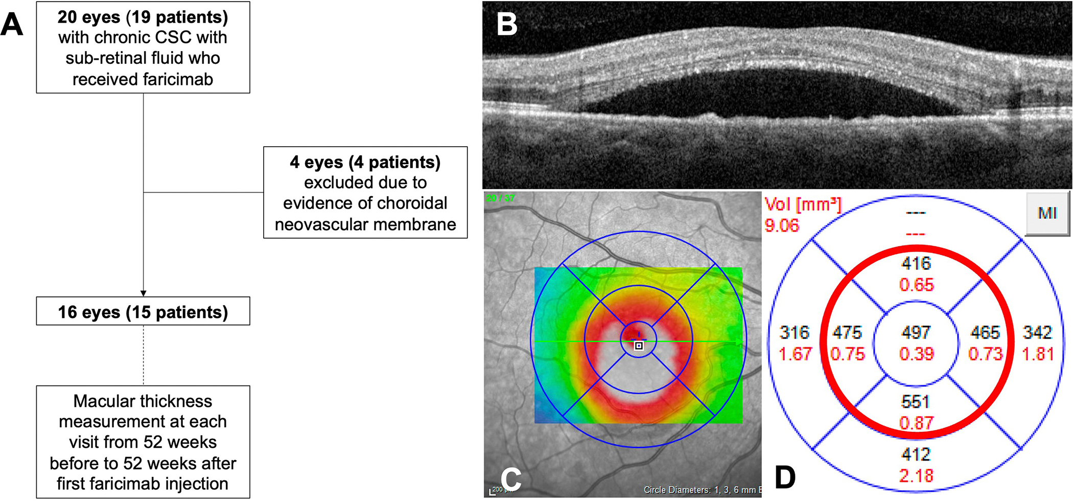
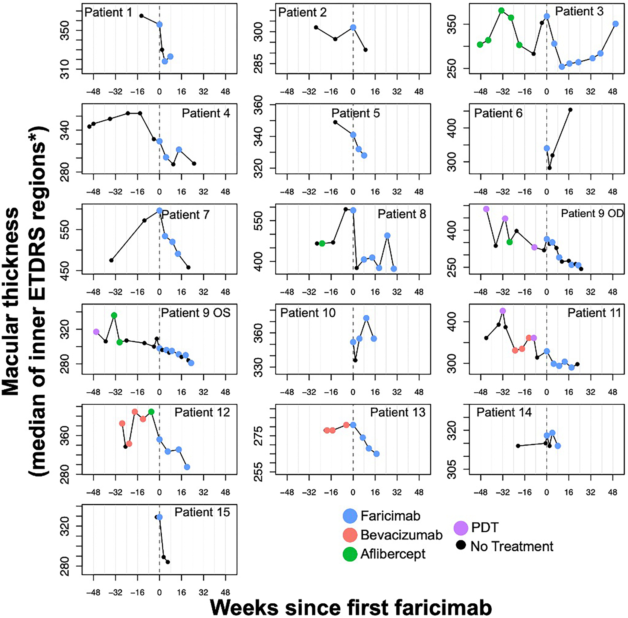
Methods: We included patients with chronic CSC (persistent or recurrent SRF for ≥6 months) who received at least one faricimab 6 mg injection between January 1 2022, and April 1 2024,. Study sites included Massachusetts Eye and Ear and University of California San Francisco. Patients with evidence of a choroidal neovascular membrane on color photos, optical coherence tomography (OCT) and/or fluorescein angiography were excluded. 16 eyes (15 patients) met the inclusion criteria. The median central macular thickness at each visit from 52 weeks before to 52 weeks after the first faricimab injection was calculated using automated Heidelberg Spectralis ETDRS subfield measurements.

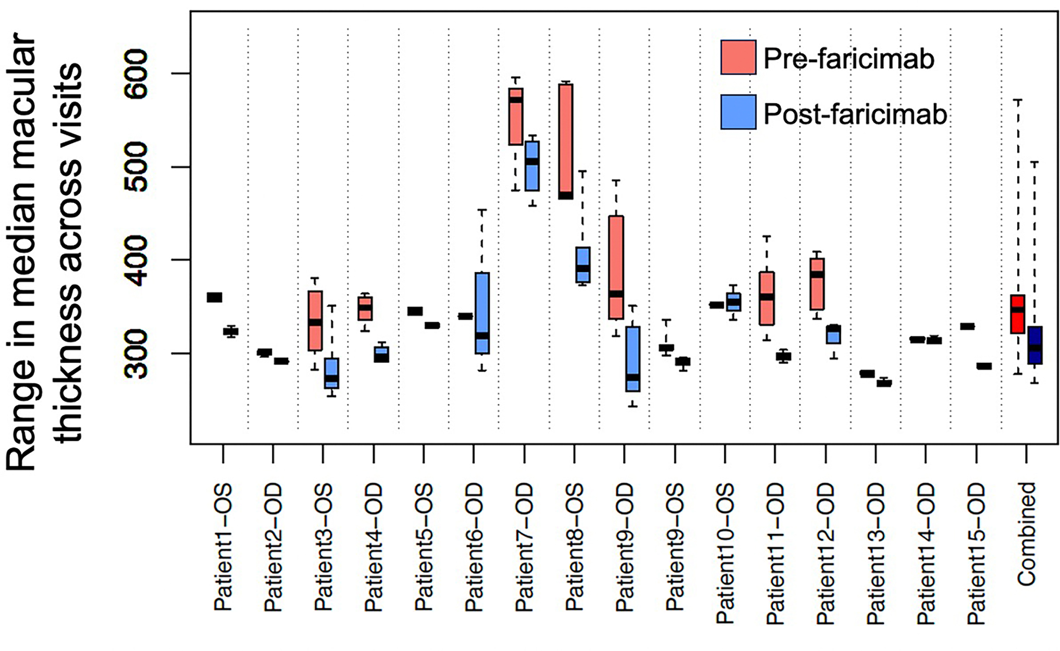
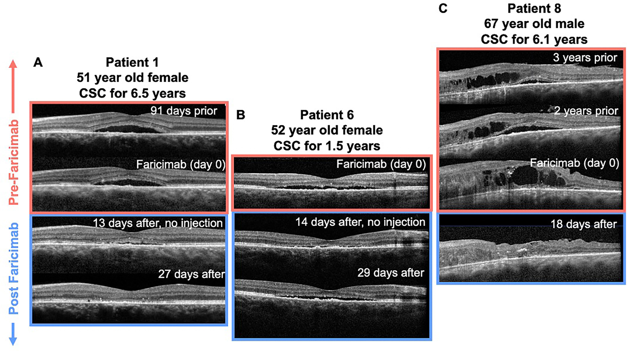
Results: Prior to treatment with faricimab, CSC had been diagnosed a median of 4.1 years (range 0.9-8) earlier and SRF (and intraretinal fluid [IRF] in a subset) had been continuously present for a median of 30 weeks (range 9-257). Decreases in macular thickness were observed in 14/16 eyes after the first faricimab injection and in 14/16 eyes in the full follow-up period compared with prior, 10 of which experienced complete resolution of SRF following the start of the first series of injections at a median of 4 weeks (range 2-25). One eye worsened after the second injection. The median improvement in macular thickness was 40 μm [range -3 to 89.5] (P = .0007). Upon review of OCT images, reductions in macular thickness were consistent with reductions in SRF and/or IRF. Visual acuity improved by 2 lines or more in 6/16 eyes.

Conclusions: In a retrospective case series of patients with chronic CSC and longstanding SRF, we observed improvement in macular thickness after intravitreal faricimab. While the small number of patients and variable natural history of CSC preclude definitive conclusions, a randomized controlled trial seems warranted.


Figures

Figure 1.
Figure 1.
Study design Panel A depicts the study flowchart. A multi-center retrospective chart review was conducted to identify patients with chronic CSC with sub-retinal fluid who received at least one faricimab 6mg injection. Study sites included Massachusetts Eye and Ear and University of California San Francisco. Patients with evidence of a choroidal neovascular membrane on color photos, optical coherence tomography (OCT) and/or fluorescein angiography were excluded. Macular thickness was quantified from OCT images taken 52 weeks before to 52 weeks after the first faricimab injection. Panel B demonstrates the confirmation of proper segmentation from Bruch’s membrane to the ILM. Panels C and D demonstrate extraction of automated macular thickness measurements (in microns) of the ETDRS central subfield and pericentral ring (inside the red circle in panel D), the median of which yielded a single thickness value per scan. ETDRS, Early Treatment of Diabetic Retinopathy Study.
Figure 2.
Figure 2.
Trajectories of individual patients before and after the first injection with faricimab Time up to 52 weeks before and up to 52 weeks after the first treatment with facirimab is shown on the x-axis, with the time of the first faricimab treatment for each patient set to 0. Median macular thickness measurement (in microns) across inner ETDRS regions is shown on the y-axis for each patient treated with at least one dose of faricimab. The y-axis scale varies between patients depending on the maximum and minimum values observed for each patient. Visits during which the patients received any form of treatment are denoted with colored circles (blue, faricimab; red, bevacizumab; green, aflibercept; pink, photodynamic therapy [PDT]), and visits during which the patients did not receive treatment are denoted with black circles. Patient 8 had two treated eyes. *Patient 13 had SRF far from the fovea and thus the superior and nasal outer ETRDS regions were used. OD, right eye; OS, left eye. ETDRS, Early Treatment of Diabetic Retinopathy Study. SRF, subretinal fluid.
Figure 3.
Figure 3.
Distributions of macular thickness before and after the first treatment with faricimab The distribution of macular thickness across visits (in μm) is shown on the y-axis for each patient before and after faricimab initiation. Macular thickness at each visit is defined as the median of the ETDRS central subfield and inner pericentral ETDRS subfields, as shown in Figure 1. Values are shown separately for measurements taken before (red) and after the first treatment with faricimab (blue). Data were included from a period ranging from 52 weeks before to 52 weeks after the first treatment with faricimab. The box and whisker plots denote the median (black line), 25th percentile (bottom of box), 75th percentile (top of box), minimum value (lower whisker) and maximum value (upper whisker) across visits for each patient. ETDRS, Early Treatment of Diabetic Retinopathy Study; OD, right eye; OS, left eye.
Figure 4.
Figure 4.
Optical coherence tomography images of patients 1, 6 and 8 before and after treatment with faricimab Panel A depicts optical coherence tomography (OCT) images from Patient 1 who had a 6.5 year history of SRF. Faricimab was administered on day 0 and the SRF resolved by day 27. Panel B depicts Patient 6 who had a 1.5 year history of SRF. Faricimab was given on day 0 (first day in our system), and the SRF improved at day 14 but returned at day 29. Panel C depicts OCT images from Patient 8 who had a 6 year history of both subretinal fluid and intraretinal fluid. 18 days after faricimab injection, SRF and IRF were dramatically improved.

Similar articles

Cited by

  • Rare genetic variation in PTPRB is associated with central serous chorioretinopathy, varicose veins and glaucoma.
    Rämö JT, Gorman BR, Weng LC, Jurgens SJ, Singhanetr P, Tieger MG, van Dijk EH, Halladay CW, Wang X, Hauser BM, Kim SH, Brinks J, Choi SH, Luo Y; FinnGen; VA Million Veteran Program; Pyarajan S, Nealon CL, Gorin MB, Wu WC, Anthony SA, Roncone DP, Sobrin L, Kaarniranta K, Yzer S, Palotie A, Peachey NS, Turunen JA, Boon CJ, Ellinor PT, Iyengar SK, Daly MJ, Rossin EJ. Rämö JT, et al. Nat Commun. 2025 May 3;16(1):4127. doi: 10.1038/s41467-025-58686-6. Nat Commun. 2025. PMID: 40319023 Free PMC article.

References

    1. Spaide RF, Campeas L, Haas A, et al. Central serous chorioretinopathy in younger and older adults. Ophthalmology. 1996;103(12):2070–2079; discussion 2079–80. - PubMed
    1. Montero JA, Ruiz-Moreno JM. Optical coherence tomography characterisation of idiopathic central serous chorioretinopathy. Br J Ophthalmol. 2005;89(5):562–564. - PMC - PubMed
    1. Mrejen S, Balaratnasingam C, Kaden TR, et al. Long-term Visual Outcomes and Causes of Vision Loss in Chronic Central Serous Chorioretinopathy. Ophthalmology. 2019;126(4):576–588. - PubMed
    1. Chan WM, Lai TYY, Lai RYK, Liu DTL, Lam DSC. Half-dose verteporfin photodynamic therapy for acute central serous chorioretinopathy: one-year results of a randomized controlled trial. Ophthalmology. 2008;115(10):1756–1765. - PubMed
    1. Chung YR, Seo EJ, Lew HM, Lee KH. Lack of positive effect of intravitreal bevacizumab in central serous chorioretinopathy: meta-analysis and review. Eye. 2013;27(12):1339–1346. - PMC - PubMed

Publication types

LinkOut - more resources