. 2025 Jan;169(1):e16217.
doi: 10.1111/jnc.16217. Epub 2024 Sep 4.

Relaxin family peptide receptor 3 (RXFP3) expressing cells in the zona incerta/lateral hypothalamus augment behavioural arousal

Affiliations

Relaxin family peptide receptor 3 (RXFP3) expressing cells in the zona incerta/lateral hypothalamus augment behavioural arousal

Brandon K Richards et al. J Neurochem. 2025 Jan.

Abstract

Fear-related psychopathologies, such as post-traumatic stress disorder, are linked to dysfunction in neural circuits that govern fear memory and arousal. The lateral hypothalamus (LH) and zona incerta (ZI) regulate fear, but our understanding of the precise neural circuits and cell types involved remains limited. Here, we examined the role of relaxin family peptide receptor 3 (RXFP3) expressing cells in the LH/ZI in conditioned fear expression and general arousal in male RXFP3-Cre mice. We found that LH/ZI RXFP3+ (LH/ZIRXFP3) cells projected strongly to fear learning, stress, and arousal centres, notably, the periaqueductal grey, lateral habenula, and nucleus reuniens. These cells do not express hypocretin/orexin or melanin-concentrating hormone but display putative efferent connectivity with LH hypocretin/orexin+ neurons and dopaminergic A13 cells. Following Pavlovian fear conditioning, chemogenetically activating LH/ZIRXFP3 cells reduced fear expression (freezing) overall but also induced jumping behaviour and increased locomotor activity. Therefore, the decreased freezing was more likely to reflect enhanced arousal rather than reduced fear. Indeed, stimulating these cells produced distinct patterns of coactivation between several motor, stress, and arousal regions, as measured by Fos expression. These results suggest that activating LH/ZIRXFP3 cells generates brain-wide activation patterns that augment behavioural arousal.

Keywords: RXFP3; arousal; defensive response; fear conditioning; hypothalamus; zona incerta.


Conflict of interest statement

Andrew J. Lawrence is the current Editor‐in‐Chief of the Journal of Neurochemistry. The authors declare no competing financial interests.

Figures

FIGURE 1
FIGURE 1
RNAscope validation of RXFP3‐Cre driver mice. (a) Representative fluorescent photomicrograph of Rxfp3 mRNA (left), Cre mRNA (middle) and merge (right) in the LH/ZI. (b) Donut graphs demonstrating the proportion of Cre‐expressing cells that express Rxfp3 and the proportion of Rxfp3 cells that co‐express Cre. Scale bar = 50 μm. n = 2 mice.
FIGURE 2
FIGURE 2
Long‐range efferent projections of LH/ZIRXFP3 cells. (a) Representative photomicrograph of eYFP‐expressing cell bodies in the ZI and LH of an RXFP3‐Cre‐eYFP reporter mouse (n = 4). (b) Diagram depicting anterograde tracing method. LH/ZIRXFP3 cells were targeted via unilateral stereotaxic delivery of a Cre‐dependent anterograde tracer virus (AAV‐DJ‐hSyn‐FLEX‐mGFP‐synaptophysin‐mRuby; n = 5) into the LH/ZI. (c) Representative fluorescent photomicrograph of anterograde tracer virus injection site in the LH/ZI. (d) Unilateral coronal diagram cascade depicting anterograde tracer injection sites across the LH/ZI. Each dot/box represents an mGFP immunoreactive cell body. Adapted from Paxinos and Franklin (2004). (e–h) Representative fluorescent photomicrographs of mGFP expression (left), mRuby expression (middle), and merge (right) of key LH/ZIRXFP3 efferent targets, including the lateral habenula (e), nucleus reuniens (f), rostral periaqueductal grey (g), and the posterior hypothalamus (h). Aq, cerebral aqueduct; LH, lateral hypothalamus; LHb, lateral habenula; pc, posterior commissure; PH, posterior hypothalamus; MHb, medial habenula; mt, mammillothalamic tract; Re, nucleus reuniens; rPAG, rostral periaqueductal grey; ZI, zona incerta. Scale bars = 100 μm.
FIGURE 3
FIGURE 3
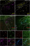
Phenotyping of LH/ZIRXFP3 cells and select LH/ZIRXFP3 efferent targets. (a, b) Representative fluorescent photomicrographs of eYFP immunoreactive neurons and hypocretin/orexin (Hcrt/Ox) (a) or melanin‐concentrating hormone (MCH) (b) immunoreactive neurons in the LH/ZI from RXFP3‐Cre‐eYFP reporter mice. (c, d) Top panels, representative fluorescent photographs of LH/ZIRXFP3 efferent targets expressing hypocretin/orexin in the LH (c) and tyrosine hydroxylase in the ZI A13 group (d). Bottom panels display zoomed‐in images of the inset box region split by fluorescent channel. The bottom right panels display merged images of the inset box region. f, fornix; Hcrt/Ox, hypocretin/orexin; MCH, melanin‐concentrating hormone; LH, lateral hypothalamus; TH, tyrosine hydroxylase; ZI, zona incerta; 3 V, third ventricle. Arrows indicate putative synapses onto hypocretin/orexin immunoreactive cells in (c), and onto tyrosine hydroxylase immunoreactive cells in (d). Scale bars = 100 μm.
FIGURE 4
FIGURE 4
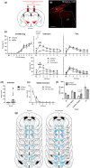
Chemogenetic activation of LH/ZIRXFP3 cells during context‐dependent conditioned fear extinction. (a) An excitatory DREADD (AAV‐hSyn‐DIO‐hM3D(Gq)‐mCherry) or control virus (AAV‐hSyn‐DIO‐mCherry) was bilaterally injected into the LH/ZI of RXFP3‐Cre mice to permit chemogenetic excitation of LH/ZIRXFP3 cells during behaviour. (b) Representative photomicrograph of hM3Dq DREADD injection site. Scale bar = 100 μm. (c) Behavioural data of freezing during fear conditioning (top left), fear extinction (top middle), and retrieval test (top right). All mice received clozapine‐N‐oxide injection (CNO; 3 mg/kg, i.p.) 30 min before the fear extinction session. Both conditioning and retrieval test days were undertaken drug‐free. Because a subset of mice displayed jumping behaviour via chemogenetic activation of LH/ZIRXFP3 cells during fear extinction, hM3Dq mice were subdivided into those that exhibited jumping behaviour (jumpers) and those that did not (non‐jumpers) for both extinction (bottom middle) and retrieval test (bottom right). (d) Graph showing that a subset of mice displayed jumping behaviour via chemogenetic activation of LH/ZIRXFP3 cells during extinction, and mCherry mice did not display jumping behaviour. (e) Jumping primarily occurred in 120‐s baseline period. Graph shows the average number of jumps during baseline and in six successive CS‐inter‐trial‐interval blocks (100 s/block). Black squares represent jumps during CS presentation (CS jumps); white squares indicate jumps during BL or the ITIs (non‐CS jumps). (f) Relative proportions of hM3Dq expression in different areas of the LH/ZI. (g) Spread of excitatory hM3Dq DREADD throughout the LH/ZI and surrounding areas for mice that displayed jumping behaviour upon chemogenetic activation during fear extinction (left, jumpers), and those that did not (right, non‐jumpers). Numbers represent the distance (mm) from Bregma. Coronal section plates adapted from Paxinos and Franklin (2004). mCherry n = 29, hM3Dq n = 32, jumpers n = 14, non‐jumpers n = 18. hM3Dq vs. mCherry, *p < 0.05, ***p < 0.001; jumpers vs. mCherry *p < 0.01, ****p < 0.0001; non‐jumpers vs. mCherry # p < 0.05. Data are presented as mean ± SEM. BL, baseline.
FIGURE 5
FIGURE 5
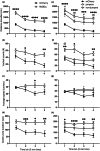
Chemogenetic activation of LH/ZIRXFP3 cells during an open‐field test. (a–d) Locomotor data showing (a) ambulatory distance, (b) vertical counts, (c) average ambulatory speed, (d) and centre zone duration of hM3Dq mice compared to mCherry mice. (e–h) Locomotor data showing (e) ambulatory distance, (f) vertical counts, (g) average ambulatory speed, (h) and centre zone duration with hM3Dq mice divided into those that displayed jumping behaviour upon chemogenetic activation of LH/ZIRXFP3 cells during fear extinction (jumpers) and those that did not (non‐jumpers). Total n = 44. mCherry n = 23, hM3Dq n = 21, jumpers n = 7, non‐jumpers n = 14. hM3Dq vs mCherry *p < 0.05; **p < 0.01; ***p < 0.001; ****p < 0.0001. Jumpers vs. non‐jumpers, @ p < 0.05. Non‐jumpers vs. mCherry, # p < 0.05, ## p < 0.01. Data are presented as mean ± SEM.
FIGURE 6
FIGURE 6
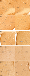
Effects of chemogenetically activating LH/ZIRXFP3 cells on Fos expression. (a–e) Representative brightfield photomicrographs of Fos expression in the cingulate cortex, secondary motor cortex (a), claustrum (b), dorsomedial striatum (c), medial preoptic area, lateral preoptic area (d), medial septum, and lateral septum (e) in chemogenetically activated mice (hM3Dq, left panels) versus controls (mCherry, right panels). AID, anterior insula, dorsal; AIV, anterior insula, ventral; cc, corpus callosum; Cg, cingulate cortex; Cl, claustrum; DEn, dorsal endopiriform cortex; DMS, dorsomedial striatum; LPO, lateral preoptic area; LS, lateral septum; LV, lateral ventricle; M2, secondary motor cortex; MPO, medial preoptic area; MS, medial septum; NAcC, nucleus accumbens core; NAcSh, nucleus accumbens shell; SHi, septohippocampal nucleus; Str, striatum. Scale bars = 100 μm.
FIGURE 7
FIGURE 7
Hierarchical clustering of Euclidean distance matrices for Fos expression between hM3Dq and mCherry groups. Modules were determined by cutting each dendrogram at 50% of the maximum height and are identified by the green squares. (a, b) Euclidean distance of each analysed brain region compared to all others in control (mCherry) mice (a) or hM3Dq mice (b). Seven modules of co‐activation were identified in controls, whereas four modules of activation were identified in the hM3Dq group. (c) Number of modules for each group after cutting the dendrogram at different percentages of the tree height. Abbreviations for regions analysed can be found in Table 2. Scale bar indicates Euclidean distance.

References

    1. Adamantidis, A. , & de Lecea, L. (2008). Physiological arousal: A role for hypothalamic systems. Cellular and Molecular Life Sciences, 65, 1475–1488. - PMC - PubMed
    1. Albert‐Gascó, H. , García‐Avilés, Á. , Moustafa, S. , Sánchez‐Sarasua, S. , Gundlach, A. L. , Olucha‐Bordonau, F. E. , & Sánchez‐Pérez, A. M. (2017). Central relaxin‐3 receptor (RXFP3) activation increases ERK phosphorylation in septal cholinergic neurons and impairs spatial working memory. Brain Structure & Function, 222, 449–463. - PubMed
    1. Anversa, R. , Campbell, E. J. , Walker, L. C. , Ch'ng, S. , Muthmainah, M. , Kremer, S. , Guimarães, A. , O'Shea, M. J. , He, S. , Dayas, C. V. , Andrews, Z. B. , Lawrence, A. J. , & Brown, R. M. (2023). A paraventricular thalamus to insular cortex glutamatergic projection gates “emotional” stress‐induced binge eating in females. Neuropsychopharmacology, 48, 1931–1940. - PMC - PubMed
    1. Arnsten, A. F. T. (2015). Stress weakens prefrontal networks: Molecular insults to higher cognition. Nature Neuroscience, 18, 1376–1385. - PMC - PubMed
    1. Bankhead, P. , Loughrey, M. B. , Fernández, J. A. , Dombrowski, Y. , McArt, D. G. , Dunne, P. D. , McQuaid, S. , Gray, R. T. , Murray, L. J. , Coleman, H. G. , James, J. A. , Salto‐Tellez, M. , & Hamilton, P. W. (2017). QuPath: Open source software for digital pathology image analysis. Scientific Reports, 7, 16878. - PMC - PubMed

Substances

LinkOut - more resources