. 2024 Dec;13(1):2401931.
doi: 10.1080/22221751.2024.2401931. Epub 2024 Sep 19.

Identification of mpox M1R and B6R monoclonal and bispecific antibodies that efficiently neutralize authentic mpox virus

Affiliations

Identification of mpox M1R and B6R monoclonal and bispecific antibodies that efficiently neutralize authentic mpox virus

Zuning Ren et al. Emerg Microbes Infect. 2024 Dec.

Abstract

In 2022, the monkeypox virus (mpox virus, MPXV) exhibited global dissemination across six continents, representing a notable challenge owing to the scarcity of targeted antiviral interventions. Passive immunotherapy, such as the use of monoclonal antibodies (mAbs) and bispecific antibodies (bsAbs), has emerged as a promising option for antiviral regimens. Here, we generated several mAbs against M1R and B6R of MPXV, and subsequently characterized the antiviral activity of these antibodies both in vitro and in vivo. Two neutralizing mAbs, M1H11 and M3B2, targeting M1R, and one B6R-specific mAb, B7C9, were identified. They exhibited varying antiviral efficacy against vaccinia virus (VACV) in vitro and in vivo. A cocktail comprising M1H11 and M3B2 demonstrated a superior protective effect in vivo. A bsAb, Bis-M1M3, was engineered by conjugating the fragment crystallizable (Fc) region of the human-mouse chimeric engineered M1H11 with the single-chain fragment variable (scFv) of M3B2. In mice challenged with MPXV, Bis-M1M3 showed a notable protective effects. Analysis of neutralization mechanism showed that these mAbs and Bis-M1M3 exerted virus-neutralizing effects before the virus infects cells. In vivo pharmacokinetic experiments showed that Bis-M1M3 has a long half-life in rhesus macaques. This study provides crucial insights for further research on broad-spectrum antiviral drugs against MPXV and other orthopoxviruses.

Keywords: Antiviral mechanism; Bispecific antibody; Monoclonal antibody; Mpox virus; Vaccinia virus.


Conflict of interest statement

No potential conflict of interest was reported by the author(s).

Figures

Figure 1.
Figure 1.
The binding, neutralizing activities, and sequences of the complementarity determining regions (CDRs) of the monoclonal antibodies (mAbs). (A–C) ELISAs for the binding activities of four M1R-targeting mAbs—M1H11, M3B2, M4B6, and M13H—and three B6R-specific mAbs—B7C9, B8A12, and B10D3—to (A) a vaccinia virus (VACV) (Tian Tan) lysate and their respective binding activities to (B) the M1R protein and (C) the B6R protein. Neutralization assays for the mAbs against the mature virion (MV) (D) or enveloped virion (EV) (E) form of VACV (Tian Tan). The error bars represent means ± SEM. (F) The binding affinities of the mAbs for the target antigens obtained through surface plasmon resonance (SPR) analysis. (G) The DNA sequences of the CDRs of three mAbs.
Figure 2.
Figure 2.
Model of the binding of M1H11 and M3B2 to M1R, and the binding of B7C9 to B6R. The structures of the MPXV proteins M1R and B6R were predicted by AlphaFold2 and the Fv domain of the monoclonal antibodies (mAbs) was predicted by SAbPred. The docked complexes were created by ZDOCK, ClusPro, and AlphaFold2 and were then further investigated manually. (A) The most plausible result for the overall architecture of M1R bound to M1H11. The structure of M1R is shown in magenta. The cysteines that form disulfide bonds are shown as sticks. The heavy and light chains of M1H11 are shown in blue and cyan, respectively. (B) The M1R-M1H11 interface. M1R and the Fv domain of M1H11 are shown in cartoon and surface representation, respectively. The residues on the heavy and light chains involved in M1R binding are shown as sticks and coloured in blue and cyan. (C) The detailed interactions between M1R and the complementarity-determining regions (CDRs) on M1H11. Residues on M1R are shown in stick representation and coloured in magenta. Residues on the heavy and light chains of M1H11 are shown in stick representation and coloured in blue and cyan. (D) The most plausible result for M1R-M3B2 binding. The structure of M1R is shown as a magenta cartoon. The cysteines that form disulfide bonds are shown as sticks. The heavy and light chains of M3B2 are coloured blue and cyan, respectively. (E) The M1R-M3B2 interface. M1R and the Fv domain of M3B2 are shown in cartoon and surface representation, respectively. The residues on the heavy and light chains involved in M1R binding are shown as sticks and coloured in blue and cyan. (F) The detailed interactions between M1R and the CDRs on M3B2. Residues on M1R are shown in stick representation and coloured in magenta. Residues on the heavy chain and light chain of M3B2 are shown in stick representation and coloured in blue and cyan. (G) The most plausible result for the overall architecture of B6R-B7C9. The structure of B6R is shown as a magenta cartoon. The cysteines that form disulfide bonds are shown as sticks. The heavy and light chains of B7C9 are coloured blue and cyan, respectively. (H) The B6R-B7C9 interface. B6R and the Fv domain of B7C9 are shown in cartoon and surface representation, respectively. The residues on the heavy and light chains involved in B6R binding are shown as sticks and coloured in blue and cyan. (I) The detailed interactions between B6R and the CDRs on B7C9. Residues on B6R are shown in stick representation and coloured in magenta. Residues on the heavy and light chains of B7C9 are shown in stick representation and coloured in blue and cyan.
Figure 3.
Figure 3.
The therapeutic effects of monoclonal antibodies (mAbs) in mice infected with the vaccinia virus (VACV) (Tian Tan). Mice (n = 8 per group) were infected with 2.5 × 105 TCID50 doses of VACV (Tian Tan) and then treated with the indicated antibodies (10 mg/kg) via intraperitoneal administration 12 h later. The lungs of mice (n = 3 per group) were sampled 2 days post-VACV (Tian Tan) challenge. (A) The body weight changes (n = 5 per group) within 10 days post-challenge. Two-way ANOVA was used for comparisons of post-challenge weight changes among the groups. “#” indicates p < 0.05 in comparison between the M1H11 & M3B2 cocktail group and other groups; “$” indicates p < 0.05 in comparison between the IgG control group and other groups. (B) Survival curves monitored within 10 days post-challenge. (C) Lung viral loads 2 days post-challenge. Error bars represent means ± SEM. One-way ANOVA was used for comparisons between the indicated antibody treatment groups and the IgG control group. **** p < 0.0001, *** p < 0.001, ** p < 0.01, * p < 0.05. (D) Haematoxylin and eosin (H&E) staining of the lungs of mice 2 days post-challenge. The yellow arrows indicate alveolar wall granulocyte infiltration, the red arrows indicate perivascular and peribronchiolar lymphocyte infiltration, and the blue arrows indicate bronchiolar epithelial cell necrosis.
Figure 4.
Figure 4.
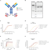
In vitro assessment of the functionality of the bispecific antibody (bsAb) Bis-M1M3. (A) A simple model of Bis-M1M3. (B) SDS–PAGE (with DTT) of Bis-M1M3, cM1H11, and cM3B2. ELISA-based analysis of the binding activities of mouse-human chimeric engineered mAbs and bsAb against (C) M1R and (D) inactivated vaccinia virus (VACV) (Tian Tan) lysate. (E) The binding affinity of Bis-M1M3 for the M1R protein obtained through SPR analysis. (F) The neutralization activities of Bis-M1M3, cM1H11, cM3B2, and the cM1H11 & cM3B2 cocktail against MPXV. The error bars represent means ± SEM.
Figure 5.
Figure 5.
The therapeutic effects of the bispecific antibody (bsAb) Bis-M1M3 in mice infected with MPXV. Mice (n = 8 per group) were infected with 5 × 105 TCID50 doses of MPXV and then treated with the indicated antibodies (5 mg/kg) via intraperitoneal administration 24 h later. The lungs of mice (n = 3 per group) were sampled 2 days post-MPXV challenge. (A) The body weight changes (n = 3 per group) within 14 days post-challenge. Two-way ANOVA was used for post-challenge comparisons of weight changes among groups. “#” indicates p < 0.05 in comparison between the Bis-M1M3 group and other groups. “&” indicates p < 0.05 in comparison between the cM1H11 & cM3B2 cocktail group and other groups. “$” indicates p < 0.05 in comparison between the IgG control group and other groups. (B) Survival curves monitored within 14 days post-challenge. (C) Lung viral loads sampled 2 days post-challenge. The error bars represent means ± SEM. One-way ANOVA was used for comparisons among the groups. **** p < 0.0001; “$” indicates p < 0.05 in comparison between the IgG control group and other groups. (D) Haematoxylin and eosin (H&E) staining of the lungs of mice 2 days post-challenge. The yellow arrows indicate alveolar wall granulocyte infiltration, the red arrows indicate perivascular and peribronchiolar lymphocyte infiltration, and the blue arrows indicate bronchiolar epithelial cell necrosis.
Figure 6.
Figure 6.
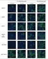
The inhibitory effects of the mouse-human chimeric engineered monoclonal antibodies (mAbs) and bispecific antibody (bsAb) against the vaccinia virus (VACV) (Tian Tan) as determined by indirect immunofluorescence analysis using confocal microscopy. Pre-incubation group: VACV (Tian Tan) and antibodies were incubated together for 1 h before cell infection. Post-infection group: Cells were first infected with VACV (Tian Tan) and then the antibodies were added. The green fluorescence indicates the presence of the VACV (Tian Tan). Nuclei were counterstained with Hoechst 33342 (with DAPI, blue). The image shows the merged FITC and DAPI signals. Magnification = ×40.
Figure 7.
Figure 7.
Serum antibody concentrations in rhesus macaques infused with Bis-M1M3. (A) Schematic diagram showing the antibody pharmacokinetics in rhesus monkeys. The rhesus macaques (n = 4 per group) were injected with a 10 mg/kg dose of antibody on Day 0, and serum was collected on Days 1, 3, 5, 7, 9, 11, and 14; the antibody concentration was determined by ELISA. (B) The half-life of Bis-M1M3 and control mAb 12G6 (an influenza antibody) in rhesus macaques was measured in serum over 14 days after the intravenous administration of a single 10 mg/kg dose of each antibody.

References

    1. Titanji BK, Tegomoh B, Nematollahi S, et al. Monkeypox: a contemporary review for healthcare professionals. Open Forum Infect Dis. 2022;9:ofac310, doi: 10.1093/ofid/ofac310 - DOI - PMC - PubMed
    1. Nuzzo JB, Borio LL, Gostin LO.. The WHO declaration of monkeypox as a global public health emergency. Jama. 2022;328:615–617. doi: 10.1001/jama.2022.12513 - DOI - PubMed
    1. 2022–2023 Mpox Outbreak Global Map . Available online: https://www.cdc.gov/poxvirus/monkeypox/response/2022/world-map.html. Accessed december 2023.
    1. Shen C, Chen J, Li R, et al. A multimechanistic antibody targeting the receptor binding site potently cross-protects against influenza B viruses. Sci Transl Med. 2017;9; doi: 10.1126/scitranslmed.aam5752 - DOI - PubMed
    1. Wu Y, Wang F, Shen C, et al. A noncompeting pair of human neutralizing antibodies block COVID-19 virus binding to its receptor ACE2. Science (New York, N.Y.). 2020;368:1274–1278. doi: 10.1126/science.abc2241 - DOI - PMC - PubMed