

doi: 10.1016/j.eats.2024.103027.
eCollection 2024 Aug.
Medial Meniscus Posterior Root Transtibial Pullout Repair With Progressively Tensioning Subcortical Fixation Button
Affiliations
- PMID: 39233796
- PMCID: PMC11369947
- DOI: 10.1016/j.eats.2024.103027
Item in Clipboard
Medial Meniscus Posterior Root Transtibial Pullout Repair With Progressively Tensioning Subcortical Fixation Button
Arthrosc Tech.
.
Abstract
We describe a surgical technique to repair medial meniscus posterior root tears through a transtibial pullout repair with a subcortical button for tibial fixation. This technique allows progressive tensioning of the repaired root without losing tension both during suturing of the knots above the button and after the procedure, owing to the specific button configuration.
© 2024 The Authors.
Conflict of interest statement
All authors (S.P., N.P., M.G., M.I., R.M.A., R.T.C., J.C.M.,) declare that they have no known competing financial interests or personal relationships that could have appeared to influence the work reported in this paper.
Figures
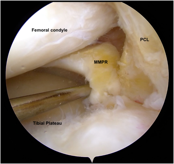
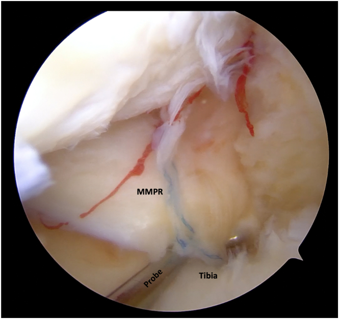
The medial meniscus posterior root (MMPR) is palpated with a probe, and the integrity is checked. Patient in supine position, right knee, arhtroscopic view from standar AL portal. (PCL, posterior cruciate ligament.)
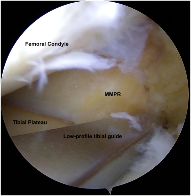
Low-profile tibial guide positioned over debrided root to reproduce its anatomic footprint. Patient in supine position, right knee, arhtroscopic view from standar AL portal. (MMPR, medial meniscus posterior root.)
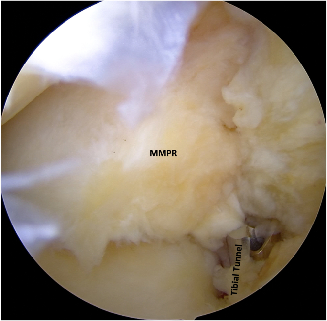
A 4.5-mm tibial tunnel is drilled into the anatomic footprint of the medial meniscus. Patient in supine position, right knee, arhtroscopic view from standar AL portal. (MMPR, medial meniscus posterior root.)
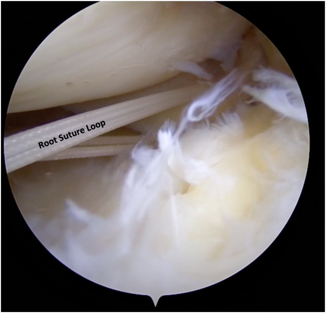
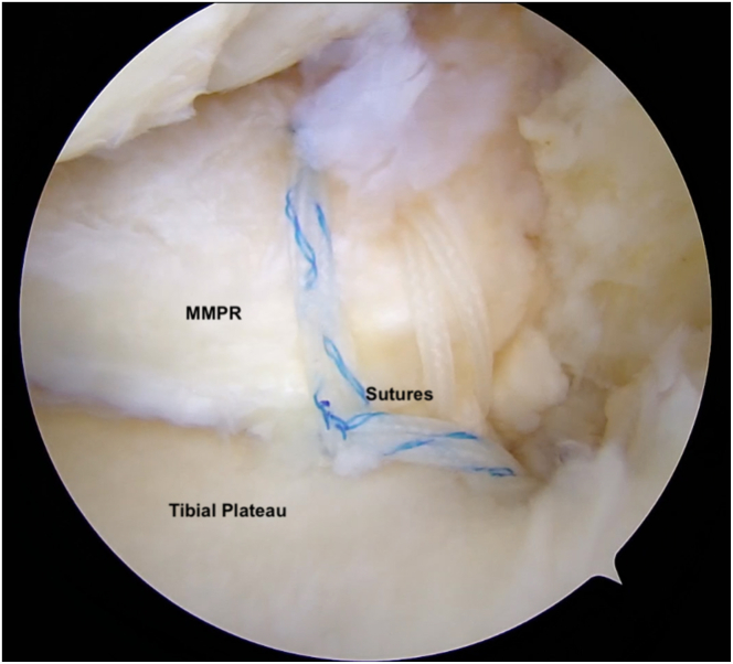
Two nonabsorbable No. 2 high-resistance sutures are passed through the medial meniscus posterior root and retrieved outside the joint. Patient in supine position, right knee, arhtroscopic view from standar AL portal.
A No. 2 Vicryl suture (Ethicon) is pulled inside the tibial tunnel into the joint and is retrieved outside the joint. Patient in supine position, right knee, arhtroscopic view from standar AL portal. (MMPR, medial meniscus posterior root.)
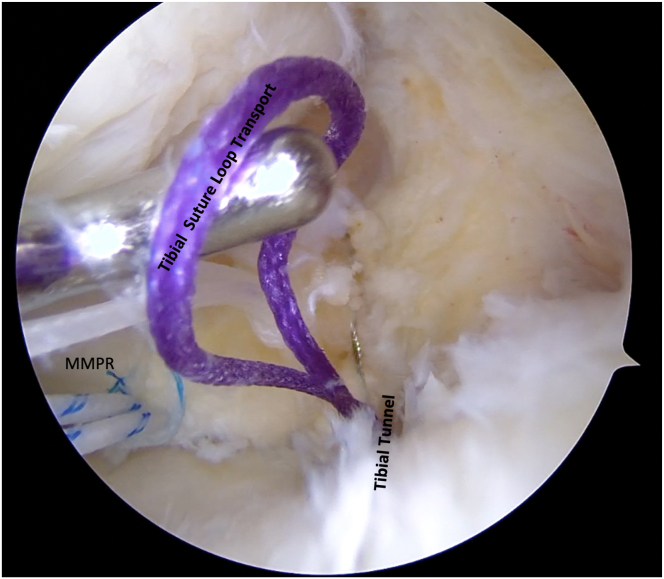
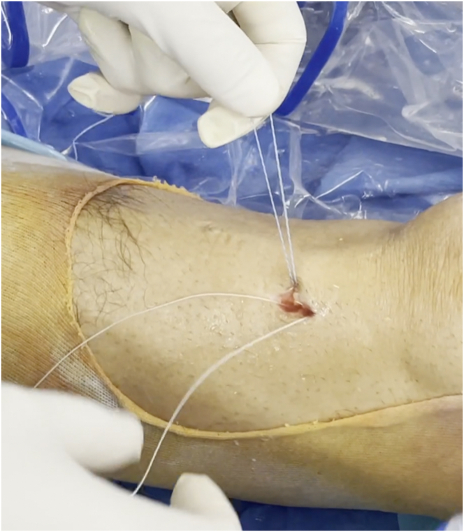
The medial meniscus posterior root (MMPR) sutures are pulled outside the joint through the tibial tunnel. Patient in supine position, right knee, arhtroscopic view from standar AL portal.
The 2 medial meniscus posterior root sutures are divided based on color. Patient in supine position, right knee, arhtroscopic view from standar AL portal.
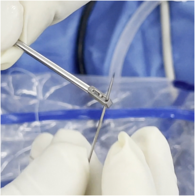
With the use of the Arthrex Biceps Button-specific needle, the 4 tails of the medial meniscus posterior root sutures are passed through the button.
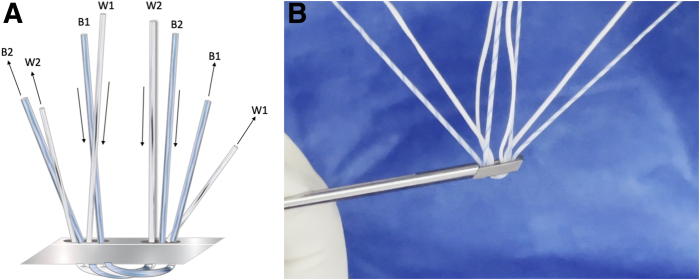
(A, B) The sutures coming from the medial meniscus posterior root are passed in the configuration shown, allowing constant tensioning of the sutures without loosening. Patient in supine position, right knee, arhtroscopic view from standar AL portal.
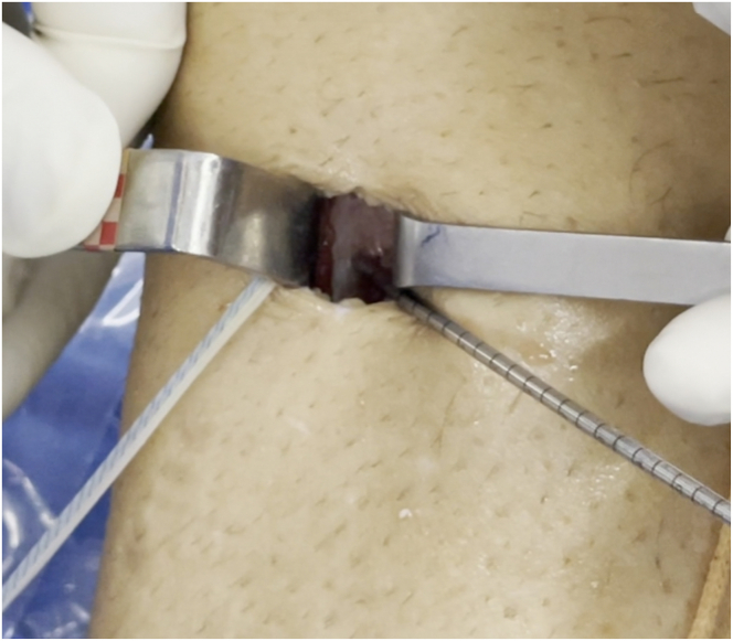
A 3.2-mm unicortical hole is drilled 2 cm below the transtibial tunnel. The sutures coming from the medial meniscus posterior root have been passed in a specific configuration that allows constant tensioning of the sutures without loosening. Patient in supine position, right knee, arhtroscopic view from standar AL portal.
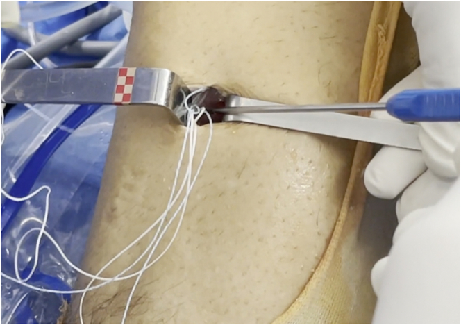
The button is inserted and impacted into the unicortical tibial hole while tension is maintained on the sutures. Patient in supine position, right knee, arhtroscopic view from standar AL portal.
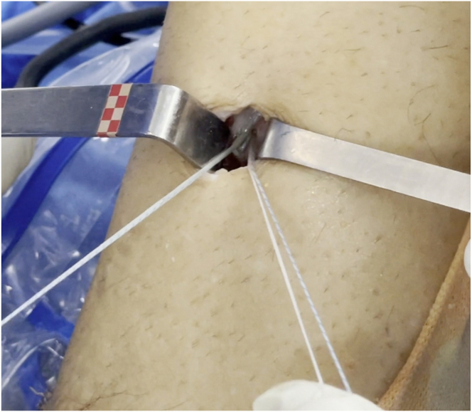
The the implant handle of the button is removed; the button is deployed in a subcortical manner, and pulling the free tails of the sutures allows the correct flip of the button. Patient in supine position, right knee, arhtroscopic view from standar AL portal.
The sutures are tensioned progressively under an arthroscopic view. Patient in supine position, right knee, arhtroscopic view from standar AL portal. (MMPR, medial meniscus posterior root.)
References
-
- Allaire R., Muriuki M., Gilbertson L., Harner C.D. Biomechanical consequences of a tear of the posterior root of the medial meniscus. Similar to total meniscectomy. J Bone Joint Surg Am. 2008;90:1922–1931. - PubMed
-
- Hein C.N., Deperio J.G., Ehrensberger M.T., Marzo J.M. Effects of medial meniscal posterior horn avulsion and repair on meniscal displacement. Knee. 2011;18:189–192. - PubMed
-
- Bernard C.D., Kennedy N.I., Tagliero A.J., et al. Medial meniscus posterior root tear treatment: A matched cohort comparison of nonoperative management, partial meniscectomy, and repair. Am J Sports Med. 2020;48:128–132. - PubMed
-
- Feucht M.J., Kühle J., Bode G., et al. Arthroscopic transtibial pullout repair for posterior medial meniscus root tears: A systematic review of clinical, radiographic, and second-look arthroscopic results. Arthroscopy. 2015;31:1808–1816. - PubMed
LinkOut - more resources
Full Text Sources
