. 2024 Aug 20;71(3):81-90.
doi: 10.5458/jag.jag.JAG-2024_0005. eCollection 2024.

Crystal Structure of Bifidobacterium bifidum Glycoside Hydrolase Family 110 α-Galactosidase Specific for Blood Group B Antigen

Affiliations

Crystal Structure of Bifidobacterium bifidum Glycoside Hydrolase Family 110 α-Galactosidase Specific for Blood Group B Antigen

Toma Kashima et al. J Appl Glycosci (1999). .

Abstract

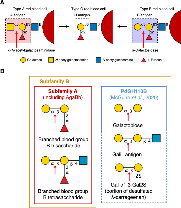
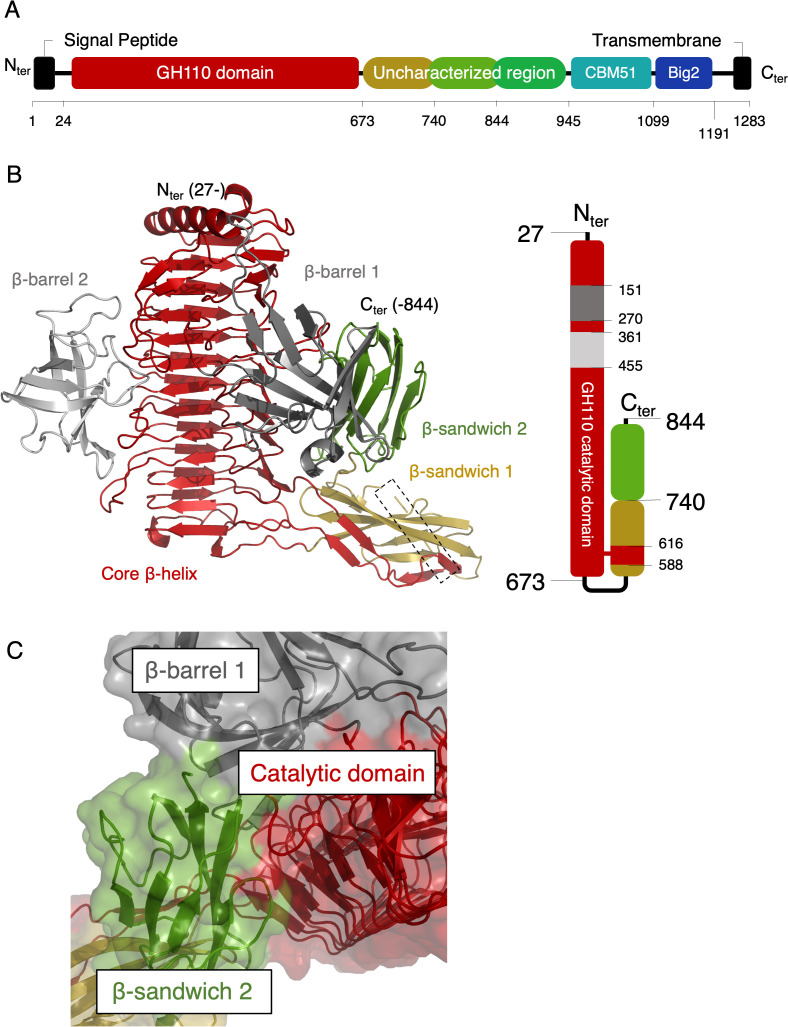
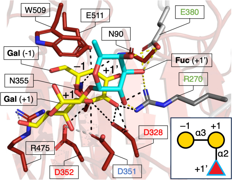
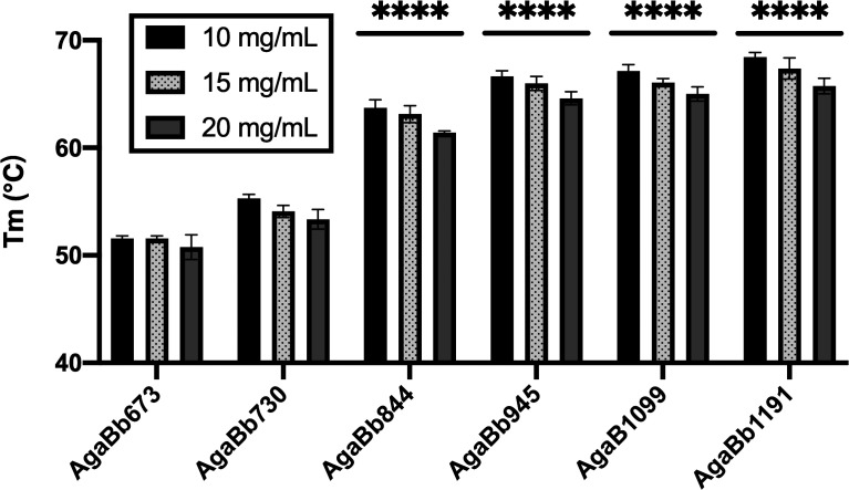
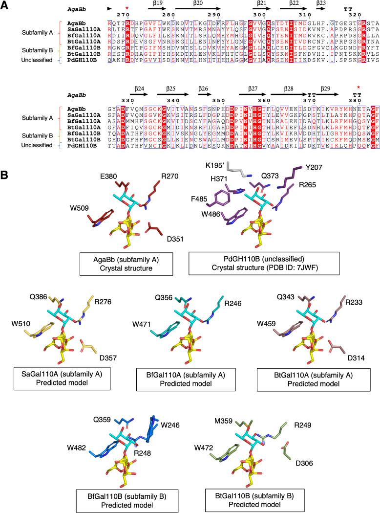
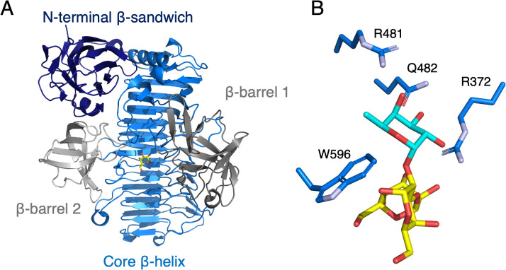
To overcome incompatibility issues and increase the possibility of blood transfusion, technologies that enable efficient conversion of A- and B-type red blood cells to the universal donor O-type is desirable. Although several blood type-converting enzymes have been identified, detailed understanding about their molecular functions is limited. α-Galactosidase from Bifidobacterium bifidum JCM 1254 (AgaBb), belonging to glycoside hydrolase (GH) 110 subfamily A, specifically acts on blood group B antigen. Here we present the crystal structure of AgaBb, including the catalytic GH110 domain and part of the C-terminal uncharacterized regions. Based on this structure, we deduced a possible binding mechanism of blood group B antigen to the active site. Site-directed mutagenesis confirmed that R270 and E380 recognize the fucose moiety in the B antigen. Thermal shift assay revealed that the C-terminal uncharacterized region significantly contributes to protein stability. This region is shared only among GH110 enzymes from B. bifidum and some Ruminococcus species. The elucidation of the molecular basis for the specific recognition of blood group B antigen is expected to lead to the practical application of blood group conversion enzymes in the future.

Keywords: Bifidobacterium; Blood group conversion; crystal structure; glycoside hydrolase; mucin; α-galactosidase.


Conflict of interest statement

Takura Wakinaka is an employee of Yamasa Corporation.

Figures

Fig. 1.
Fig. 1.. Enzymatic blood group conversion and substrate specificities of GH110.
(A) Glycan structure of blood group epitopes and the enzymes implicated in the conversion of blood group A and B epitopes into universal O blood group (Prepared based on Rahfeld et al., 2019) [6]. (B) Differences in substrate specificity between GH110 subfamilies. Cleavage sites of the enzymes are indicated with red arrows. Substrates that PdGH110B is expected to cleave, but remain to be experimentally verified are indicated with a dashed line box.
Fig. 2.
Fig. 2.. Domain organization of AgaBb and crystal structure of AgaBb844.
(A) Domain organization of AgaBb. GH110, glycoside hydrolase family 110. AgaBb, α-galactosidase from Bifidobacterium bifidum. (B) The overall structure (left) and domain architecture (right) of AgaBb. The monomer is colored to show the core β-helix (red), β-barrel 1 (dark gray), β-barrel 2 (light gray), β-sandwich 1 (gold), and β-sandwich 2 (green). The disordered region (732-739) is indicated in a dotted box. (C) Surface representation of the β-sandwich 2 domain showing extensive interactions with the core β-helix and β-barrel 1 domain.
Fig. 3.
Fig. 3.. Structure of the active site of AgaBb with modeled blood group B trisaccharide.
The protein domains are colored as described in Fig. 2. A schematic representation of blood group B trisaccharide is shown in the lower right panel. Gal (yellow) and Fuc (cyan) are shown in sticks and their corresponding subsites in the active site are indicated. The general acid (D351) and base residue candidates (D328 and D352) are indicated by blue and red characters, respectively. Protein residues constituting the +1' subsite are indicated by green characters. Polar contacts with the sugar moieties in −1, +1, and +1' subsites are indicated with black, gray, and yellow dashed lines, respectively.
Fig. 4.
Fig. 4.. Protein thermostability of C-terminal deletants of AgaBb.
Melting temperature (Tm, in °C) of the protein constructs of different lengths at different protein concentrations (10, 15, and 20 mg/mL) were determined by thermal shift assay. Data were collected in triplicate and plotted as the mean ± standard deviation (SD). The average Tm of the three concentrations of the constructs was compared to that of AgaBb673 using the two-way ANOVA test (****, P < 0.0001). Comparison of the average Tm of the constructs with that of AgaBb730 also displayed similar significance (not shown).
Fig. 5.
Fig. 5.. Comparison of the putative fucose recognition site in AgaBb with that of previously characterized GH110 enzymes.
(A) Multiple amino-acid sequence alignment. The residues implicated in the fucose recognition in AgaBb are indicated with red asterisks above the sequences. (B) Putative +1' subsite of AgaBb and GH110 enzymes in subfamilies A and B. The structures other than AgaBb and PdGH110B are predicted models. Residues within 4 Å distance from the fucose are shown. The protein names, and sequence identities and Cα RMSD values to AgaBb are as follows: PdGH110B (27 %, 1.17 Å), α-galactosidase from P. distincta; SaGal110A (46 %, 0.68 Å), α-galactosidase from S. avermitilis; BfGal110A (32 %, 0.83 Å) and BfGal110B (38 %, 0.70 Å), α-galactosidases from B. fragilis; BtGal110A (31 %, 1.22 Å) and BtGal110B (30 %, 0.94 Å), α-galactosidases from B. thetaiotaomicron.
Fig. 6.
Fig. 6.. The crystal structure A. muciniphila GH110 α-galactosidase (AmGH110).
(A) The overall structure. (B) Putative +1' subsite. Shown as in Fig. 5B. The structure available on the database (PDB ID: 8PVS) is shown.

Similar articles

Cited by

References

    1. Rydberg L. ABO-incompatibility in solid organ transplantation. Transfus Med. 2001; 11: 325-42. - PubMed
    1. Galili U. Xenotransplantation and ABO incompatible transplantation: The similarities they share. Transfus Apher Sci. 2006; 35: 45-58. - PubMed
    1. Mitra R, Mishra N, Rath GP. Blood groups systems. Ind J Anaesth. 2014; 58: 524-8. - PMC - PubMed
    1. Rossez Y, Maes E, Lefebvre Darroman T, Gosset P, Ecobichon C, Joncquel Chevalier Curt M, et al. Almost all human gastric mucin O-glycans harbor blood group A, B or H antigens and are potential binding sites for Helicobacter pylori. Glycobiology. 2012; 22: 1193-206. - PubMed
    1. Misevic G, Misevic G. ABO blood group system. Blood Genom. 2018; 2: 71-84.

LinkOut - more resources