. 2024 Aug 21:12:1456416.
doi: 10.3389/fcell.2024.1456416. eCollection 2024.

Comparative study of systemic and local delivery of mesenchymal stromal cells for the treatment of chronic kidney disease

Affiliations

Comparative study of systemic and local delivery of mesenchymal stromal cells for the treatment of chronic kidney disease

Emil Gregersen et al. Front Cell Dev Biol. .

Abstract

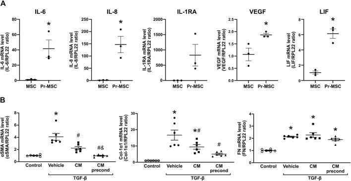
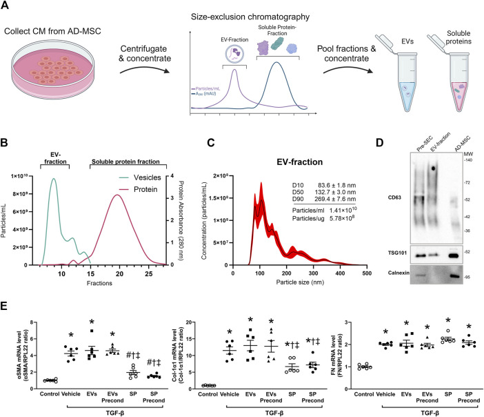
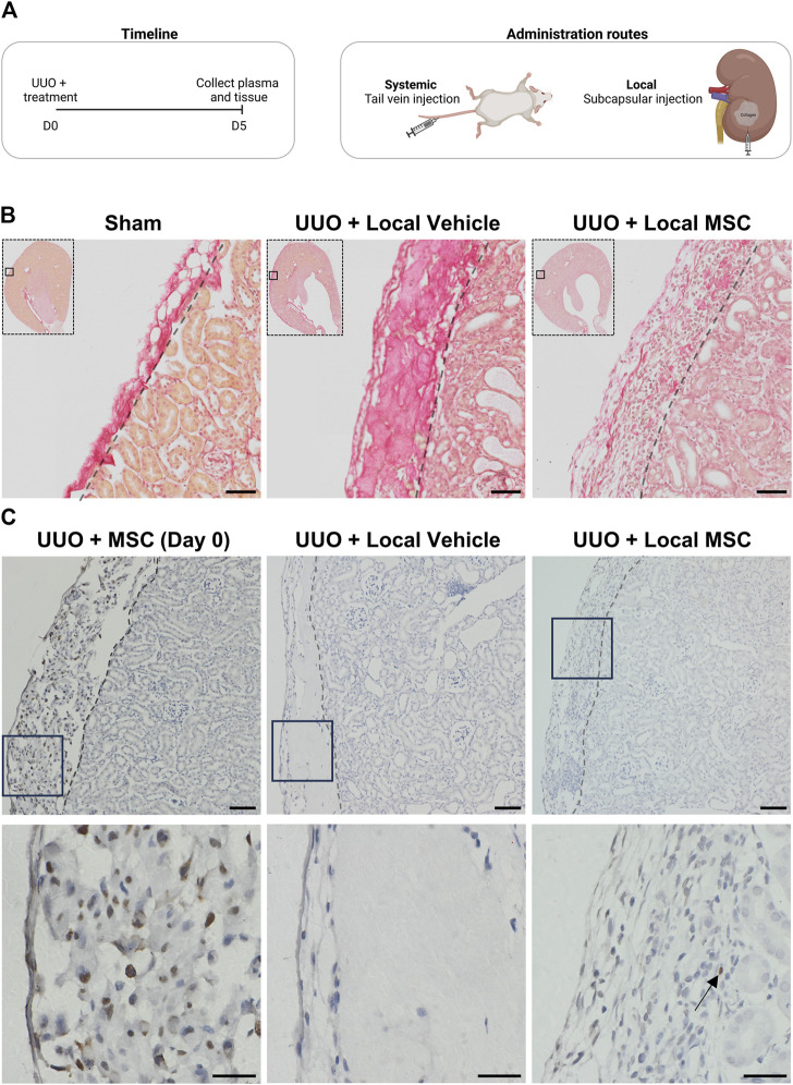
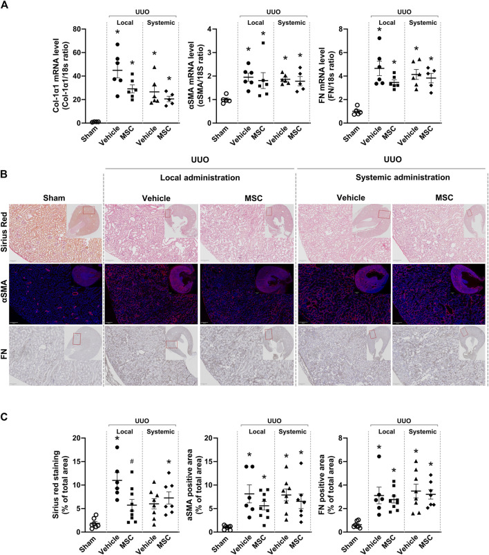
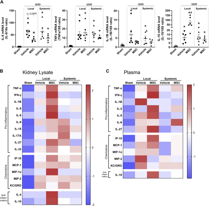
Renal fibrosis, characterized by excessive extracellular matrix accumulation, leads to a progressive decline of renal function and is a common endpoint of chronic kidney disease (CKD). Current treatments primarily focus on managing underlying diseases, offering limited direct intervention for the fibrotic process. This study explores the anti-fibrotic potential of human adipose-derived mesenchymal stromal cells (MSCs) and their derived extracellular vesicles (EVs) in the context of CKD, emphasizing the effects of systemic versus local delivery methods. Preconditioned MSCs (Pr-MSCs) were treated with TNF-α and IFN-γ to enhance their immunomodulatory capabilities, and demonstrated significant anti-fibrotic effects in vitro, reducing mRNA expression of fibrosis markers in TGF-β stimulated HKC-8 cells. Our in vivo findings from a murine unilateral ureteral obstruction (UUO) model of CKD showed that local deliveries of Pr-MSCs reduced collagen deposition and increased expression of the anti-inflammatory cytokine IL-10. Systemic administration of Pr-MSCs did not show any significant effect on UUO-induced injury. In addition, EVs did not replicate the anti-fibrotic effects observed with their parent cells, suggesting that soluble proteins or metabolites secreted by Pr-MSCs might be the primary mediators of the anti-fibrotic and immunomodulatory effects. This study provides critical insights into the therapeutic efficacy of MSCs, highlighting the importance of delivery methods and the potential of preconditioning strategies in enhancing MSC-based therapies for renal fibrosis.

Keywords: chronic kidney disease; delivery; fibrosis; inflammation; mesenchymal stem cells; mesenchymal stromal cells (MSCs).


Conflict of interest statement

The authors declare that the research was conducted in the absence of any commercial or financial relationships that could be construed as a potential conflict of interest.

Figures

FIGURE 1
FIGURE 1
Mitigating TGF-β-induced fibrosis in HKC-8 cells by conditioned media (CM) from adipose-derived mesenchymal stromal cells (MSCs). (A) Gene expression analysis of MSCs, that have been preconditioned (Pr-MSC) overnight with 10 ng/mL of tumor necrosis factor-α (TNF-α) and interferon-γ (IFN-γ) or left without preconditioning. Expression levels were normalized to RPL22 and compared to the baseline expression. Statistical analysis: Unpaired t-test. *P < 0.05. (B) HKC-8 cells were allowed to adhere and subjected to overnight serum-free incubation. Subsequently, fibrosis was induced with transforming growth factor-β (TGF-β). Concurrently, cells were treated for 48 h with CM from MSCs without preconditioning, MSCs preconditioned with TNF-α and IFN-γ (CM precond), or a vehicle control (advanced MEM). Gene expression levels of α-smooth muscle actin (αSMA), Collagen 1a1 (Col-1a1), and Fibronectin (FN) were evaluated using qPCR and normalized to the control gene RPL22. Statistical analysis: One-way ANOVA followed by Tukey’s multiple comparison. *P < 0.05 vs. control, #P < 0.05 vs. vehicle, & p < 0.05 vs. CM. Data are presented as mean ± SEM.
FIGURE 2
FIGURE 2
Isolation and characterization of extracellular vesicles (EVs) and soluble proteins (SPs) from adipose-derived mesenchymal stromal cells (AD-MSC). (A) Schematic representation depicting the isolation of EV and soluble protein isolation from conditioned media (CM) of MSCs using size-exclusion chromatography (SEC). Following SEC, both the EV and soluble protein fractions were concentrated to the same volume, resulting in the same concentration factor. (B) Actual elution profile of EVs and soluble proteins post-SEC, utilizing a qEVoriginal 70 nm column. Fractions of 0.5 mL were collected after the void volume (3 mL). Nanoparticle tracking analysis (NTA; depicted by the green line) was employed to estimate particle numbers, whereas protein concentrations (indicated by the red line) were quantified through absorbance measurements at 280 nm. Fractions 6–10 were combined to form the EV-enriched fraction, while fractions 14–29 were pooled as the soluble protein fraction. (C) Particle size and concentration of EV-fraction was analyzed using NTA. The black line represents the mean concentration, while the red line indicates the standard error of the mean. In this example, EV-fraction was concentrated around 100 times compared to CM collected from MSC. (D) Western blot analysis assessing the presence of CD63, TSG101, and calnexin in both EV and MSC lysates. EV lysates were acquired from EVs isolated by ultracentrifugation of CM prior to SEC, as well as from the EV-fraction obtained post-SEC. (E) HKC-8 cells were allowed to adhere and subjected to overnight serum-free incubation. Subsequently, fibrosis was induced with transforming growth factor-β (TGF-β). Concurrently, cells were treated for 48 h with EVs or SPs from MSCs without preconditioning, MSCs preconditioned with TNF-α and IFN-γ (precond), or PBS as vehicle control. Gene expression levels of α-smooth muscle actin (αSMA), Collagen 1α1 (Col-1α1), and Fibronectin (FN) were evaluated using qPCR and normalized to the control gene RPL22. Statistical analysis: One-way ANOVA followed by Tukey’s multiple comparison. *P < 0.05 vs. control, #P < 0.05 vehicle, P < 0.05 vs. EVs, P < 0.05 vs. precond EVs.
FIGURE 3
FIGURE 3
Evaluating systemic and local delivery of adipose-derived mesenchymal stromal cells (MSC) in a murine chronic kidney disease model. (A) Experimental timeline for murine in vivo study. Mice were subjected to unilateral ureteral obstruction (UUO) and received treatment on Day 0. On Day 5, the animals were euthanized, and tissue samples were collected for analysis. Two treatment routes were employed: (1) systemic administration of three million MSCs in PBS via tail vein injection, and (2) local delivery of three million MSCs embedded in a collagen hydrogel matrix through subcapsular injection as well as their vehicle alone. (B) Kidneys from mice subjected to UUO and treated locally with either vehicle or MSC, as well as from sham-operated mice, were assessed using Sirius red staining. Scale bar = 50 µm (C) HNA-stained kidney sections (x10 on top and x40 below) from UUO mice include: (1) Mice injected subcapsularly with MSCs in collagen matrix, sacrificed 3 h post-operation (Day 0), (2) UUO mice treated with subcapsular injected collagen matrix alone and harvested at day 5 (UUO + Vehicle), and (3) UUO mice injected subcapsularly with MSCs in collagen and harvested at day 5 (UUO + MSC). For x10 scale bar = 50 µm. For x40 scale bar = 20 µm. Arrows show the location of a single nucleus stained in the UUO + MSC group on day 5.
FIGURE 4
FIGURE 4
Local delivery of adipose-derived mesenchymal stromal cells (MSCs) decreases collagen deposition in mice subjected to unilateral ureteral obstruction (UUO). UUO mice received either systemic treatment via tail vein injection of MSCs or local treatment through subcapsular injection of MSCs. On Day 5 post-treatment, the animals were euthanized, and tissue samples were harvested for subsequent analysis. (A) Relative gene expression levels of, Collagen Type I α1 Chain (Col-1α1), αSMA, and fibronectin (FN) were measured by qPCR, with normalization to 18s expression. Statistical test: One-way ANOVA followed by Tukey’s multiple comparison. *P < 0.05 vs. sham, #P < 0.05 vs. vehicle local. (B) Representative pictures of Sirius Red staining targeting collagen, immunofluorescent staining targeting αSMA and immunostaining of FN. Scale bar = 100 µm. (C) Quantification of Sirius Red stained, αSMA excluding signals originating from arterial structures and FN, on whole kidney scans excluding outermost cortex and inner medulla. Statistical analysis: One-way ANOVA followed by Tukey’s multiple comparison. *P < 0.05 vs. sham, #P < 0.05 vs. vehicle controls. Data are presented as mean ± SEM.
FIGURE 5
FIGURE 5
Local delivery of adipose-derived mesenchymal stromal cells (MSCs) increases pro- and anti-inflammatory factors in mice subjected to unilateral ureteral obstruction (UUO). UUO mice received either systemic treatment via tail vein injection of MSCs or local treatment through subcapsular injection of MSCs. On Day 5 post-treatment, the animals were euthanized, and tissue samples were harvested for subsequent analysis. (A) Relative gene expression levels of IL-6, TNF-α, IL-1β, and IL-10 were measured by qPCR, with normalization to 18s expression. Statistical test: One-way ANOVA followed by Tukey’s multiple comparison. *P < 0.05 vs. sham, #P < 0.05 vs. vehicle. Data are presented as mean ± SEM. (B, C) Heatmap visualizing the relative protein levels of cytokines and chemokines in kidney lysate and plasma. Data is presented as z-score with the scale bar to the right indicating the range.

Similar articles

References

    1. Aabling R. R., Alstrup T., Kjær E. M., Poulsen K. J., Pedersen J. O., Revenfeld A. L., et al. (2023). Reconstitution and post-thaw storage of cryopreserved human mesenchymal stromal cells: pitfalls and optimizations for clinically compatible formulants. Regen. Ther. 23, 67–75. 10.1016/j.reth.2023.03.006 - DOI - PMC - PubMed
    1. Aggarwal S., Pittenger M. F. (2005). Human mesenchymal stem cells modulate allogeneic immune cell responses. Blood 105 (4), 1815–1822. 10.1182/blood-2004-04-1559 - DOI - PubMed
    1. Almeida A., Lira R., Oliveira M., Martins M., Azevedo Y., Silva K. R., et al. (2022). Bone marrow-derived mesenchymal stem cells transplantation ameliorates renal injury through anti-fibrotic and anti-inflammatory effects in chronic experimental renovascular disease. Biomed. J. 45 (4), 629–641. 10.1016/j.bj.2021.07.009 - DOI - PMC - PubMed
    1. Caplan H., Olson S. D., Kumar A., George M., Prabhakara K. S., Wenzel P., et al. (2019). Mesenchymal stromal cell therapeutic delivery: translational challenges to clinical application. Front. Immunol. 10, 1645. 10.3389/fimmu.2019.01645 - DOI - PMC - PubMed
    1. Carceller M. C., Guillén M. I., Gil M. L., Alcaraz M. J. (2021). Extracellular vesicles do not mediate the anti-inflammatory actions of mouse-derived adipose tissue mesenchymal stem cells secretome. Int. J. Mol. Sci. 22 (3), 1375–1414. 10.3390/ijms22031375 - DOI - PMC - PubMed

LinkOut - more resources