. 2024 Nov;11(41):e2309697.
doi: 10.1002/advs.202309697. Epub 2024 Sep 5.

Targeting Dual Immune Checkpoints PD-L1 and HLA-G by Trispecific T Cell Engager for Treating Heterogeneous Lung Cancer

Affiliations

Targeting Dual Immune Checkpoints PD-L1 and HLA-G by Trispecific T Cell Engager for Treating Heterogeneous Lung Cancer

Yu-Chuan Lin et al. Adv Sci (Weinh). 2024 Nov.

Abstract

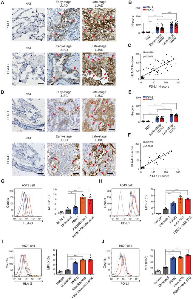
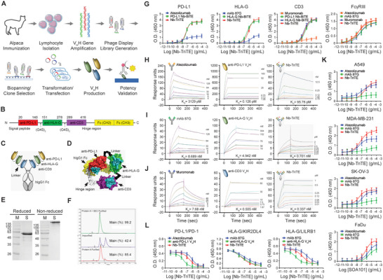
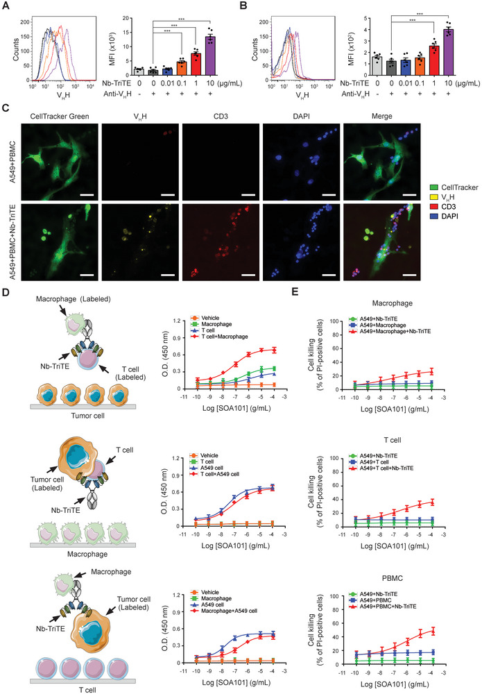
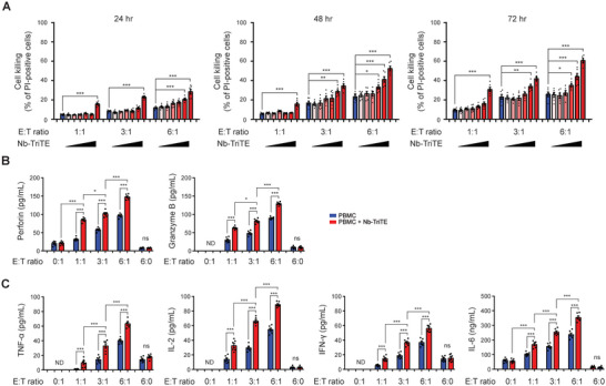
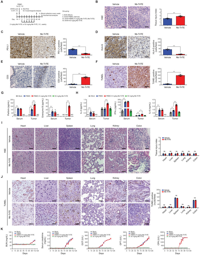
Immunotherapy targeting immune checkpoints (ICPs), such as programmed death-ligand-1 (PD-L1), is used as a treatment option for advanced or metastatic non-small cell lung cancer (NSCLC). However, overall response rate to anti-PD-L1 treatment is limited due to antigen heterogeneity and the immune-suppressive tumor microenvironment. Human leukocyte antigen-G (HLA-G), an ICP as well as a neoexpressed tumor-associated antigen, is previously demonstrated to be a beneficial target in combination with anti-PD-L1. In this study, a nanobody-based trispecific T cell engager (Nb-TriTE) is developed, capable of simultaneously binding to T cells, macrophages, and cancer cells while redirecting T cells toward tumor cells expressing PD-L1- and/or HLA-G. Nb-TriTE shows broad spectrum anti-tumor effects in vitro by augmenting cytotoxicity mediated by human peripheral blood mononuclear cells (PBMCs). In a humanized immunodeficient murine NSCLC model, Nb-TriTE exhibits superior anti-cancer potency compared to monoclonal antibodies and bispecific T cell engagers. Nb-TriTE, at the dose with pharmacoactivity, does not induce additional enhancement of circulating cytokines secretion from PMBCs. Nb-TriTE effectively prolongs the survival of mice without obvious adverse events. In conclusion, this study introduces an innovative therapeutic approach to address the challenges of immunotherapy and the tumor microenvironment in NSCLC through utilizing the dual ICP-targeting Nb-TriTE.

Keywords: human leukocyte antigen‐G (HLA‐G); immune checkpoint (ICP); nanobody‐based trispecific T cell engager (Nb‐TriTE); non‐small cell lung cancer (NSCLC); programmed death‐ligand 1 (PD‐L1).


Conflict of interest statement

Y.C.L., J.H.C.H., and H.C.C. are employed by Shine‐On BioMedical Co., Ltd. during the conduct of this study. C.C.H. and S.C.C. are employed and owned equity interests in Shine‐On BioMedical Co., Ltd. during the conduct of this study. The other authors declare no direct conflicts of interest.

Figures

Figure 1
Figure 1
PD‐L1 and HLA‐G are highly expressed in refractory NSCLC. A) Programmed death‐ligand 1 (PD‐L1) and human leukocyte antigen‐G (HLA‐G) staining were performed on human LUAD tissue slices from different stages. Paired normal adjacent noncancerous tissues (NAT) were served as a control. B) Quantitative analysis for PD‐L1 and HLA‐G staining are shown as H‐scores respectively. Data are means ± SEM of 44 independent samples. C) The correlation between PD‐L1 and HLA‐G expression in LUAD was evaluated and quantified using Pearson's correlation coefficient (R). In parallel, D) PD‐L1 and HLA‐G staining were performed on human LUSC tissue slices from different stages. Paired NAT were served as a control. E) Quantitative analysis for PD‐L1 and HLA‐G staining are shown as H‐scores respectively. Data are means ± SEM of 22 independent samples. F) The correlation between PD‐L1 and HLA‐G expression in LUSC was evaluated and quantified using Pearson's correlation coefficient (R). G)The expression of HLA‐G on A549 and I) H520 cell membrane was examined by flow cytometric analysis, following a 24‐h treatment with PBMC, Atezolizumab, or PBMC + Atezolizumab. Quantitative results are shown as mean fluorescence intensity (MFI). Data represent means ± SEM of six independent experiments. H) The expression of PD‐L1 on A549 and J) H520 cell membrane was examined by flow cytometric analysis, following a 24‐h treatment with PBMC, HLA‐G monoclonal antibody clone 87G (mAb 87G), or PBMC + mAb 87G. Quantitative results are shown as MFI. Data represent means ± SEM of six independent experiments. *< 0.05; *< 0.01; ***< 0.001. NSCLC, non‐small cell lung cancer; LUAD, lung adenocarcinoma; LUSC, lung squamous cell carcinoma.
Figure 2
Figure 2
Characterization of Nb‐TriTE. A) Schematic representation for the selection and identification process of VHH nanobodies in this study. B) The construction of Nb‐TriTE was engineered with three VHH nanobodies specific to anti‐PD‐L1, anti‐HLA‐G, and CD3ɛ, respectively, following a secreatable signal peptide. These three VHH fragments were connected through two flexible GS linkers (G4S)2 and followed by the human IgG1 Fc regions. C) Schematic diagram showing the components and conformation of the developed Nb‐TriTE. D) The 3D structure of Nb‐TriTE was predicted by the Alphafold2 software. E) Reduced or non‐reduced sodium dodecyl sulphate‐polyacrylamide gel electrophoresis was performed to confirm the molecular weight of Nb‐TriTE. F) The purity of Nb‐TriTE was determined by size exclusion chromatography using the Superdex 200 Increase columns. G) Binding ability of Nb‐TriTE and Nb‐BiTE to specific antigens, including human PD‐L1, HLA‐G, and CD3, and the interaction between the Fc region of Nb‐TriTE and Fc gamma receptor III (FcγRIII) were assessed and compared to that of monoclonal antibodies using ELISA. The antibody‐antigen interactions were analyzed by BIAcore SPR biosensor. Sensor chips were coated with H) rhPD‐L1, I) rhHLA‐G, or J) rhCD3, followed by binding with commercial monoclonal antibodies including Atezolizumab targeting PD‐L1, mAb 87G targeting HLA‐G, and Muromonab targeting CD3, the individual VHH components of Nb‐TriTE, or Nb‐TriTE. Results are presented as sensorgrams with the equilibrium dissociation constant (KD ). K) Solid tumor cell lines including A549, MDA‐MB‐231, SK‐OV‐3, and FaDu cells were individually seeded onto 96‐well plates. These cells were then incubated with the commercial antibodies Atezolizumab, mAb 87G, and Nb‐TriTE, respectively. The binding affinity of Atezolizumab, mAb 87G, and Nb‐TriTE toward to these cancer cells was analyzed. L) Nb‐TriTE combined with recombinant human PD‐1, KIR2DL4, LILRB1 proteins, followed by added into 96‐well plates that were pre‐coated with PD‐1 or HLA‐G antigen. The ability of Nb‐TriTE competing the binding of PD‐L1/PD‐1, HLA‐G/KIR2DL4, or HLA‐G/KIR2DL4 was examined. The data represented are the means ± SEM of six independent experiments.
Figure 3
Figure 3
Nb‐TriTE simultaneously connects tumor cells with T cells, further enhancing the cell killing activity of T cells and immune cells in PBMCs. A) A549 cells, with a density of 1 × 10⁵ cells mL−1, and B) PBMCs, with a density of 3 × 10⁵ cells mL−1 were independently incubated with 0, 0.01, 0.1, 1, or 10 µg mL−1 Nb‐TriTE, followed by incubation with anti‐camelid VHH‐iFluor 555 Ab. The binding levels of Nb‐TriTE were determined by flow cytometric analysis, and quantitative results are represented as mean fluorescence intensity (MFI). The data presented are the means ± SEM of six independent samples. C) A549 cells were pre‐labeled with CellTracker Green and seeded into 12‐well plates, then followed by incubation with PBMCs in the presence or absence of Nb‐TriTE. Immunocytochemistry was performed to examine the association between A549 cells, CD3+ T cells, and Nb‐TriTE using fluorescence microscopy. The original magnification is ×400. The scale bar represents 50 µm. D) The schematic diagrams show the experimental strategy for evaluating the linkage between A549 cells, T cells, macrophages, and Nb‐TriTE through cell‐based ELISA. HRP‐labeled E‐cadherin+ A549 cells with HRP‐labeled CD3+ T cells, HRP‐labeled E‐cadherin+ A549 cells with HRP‐labeled CD68+ macrophages, and HRP‐labeled CD3+ T cells with HRP‐labeled CD68+ macrophages were incubated with Nb‐TriTE at a series concentration and then sequentially introduced into culture plates pre‐seeded with macrophages, T cells, and A549 cells at a density of 1 × 10⁵ cells mL−1. After incubation with 3,3′,5,5′‐tetramethylbenzidine, the reaction was terminated using 2N sulphuric acid, and then the absorbance at 450 nm was determined using ELISA reader. E) A549 cells were seeded into 24‐well plates at a density of 4 × 10⁴ cells mL−1 and then stained with CellTracker Green. A549 cells were then cocultured with macrophages, T cells, and PBMC, respectively, in the presence of Nb‐TriTE at a range of concentrations for 24 h. Thereafter, the cocultured cells were stained with propidium iodide (2 µg mL−1), and the dead cells were detected using flow cytometer. ***< 0.001.
Figure 4
Figure 4
Nb‐TriTE enhances the cell‐killing capacity and cytokine release of PBMCs. A) A549 cells at 4 x 10⁴ were stained with CellTracker Green followed by cocultured with PBMCs at E:T ratios of 1:1, 1:3, and 6:1 in the presence of Nb‐TriTE at concentrations of 0, 0.01, 0.1, 1, 10, and 100 µg mL−1, respectively. After coculturing for 24, 48, and 72 h, the cell‐killing ability was evaluated by propidium iodide staining‐based cell death detection and analyzed by flow cytometer. Data represent means ± SEM of six independent samples. A549 cells at 1 × 10⁴ were then incubated with 10 µg mL−1 Nb‐TriTE and PBMCs at E:T ratios of 0:1, 1:1, 3:1, 6:1, and 6:0 for 48 h. The release of B) cytotoxic molecules, perforin and granzyme B, and C) cytokines including TNF‐α, IL‐2, IFN‐γ, and IL‐6 in the supernatants were assessed via ELISA. Quantitative results were means ± SEM of six independent samples for each group. *< 0.05; **< 0.01; ***< 0.001. ND, not detected; ns, not significant.
Figure 5
Figure 5
Potent cell‐killing activity of PBMCs in combination with Nb‐TriTE against PD‐L1‐ or HLA‐G‐overexpressing cancer cells. PD‐L1 and HLA‐G levels in mock cells and cells overexpressing PD‐L1 (ovPD‐L1), HLA‐G (ovHLA‐G), and both PD‐L1 and HLA‐G (ovPD‐L1/ovHLA‐G) in A549 cells or H520 cells were determined and compared by A,D) western blot and B,E) flow cytometry analysis using specific antibodies. Flow cytometric data are represented as histogram overlays. C,F) Mock, ovPD‐L1, ovHLA‐G, and ovPDL‐1/ovHLA‐G A549 cells or H520 cells (4 × 104) were stained with CellTracker Green followed by incubation with Nb‐TriTE (10 µg mL−1), PBMCs (E:T = 3:1), or PBMCs combined with Nb‐TriTE for 48 h. The cell‐killing ability was then evaluated by PI staining‐based cell death detection via flow cytometer analysis. Data represent the means ± SEM of six independent samples. G) The expression levels of PD‐L1 and H) HLA‐G on cell surface of MDA‐MB‐231, U‐87 MG, SK‐OV‐3, and FaDu cells were detected by flow cytometry. I) Cancer cell lines, including MDA‐MB‐231, U‐87 MG, SK‐OV‐3, and FaDu cells at 4 × 104, were labeled with CellTracker Green followed by incubation with Nb‐TriTE (10 µg mL−1), PBMCs (E:T = 3:1), or PBMCs combined with Nb‐TriTE for 48 h. Cell‐killing ability was assessed by propidium iodide staining‐based cell death detection. Data represent the means ± SEM of six independent samples. ***< 0.001.
Figure 6
Figure 6
Nb‐TriTE suppresses NSCLC tumor growth and extends survival in humanized xenograft mice. Treatment protocols for animal studies comparing A) the anti‐cancer efficacy of Nb‐TriTE and commercial monoclonal antibodies, D) dosage‐regimen, G) dosage‐ranging, and J) ICP heterogenicity of tumor cells. B,E,H,K) Tumor size was monitored and recorded weekly using the representative in vivo imaging system (IVIS). Tumor changes were quantified as tumor growth inhibition (TGI). C,F,I,L) The survival rate of mice was plotted using the Kaplan–Meier method. Mice were considered dead when bioluminescence reached or exceeded 1.5 × 10⁷. The data is presented as the means ± SEM from five to six independent samples. ovA549, A549 cells overexpressing both PD‐L1 and HLA‐G. *p < 0.05; **p < 0.01; ***p < 0.001.
Figure 7
Figure 7
Nb‐TriTE redirects T cells targeting tumor sites without apparent toxicities in humanized NSCLC xenograft mice. A) Treatment protocol for safety experiments. NSG mice were intravenous injected with PMBCs on day −7, followed by intravenous injection with Nb‐TriTE once weekly for six weeks. Mice were sacrificed on day 42 after the initial treatment of Nb‐TriTE. B) H&E staining, and IHC staining for C) PD‐L1, D) HLA‐G, E) CD3, and F) TUNEL staining were performed on the remaining inoculated tumor sections. Mice serum was collected before being sacrificed on day 42. The secretion levels of G) perforin and granzyme B, and H) cytokines including IL‐2, IFN‐γ, and IL‐6 in mice serum and tumor sites were assessed using ELISA. I) H&E stain evaluated the morphological changes in normal tissues and quantified as tissue injury index form six separate samples. J) The apoptotic cells in normal tissues were evaluated by TUNEL staining and shown as positive stained nuclei per field. K) Blood urea nitrogen (BUN), serum creatinine, glutamate‐oxaloacetate transaminase (GOT), glutamate‐pyruvate transaminase (GPT), and creatine‐phospho‐kinase (CPK) were assessed on day 42 after the initial treatment of Nb‐TriTE. The original magnification was ×400. The scale bar in each image represents 50 µm. *p < 0.05; **p < 0.01; ***p < 0.001. ND, not detected; ns, not significant.

Similar articles

Cited by

References

    1. Thandra K. C., Barsouk A., Saginala K., Aluru J. S., Barsouk A., Contemp Oncol 2021, 25, 45. - PMC - PubMed
    1. a) Heldwein M. B., Schlachtenberger G., Doerr F., Menghesha H., Bennink G., Schroeder K. M., Schaefer S. C., Wahlers T., Hekmat K., Surg Oncol 2022, 40, 101674; - PubMed
    2. b) Oser M. G., Niederst M. J., Sequist L. V., Engelman J. A., Lancet Oncol. 2015, 16, e165. - PMC - PubMed
    1. a) Noreldeen H. A. A., Liu X., Xu G., J. Sep. Sci. 2020, 43, 120; - PubMed
    2. b) Wang C., Qiao W., Jiang Y., Zhu M., Shao J., Wang T., Liu D., Li W., J. Cell. Physiol. 2020, 235, 4913. - PMC - PubMed
    1. a) Pennock G. K., Chow L. Q., Oncologist 2015, 20, 812; - PMC - PubMed
    2. b) Marshall H. T., Djamgoz M. B. A., Front Oncol 2018, 8, 315; - PMC - PubMed
    3. c) Jafari S., Molavi O., Kahroba H., Hejazi M. S., Maleki‐Dizaji N., Barghi S., Kiaie S. H., Jadidi‐Niaragh F., Cell. Mol. Life Sci. 2020, 77, 3693; - PMC - PubMed
    4. d) Ogasawara K., Newhall K., Maxwell S. E., Dell'Aringa J., Komashko V., Kilavuz N., Delarue R., Czuczman M., Sternas L., Rose S., Beach C. L., Novick S., Zhou S., Palmisano M., Li Y., Clin. Pharmacokinet. 2020, 59, 217. - PMC - PubMed
    1. a) Wang Z., Wu X., Cancer Med. 2020, 9, 8086; - PMC - PubMed
    2. b) Osipov A., Murphy A., Zheng L., Adv. Cancer Res. 2019, 143, 63. - PubMed