. 2024 Oct 8;68(10):e0075324.
doi: 10.1128/aac.00753-24. Epub 2024 Sep 5.

IMT-P8 potentiates Gram-positive specific antibiotics in intrinsically resistant Gram-negative bacteria

Affiliations

IMT-P8 potentiates Gram-positive specific antibiotics in intrinsically resistant Gram-negative bacteria

Vidhu Singh et al. Antimicrob Agents Chemother. .

Abstract

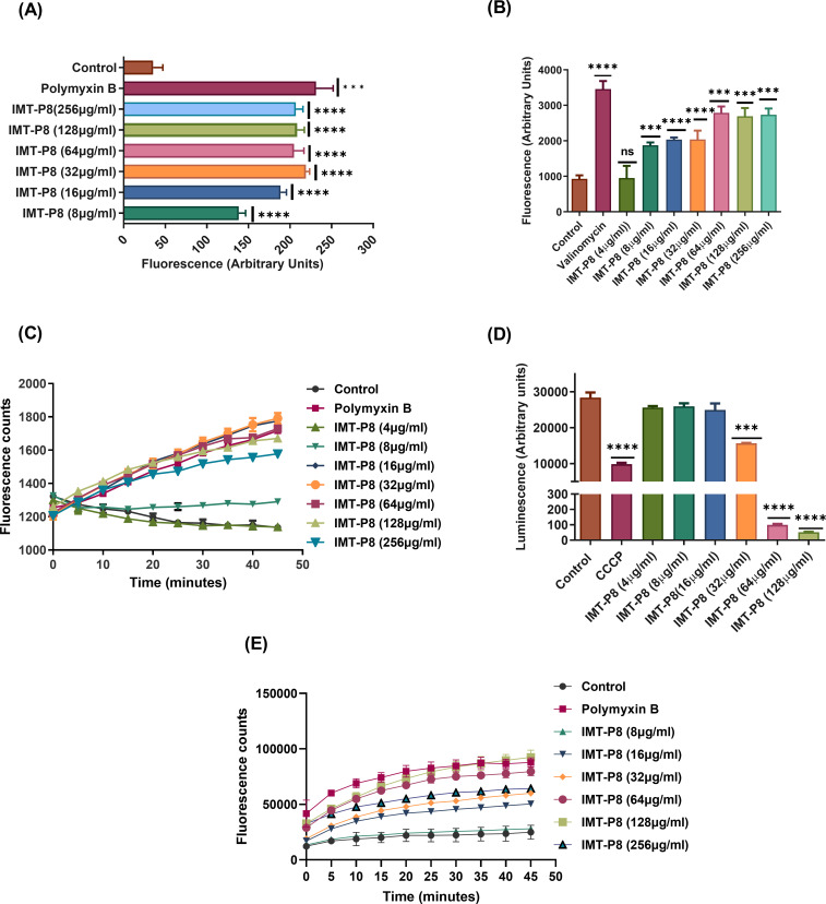
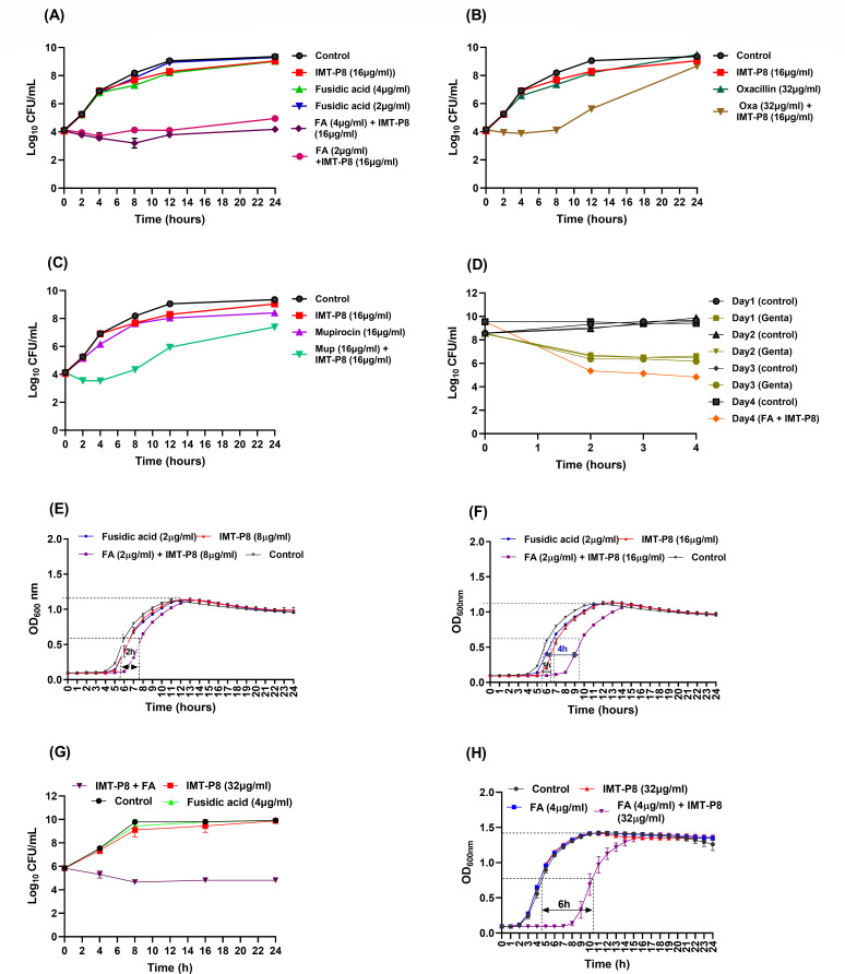
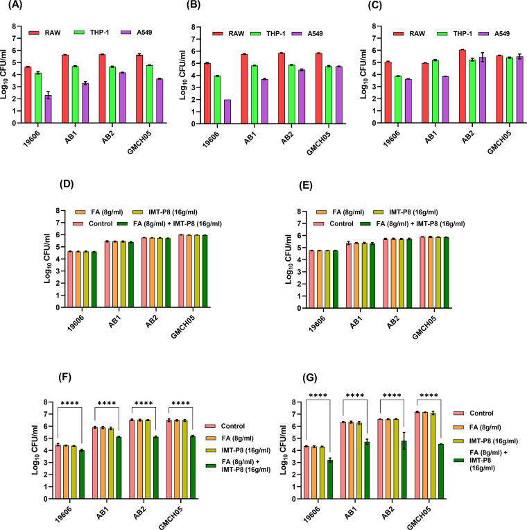
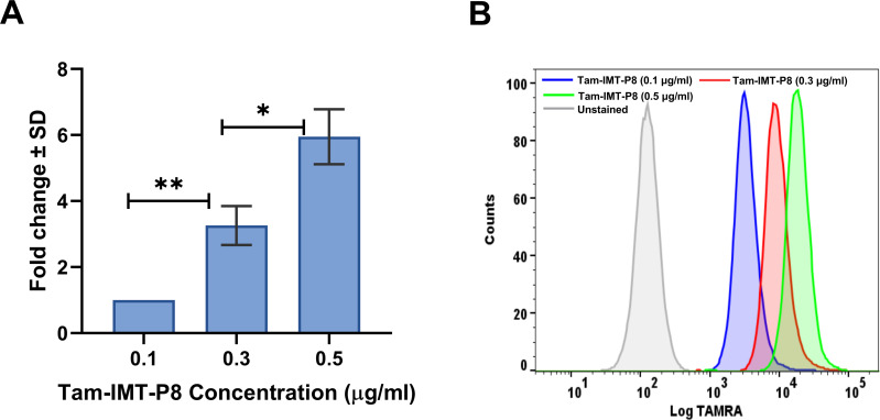
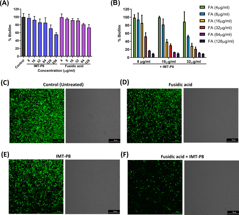
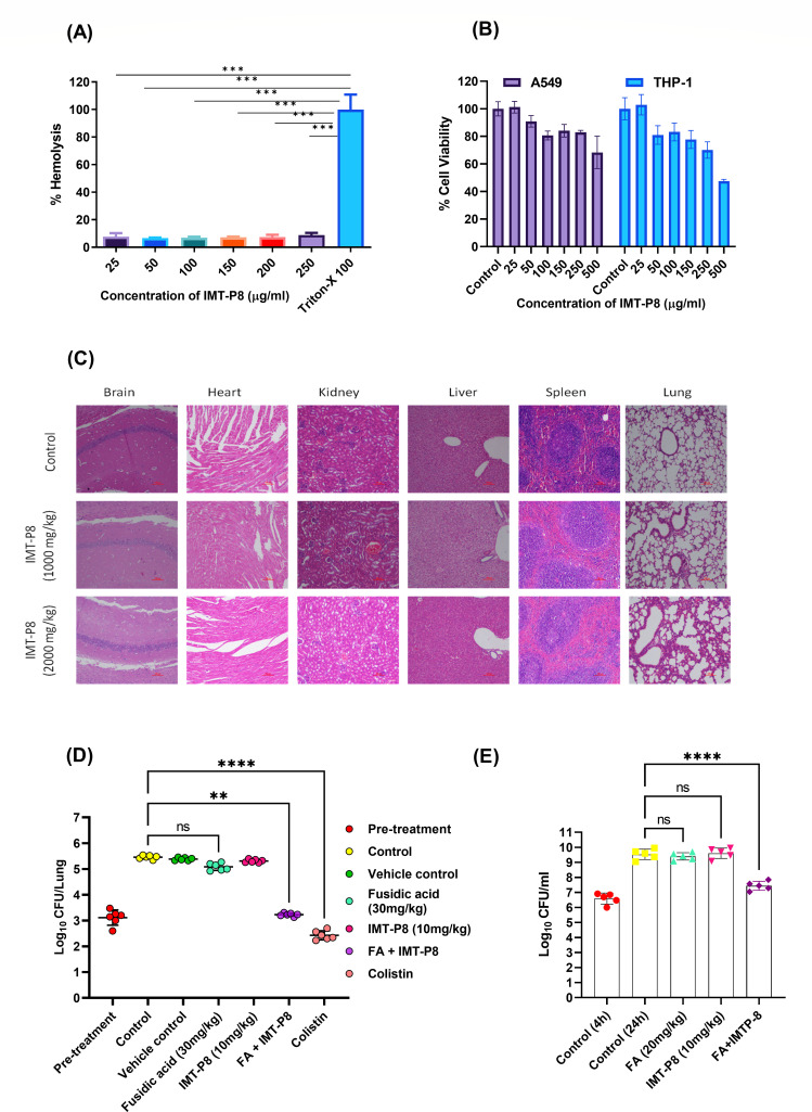
Gram-negative bacteria (GNB) pose a major global public health challenge as they exhibit a remarkable level of resistance to antibiotics. One of the factors responsible for promoting resistance against a wide range of antibiotics is the outer membrane (OM) of Gram-negative bacteria. The OM acts as a barrier that prevents the entry of numerous antibiotics by reducing their influx (due to membrane impermeability) and enhancing their efflux (with the help of efflux pumps). Our study focuses on analyzing the effect of IMT-P8, a cell-penetrating peptide (CPP), to enhance the influx of various Gram-positive specific antibiotics in multi-drug resistant Gram-negative pathogens. In the mechanistic experiments, IMT-P8 permeabilizes the OM at the same concentrations at which it enhances the activity of various antibiotics against GNB. Cytoplasmic membrane permeabilization was also observed at these concentrations, indicating that IMT-P8 acts on both the outer and cytoplasmic membranes. IMT-P8 interferes with the intrinsic resistance mechanism of GNB and has the potential to make Gram-positive specific antibiotics effective against GNB. IMT-P8 extends the post-antibiotic effect and in combination with antibiotics shows anti-persister activity. The IMT-P8/fusidic acid combination is effective in eliminating intracellular pathogens. IMT-P8 with negligible toxicity displayed good efficacy in murine lung and thigh infection models. Based on these findings, IMT-P8 is a potential antibiotic adjuvant to treat Gram-negative bacterial infections that pose a health hazard.

Keywords: A. baumannii; Gram-negative bacteria; antibiotic potentiation; cell-penetrating peptide; combination therapy; membrane permeabilization; multi-drug resistance.


Conflict of interest statement

The authors declare no conflict of interest.

Figures

Fig 1
Fig 1
(A) Outer membrane permeabilization assay using NPN was performed on A. baumannii ATCC 19606. Polymyxin B was taken as a positive control. (B) Effect of IMT-P8 on the cytoplasmic membrane depolarization of A. baumannii ATCC 19606 using DiSC3 dye. Valinomycin (K+ ionophore) was included as a positive control. (C) Membrane permeabilization assay using PI dye was performed on A. baumannii ATCC 19606. Polymyxin B was included as a positive control. (D) Effect of IMT-P8 on intracellular ATP levels of A. baumannii ATCC 19606 using a luciferin-luciferase bioluminescence assay. CCCP (a known H+ ionophore) was included as a positive control. (E) The effect of IMT-P8 on the membrane permeabilization assay using PI dye was performed on E. coli ATCC 25922. The analysis was carried out using an unpaired t-test. ****P < 0.0001, ***P < 0.001, and **P < 0.01 indicated the significant difference. All experiments were performed in triplicate, and the data were plotted as mean ± SD of three replicates.
Fig 2
Fig 2
Time kill curves of A. baumannii ATCC 19606 showing the bacteriostatic effect of (A) fusidic acid (2 and 4 µg/mL) in the presence of IMT-P8 (16 µg/mL), (B) oxacillin (32 µg/mL) in combination with IMT-P8 (16 µg/mL), and (C) mupirocin (16 µg/mL) in combination with IMT-P8 (16 µg/mL). The experiment was performed in triplicate, and each time point represents the mean ± SD of three readings. (D) A. baumannii ATCC 19606 persister cells were developed in the presence of gentamicin (20× MIC). These persister cells were treated with fusidic acid (16 µg/mL) and IMT-P8 (32 µg/mL) alone and in combination for 4 h. The experiment was performed in triplicate, and each time point represents the mean ± SD of three readings. Post-antibiotic effect of (E) fusidic acid (2 µg/mL) + IMT-P8 (8 µg/mL) and (F) fusidic acid (2 µg/mL) + IMT-P8 (16 µg/mL) in A. baumannii ATCC 19606. (G) Time kill curve of E. coli ATCC 25922 showing the bacteriostatic effect of fusidic acid (4 µg/mL) in the presence of IMT-P8 (32 µg/mL). (H) Post-antibiotic effect of fusidic acid (4 µg/mL) + IMT-P8 (32 µg/mL) in E. coli ATCC 25922. Each time point represents the mean ± SD of three readings.
Fig 3
Fig 3
Intracellular invasion and proliferation of four different strains of A. baumannii (ATCC 19606, clinical strains AB1, AB2, and GMCH05) in three different cell lines. viz., RAW macrophage cell line, PMA-differentiated THP-1 macrophage cell line, and lung epithelial A549 cell line at different time points: (A) 2 h post-infection, (B) 4 h post-infection, and (C) 8 h post-infection. The efficacy of IMT-P8, fusidic acid alone, and in combination to kill intracellular bacteria at different time points: (D) 2 h post-treatment, (E) 4 h post-treatment, (F) 8 h post-treatment, and (G) 12 h post-treatment, respectively. All the experiments were performed in triplicate, and data correspond to mean ± SD from three replicates. Two-way analysis of variance (ANOVA) followed by Dunnett’s test for multiple comparisons, N = 2 independent experiments with triplicates, ****P < 0.0001 indicated the significant difference, and P > 0.05 was considered as nonsignificant (ns).
Fig 4
Fig 4
Flow cytometric analysis of TAMRA-labeled IMT-P8 uptake into the THP-1 cells at 1 h. (A) Data are shown in the bar graph, and (B) overlay histogram is presented as mean fluorescence intensity (MFI) of intracellular TAMRA-labeled IMT-P8 and log fluorescence intensity of intracellular TAMRA--labeled IMT-P8 signal, respectively, in THP-1 cells. Data in the bar graph are presented as a representative plot of background-corrected MFI ± SD from three independent experiments (N = 3, *P < 0.01, **P < 0.001). The analysis was carried out using an unpaired t-test.
Fig 5
Fig 5
(A) Effect of IMT-P8 and fusidic acid alone on inhibition of A. baumannii ATCC 19606 biofilm; CV staining was used to determine the biomass of biofilm. (B) Effect of IMT-P8 and fusidic acid combination on inhibition of biofilm formation was assessed using CV assay. The data correspond to mean ± SD from three replicates. Confocal imaging of pre-formed biofilm of A. baumannii showing the effect of fusidic acid and IMT-P8 combination on eradication of biofilm. (C) Untreated control. (D) Fusidic acid (32 µg/mL). (E) IMT-P8 (16 µg/mL). (F) Fusidic acid + IMT-P8. The effect of fusidic acid and IMT-P8 alone or in combination on preformed biofilm eradication was assessed using confocal laser scanning microscopy (60×); the biofilms were stained with SYTO 9 (green). A. baumannii cells within biofilm give green fluorescence. The images are representatives of two independent experiments.
Fig 6
Fig 6
(A) Hemolytic effect of IMT-P8 (25–250 µg/mL) on rabbit erythrocytes. Triton X-100 (0.1%) was included as a positive control. The analysis was carried out using an unpaired t-test. ****P < 0.0001, ***P < 0.001, and **P < 0.01 indicated the significant difference. The data were plotted as mean ± SD of three replicates. (B) In vitro mammalian toxicity of IMT-P8 in A549 and THP-1 cell lines. The data correspond to the mean ± SD of three replicates. (C) Histopathology of six major organs of mice after the subcutaneous administration of two different doses (1000, 2000 mg/kg) of IMT-P8. (D and E) In vivo efficacy of fusidic acid and IMT-P8 combination in the (D) neutropenic murine lung infection model and (E) neutropenic murine thigh infection model. CFU/ lung and CFU/thigh were plotted as individual points, and error bars represent the SD within an experimental group. The Kruskal-Wallis test was used for analysis. ****P < 0.0001, ***P < 0.001, and **P < 0.01 indicated the significant difference, and P > 0.05 was considered as ns.

Similar articles

Cited by

References

    1. Antimicrobial Resistance Collaborators . 2022. Global burden of bacterial antimicrobial resistance in 2019: a systematic analysis. Lancet 399:629–655. doi:10.1016/S0140-6736(21)02724-0 - DOI - PMC - PubMed
    1. Amann S, Neef K, Kohl S. 2019. Antimicrobial resistance (AMR). Eur J Hosp Pharm 26:175–177. doi:10.1136/ejhpharm-2018-001820 - DOI - PMC - PubMed
    1. Boucher HW, Talbot GH, Bradley JS, Edwards JE, Gilbert D, Rice LB, Scheld M, Spellberg B, Bartlett J. 2009. Bad bugs, no drugs: no ESKAPE! An update from the infectious diseases society of America. Clin Infect Dis 48:1–12. doi:10.1086/595011 - DOI - PubMed
    1. Wesseling CMJ, Martin NI. 2022. Synergy by perturbing the Gram-negative outer membrane: opening the door for Gram-positive specific antibiotics. ACS Infect Dis 8:1731–1757. doi:10.1021/acsinfecdis.2c00193 - DOI - PMC - PubMed
    1. Lewis K. 2013. Platforms for antibiotic discovery. Nat Rev Drug Discov 12:371–387. doi:10.1038/nrd3975 - DOI - PubMed

MeSH terms

Substances

LinkOut - more resources