. 2024 Sep 25;9(9):e0046524.
doi: 10.1128/msphere.00465-24. Epub 2024 Sep 5.

The three Plasmodium falciparum Aurora-related kinases display distinct temporal and spatial associations with mitotic structures in asexual blood stage parasites and gametocytes

Affiliations

The three Plasmodium falciparum Aurora-related kinases display distinct temporal and spatial associations with mitotic structures in asexual blood stage parasites and gametocytes

Matthias Wyss et al. mSphere. .

Abstract

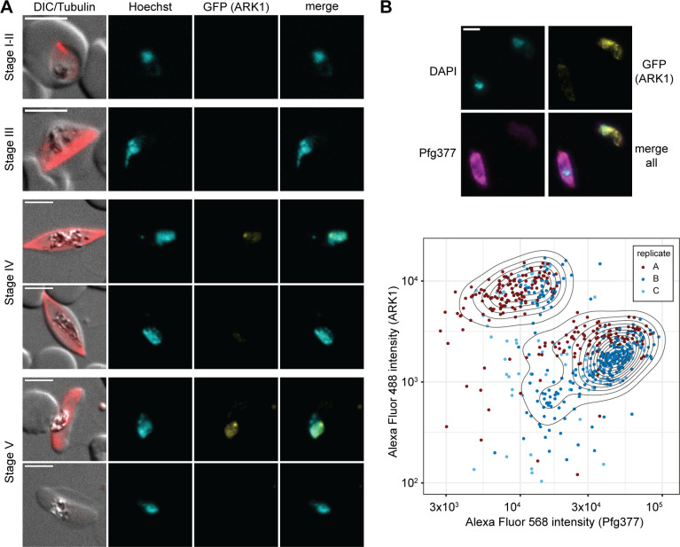
Aurora kinases are crucial regulators of mitotic cell cycle progression in eukaryotes. The protozoan malaria parasite Plasmodium falciparum replicates via schizogony, a specialized mode of cell division characterized by consecutive asynchronous rounds of nuclear division by closed mitosis followed by a single cytokinesis event producing dozens of daughter cells. P. falciparum encodes three Aurora-related kinases (PfARKs) that have been reported essential for parasite proliferation, but their roles in regulating schizogony have not yet been explored in great detail. Here, we engineered transgenic parasite lines expressing GFP-tagged PfARK1-3 to provide a systematic analysis of their expression timing and subcellular localization throughout schizogony as well as in the non-dividing gametocyte stages, which are essential for malaria transmission. We demonstrate that all three PfARKs display distinct and highly specific and exclusive spatiotemporal associations with the mitotic machinery. In gametocytes, PfARK3 is undetectable, and PfARK1 and PfARK2 show male-specific expression in late-stage gametocytes, consistent with their requirement for endomitosis during male gametogenesis in the mosquito vector. Our combined data suggest that PfARK1 and PfARK2 have non-overlapping roles in centriolar plaque maturation, assembly of the mitotic spindle, kinetochore-spindle attachment and chromosome segregation, while PfARK3 seems to be exquisitely involved in daughter cell cytoskeleton assembly and cytokinesis. These important new insights provide a reliable foundation for future research aiming at the functional investigation of these divergent and possibly drug-targetable Aurora-related kinases in mitotic cell division of P. falciparum and related apicomplexan parasites.IMPORTANCEMalaria parasites replicate via non-conventional modes of mitotic cell division, such as schizogony, employed by the disease-causing stages in the human blood or endomitosis during male gametogenesis in the mosquito vector. Understanding the molecular mechanisms regulating cell division in these divergent unicellular eukaryotes is not only of scientific interest but also relevant to identify potential new antimalarial drug targets. Here, we carefully examined the subcellular localization of all three Plasmodium falciparum Aurora-related kinases (ARKs), distantly related homologs of Aurora kinases that coordinate mitosis in model eukaryotes. Detailed fluorescence microscopy-based analyses revealed distinct, specific, and exclusive spatial associations for each parasite ARK with different components of the mitotic machinery and at different phases of the cell cycle during schizogony and gametocytogenesis. This comprehensive set of results closes important gaps in our fragmentary knowledge on this important group of kinases and offers a valuable source of information for future functional studies.

Keywords: Aurora-related kinase; Plasmodium falciparum; cell division; gametocytes; malaria; mitosis; schizogony.


Conflict of interest statement

The authors declare no conflict of interest.

Figures

Fig 1
Fig 1
PfARK1 is expressed in a subset of nuclei during schizogony. (A) Localization of PfARK1–GFP throughout intra-erythrocytic asexual development as assessed by live cell fluorescence microscopy. Numbers in brackets indicate the age range (hours post invasion) of the synchronous parasite samples imaged at consecutive time points across intra-erythrocytic development. R, ring stage; T, trophozoite; S, schizont; seg., segmented. (B, C) Localization of PfARK1-GFP, centrin, and α/β-tubulin (B) or CENH3 (C) in schizonts as assessed by IFAs. Representative images are shown in each panel. DNA was stained with Hoechst (live) or 4′,6-diamidino-2-phenylindole (DAPI) (IFA). DIC, differential interference contrast. A1, PfARK1; CEN, centrin; TUB, α/β-tubulin; CH3, CENH3; i/ii, zoom selections. Scale bar = 5 µm.
Fig 2
Fig 2
PfARK2 is expressed in all nuclei during schizogony. (A) Localization of PfARK2–GFP throughout intra-erythrocytic asexual development as assessed by live cell fluorescence microscopy. Numbers in brackets indicate the age range (hours post invasion) of the synchronous parasite samples imaged at consecutive time points across intra-erythrocytic development. R, ring stage; T, trophozoite; S, schizont; seg., segmented. (B, C) Localization of PfARK2–GFP, centrin, and α/β-tubulin (B) or CENH3 (C) in schizonts as assessed by IFAs. Representative images are shown in each panel. DNA was stained with Hoechst (live) or DAPI (IFA). DIC, differential interference contrast. A2, PfARK2; CEN, centrin; TUB, α/β-tubulin; CH3, CENH3; i, zoom selections. Scale bar = 5 µm.
Fig 3
Fig 3
PfARK3 expression is restricted to late-stage schizonts. (A) Localization of PfARK3–GFP throughout intra-erythrocytic asexual development as assessed by live cell fluorescence microscopy. Numbers in brackets indicate the age range (hours post invasion) of the synchronous parasite samples imaged at consecutive time points across intra-erythrocytic development. R, ring stage; T, trophozoite; S, schizont; seg., segmented. (B, C) Localization of PfARK3–GFP, centrin, and CENH3 in late-stage schizonts as assessed by IFAs. Representative images are shown in each panel. DNA was stained with Hoechst (live) or DAPI (IFA). DIC, differential interference contrast. A3, PfARK3; CEN, centrin; CH3, CENH3; i, zoom selections. Scale bar = 5 µm.
Fig 4
Fig 4
PfARK1 shows male-specific expression in late-stage gametocytes. (A) PfARK1–GFP expression in gametocytes was assessed by live cell fluorescence microscopy. Representative images are shown. DNA was stained with Hoechst. MTs were stained with SPY555-tubulin. Stages of gametocyte development are indicated. Representative images are shown. DIC, differential interference contrast. Scale bar = 5 µm. (B) Top: localization of PfARK1–GFP (yellow) and Pfg377 (magenta) in stage V gametocytes as assessed by IFA. Representative images are shown. DNA was stained with DAPI. Scale bar = 5 µm. Bottom: quantification of PfARK1–GFP (Alexa Fluor 488) and Pfg377 (Alexa Fluor 568) IFA fluorescence signal intensities in single stage V gametocytes. The assay was performed in three independent biological replicates (251, 259, and 137 cells analyzed per replicate). Dots in the contour plot represent the fluorophore intensities measured for individual cells.
Fig 5
Fig 5
PfARK2 is expressed throughout gametocytogenesis with male-specific expression observed in late-stage gametocytes. (A) PfARK2–GFP expression in gametocytes was assessed by live cell fluorescence microscopy. Representative images are shown. DNA was stained with Hoechst. MTs were stained with SPY555-tubulin. Stages of gametocyte development are indicated. Representative images are shown. DIC, differential interference contrast. Scale bar = 5 µm. (B) Top: localization of PfARK2–GFP (yellow) and Pfg377 (magenta) in stage V gametocytes as assessed by IFA. Representative images are shown. DNA was stained with DAPI. Scale bar = 5 µm. Bottom: quantification of PfARK2–GFP (Alexa Fluor 488) and Pfg377 (Alexa Fluor 568) IFA fluorescence signal intensities in single stage V gametocytes. The assay was performed in three independent biological replicates (231, 170, and 242 cells analyzed per replicate). Dots in the contour plot represent the fluorophore intensities measured for individual cells.
Fig 6
Fig 6
PfARK1 and PfARK2 localize to the intranuclear body in male late-stage gametocytes. (A, B) Localization of PfARK1–GFP and centrin (A) or PfARK2–GFP and centrin (B) in female and male stage IV gametocytes as assessed by IFAs. MTs were stained with antibodies against α/β-tubulin. Representative images are shown in each panel. DNA was stained with DAPI. DIC, differential interference contrast. A1/2, PfARK1/PfARK2; CEN, centrin; i, zoom selections. Scale bar = 5 µm.

Similar articles

Cited by

References

    1. Simon CS, Stürmer VS, Guizetti J. 2021. How many is enough? - challenges of multinucleated cell division in malaria parasites. Front Cell Infect Microbiol 11:658616. doi:10.3389/fcimb.2021.658616 - DOI - PMC - PubMed
    1. Milner DA. 2018. Malaria pathogenesis, p 8. Cold Spring Harb Perspect Med. - PMC - PubMed
    1. Ngotho P, Soares AB, Hentzschel F, Achcar F, Bertuccini L, Marti M. 2019. Revisiting gametocyte biology in malaria parasites. FEMS Microbiol Rev 43:401–414. doi:10.1093/femsre/fuz010 - DOI - PMC - PubMed
    1. Kafsack BFC, Rovira-Graells N, Clark TG, Bancells C, Crowley VM, Campino SG, Williams AE, Drought LG, Kwiatkowski DP, Baker DA, Cortés A, Llinás M. 2014. A transcriptional switch underlies commitment to sexual development in malaria parasites. Nature New Biol 507:248–252. doi:10.1038/nature12920 - DOI - PMC - PubMed
    1. Sinha A, Hughes KR, Modrzynska KK, Otto TD, Pfander C, Dickens NJ, Religa AA, Bushell E, Graham AL, Cameron R, Kafsack BFC, Williams AE, Llinas M, Berriman M, Billker O, Waters AP. 2014. A cascade of DNA-binding proteins for sexual commitment and development in Plasmodium. Nature New Biol 507:253–257. doi:10.1038/nature12970 - DOI - PMC - PubMed

LinkOut - more resources