Randomized Controlled Trial
. 2024 Oct 7;221(10):e20240604.
doi: 10.1084/jem.20240604. Epub 2024 Sep 5.

Use of 3M-052-AF with Alum adjuvant in HIV trimer vaccine induces human autologous neutralizing antibodies

Affiliations
Randomized Controlled Trial

Use of 3M-052-AF with Alum adjuvant in HIV trimer vaccine induces human autologous neutralizing antibodies

William O Hahn et al. J Exp Med. .

Abstract

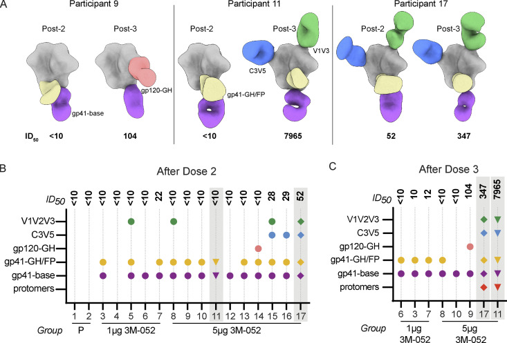
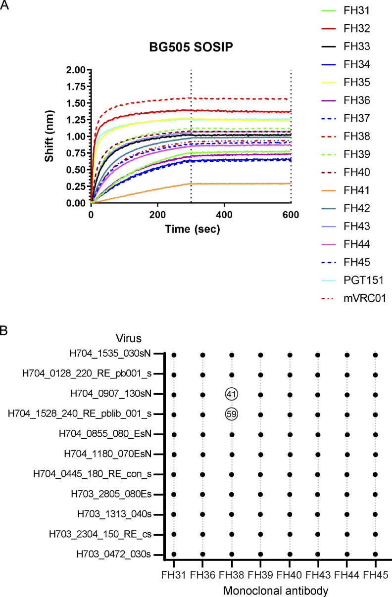
Stabilized trimers preserving the native-like HIV envelope structure may be key components of a preventive HIV vaccine regimen to induce broadly neutralizing antibodies (bnAbs). We evaluated trimeric BG505 SOSIP.664 gp140 formulated with a novel TLR7/8 signaling adjuvant, 3M-052-AF/Alum, for safety, adjuvant dose-finding, and immunogenicity in a first-in-healthy adult (n = 17), randomized, and placebo-controlled trial (HVTN 137A). The vaccine regimen appeared safe. Robust, trimer-specific antibody, and B cell and CD4+ T cell responses emerged after vaccination. Five vaccinees developed serum autologous tier 2 nAbs (ID50 titer, 1:28-1:8647) after two to three doses targeting C3/V5 and/or V1/V2/V3 Env regions by electron microscopy and mutated pseudovirus-based neutralization analyses. Trimer-specific, B cell-derived monoclonal antibody activities confirmed these results and showed weak heterologous neutralization in the strongest responder. Our findings demonstrate the clinical utility of the 3M-052-AF/Alum adjuvant and support further improvements of trimer-based Env immunogens to focus responses on multiple broad nAb epitopes.


Conflict of interest statement

Disclosures: H. Janes reported grants from the National Institutes of Health during the conduct of the study and grants from the National Institutes of Health outside the submitted work. M. Tomai reported being an employee at 3M Company when this research was started and having received stock options while working at 3M. M. Tomai also received options from 3M while these studies were being performed. M. Tomai is now a contract employee for Solventum, which owns the vaccine adjuvant 3M-052 that was used in this study. L. Corey reported grants from NIAID/National Institutes of Health during the conduct of the study. S. Edupuganti reported grants from HIV Vaccine Trials Network during the conduct of the study. I. Frank reported grants from Gilead Sciences and Johnson and Johnson during the conduct of the study and personal fees from GlaxoSmithKline and Johnson and Johnson outside the submitted work. R.W. Sanders and J.P. Moore reported a patent to EP2765138A3/US20140212458 issued. A.B. Ward reported personal fees from Third Rock Ventures outside the submitted work. N. Rouphael reported grants from Emory University during the conduct of the study; and that Emory receives funds for N. Rouphael to conduct research from Sanofi, Lilly, Merck, Quidel, Immorna, Vaccine Company, and Pfizer. N. Rouphael served on selected advisory boards for Sanofi, Seqirus, Pfizer and Moderna and is a paid clinical trials safety consultant for ICON, CyanVac, Imunon, and EMMES. Fred Hutch Cancer Center receives funds for M.J. McElrath to conduct research from Janssen, Sanofi, Regeneron, and Moderna. No other disclosures were reported.

Figures

Figure 1.
Figure 1.
Local and systemic reactogenicity associated with BG505 SOSIP.664 with 3M-052-AF/Alum. The maximum local (left) and systemic (right) events reported within 7 days after the first, second, and optional third dose (indicated by *) of BG505 SOSIP.664 gp140 with 3M-052-AF (at 1 or 5 µg) + Alum or in the placebo group. Reactions were graded as mild (yellow), moderate (green), or severe (blue). The number of participants in each group is listed above.
Figure 2.
Figure 2.
Vaccine-induced serum binding antibodies, tier 2 neutralizing antibodies, and circulating memory IgG+ B cells. (A) BG505 SOSIP.664 gp140–specific responses were measured by BAMA in serum preimmunization and 2 wk following the indicated dose of vaccine. IgG binding magnitude AUC values are shown against BG505 SOSIP.664 gp140. (B) ID50 neutralizing antibody titers against the autologous tier 2 HIV-1 BG505/T332N using the TZMbl neutralization assay preimmunization and after the indicated vaccine dose. (C) Percent of IgG+ B cells that bind BG505 SOSIP.664 gp140 fluorescent probe as assessed by flow cytometry. B cells were identified using doublet exclusion, lymphocyte scatter profile, viability dye, and stained negative for lineage markers CD3, CD56, and CD14; positive for CD19 and CD20. IgG+ B cells are gated as IgD and IgG+. For the x axis, “Pre” is the Month 0 sample taken before vaccination, and “P” represents the placebo group. The dark red lines indicate responses of the same two individuals with the highest neutralizing titers after the third dose, shown in B (participant 11 indicated by ▼ and Participant 17 indicated by ♦). Filled symbols indicate positive responses, and the numbers at the top of the graph represent the fraction of participants who had a positive response at the indicated timepoint out of total participants.
Figure S1.
Figure S1.
Antibody and CD8+ T cell responses. (A) Serum gp41-specific IgG responses were measured by BAMA in serum 2 wk following the indicated dose of vaccine, and the response magnitude is shown as AUC values. (B) ID50 neutralizing antibody titers against the tier 1 virus MW965.26 using the TZMbl neutralization assay after the indicated vaccine dose. The participants indicated by darker shading are the same two individuals with the highest neutralizing antibody titers after the third dose (Participant 11 indicated by ▼ and Participant 17 indicated by ♦). (C) Percent of CD8+ T cells expressing IFN-γ and/or IL-2 in response are shown to synthetic BG505 Env peptides spanning the gp120 and gp41 ectodomain using an ex vivo intracellular cytokine staining assay. Positive responses are shown with filled symbols, and the numbers at the top of the graph represent the fraction of participants who had a positive response at the indicated timepoint. (D) Participant 17 neutralizing antibody titers pseudoviruses with mutations introduced at the indicated positions and color coded as in A. “Pan KO mutant” includes the following mutations: T332N, T465N, S241N, 133aA, and 141aN. Open bars represent titers that were not impacted by the mutations (<threefold reduction relative to wild-type virus).
Figure 3.
Figure 3.
Antigen-specific CD4+ T cell responses elicited by BG505 SOSIP.664 gp140 with either 1 or 5 µg of 3M-052-AF/Alum. (A) Percent of CD4+ T cells expressing IFN-γ and/or IL-2 in response to synthetic BG505 Env peptides spanning the gp120 and gp41 ectodomain using an ex vivo intracellular cytokine staining assay. Positive responses are shown with filled symbols, and numbers at the top of the graph represent the fraction of participants who had a positive response at the indicated timepoint. (B) Cluster analysis of CD4+ T cells from all treatment samples was combined. CD4+ T cells expressing at least one of seven of the functional markers (IFN-γ, IL-2, TNF, CD154, IL-4, IL-5/13, and IL-17a) were extracted and used as input. 18 clusters were identified. (C) Data for six clusters with positive responses (see Materials and methods) were graphed as background-subtracted percentages of CD4+ T cells for the total Env response (gp120 and gp41). The dark red lines indicate CD4+ T cell responses in the two participants with the highest neutralizing titers (Fig. 2 B) after the third dose.
Figure S2.
Figure S2.
EM serum polyclonal antibody epitope mapping data. Segmented maps representing polyclonal antibody responses detected against BG505 SOSIP antigen for each participant after two or three doses. Participant numbers are the same as Fig. 4. Treatment group is denoted next to each participant number (P: placebo, 1 µg 3M-052, 5 µg 3M-052). Polyclonal epitopes are colored as defined for Participant 17 (after dose 2) and Participant 9 (after dose 3). Representative maps have been deposited to the Electron Microscopy Data Bank under the listed accession codes: antibodies with red letter “a” are found in the main Electron Microscopy Data Bank entry map; antibodies with letters “b,” “c,” or “d” can be found as “additional maps” under the Download tab of the respective entry on https://www.emdataresource.org/.
Figure 4.
Figure 4.
Three vaccine doses induce serum antibodies recognizing a wider range of Env epitopes, including V1/V2/V3 regions in selected participants with autologous neutralizing antibodies. (A) Representative EMPEM segmented 3D maps of participants after the second and third doses. Antibody Fab colored by epitope; trimeric Env in gray. (B and C) Epitope regions recognized by serum polyclonal antibodies using EMPEM after the second dose (B) and third dose (C). ID50 neutralizing titers to BG505/T332N are listed at the top. The x-axis indicates the participant numbers and dose of 3M-052-AF. Colors correspond to epitope regions illustrated in A. Grey shading indicates the two participants with the highest neutralizing titers after the third dose (see Fig. 2 B), represented by a triangle and diamond. P, placebo.
Figure 5.
Figure 5.
Mapping BG505 SOSIP.664 gp140 epitope regions recognized by serum and monoclonal neutralizing antibody responses. (A) Schematic representation of the epitope surface targeted by polyclonal serum antibodies to BG505 SOSIP.664 gp140, as determined by published cryoEM structures (Antanasijevic et al., 2021). The locations of mutations used in pseudovirus-based epitope mapping are marked at indicated positions. (B) Serum antibody neutralization titers for Participant 11 were mapped using pseudoviruses with mutations introduced at indicated positions and color-coded as in A. “Pan KO mutant” includes the following mutations: T332N, T465N, S241N, 133aA, and 141aN. Open bars represent titers that were not impacted by the mutations (<threefold reduction relative to wild-type BG505 virus). (C) IC50 of monoclonal antibodies against BG505/T332N isolated from Participant 11 after the third dose. Circled points indicate samples also analyzed by nsEM. (D) nsEM 3D maps of indicated monoclonal antibodies in complex with BG505 SOSIP.664 gp140, with % somatic hypermutation of heavy and light chains indicated.
Figure S3.
Figure S3.
Monoclonal antibody binding and neutralization. (A) Biolayer interferometry of monoclonal antibodies to test for binding to BG505 SOSIP.664. VRC01 and PGT151 are monoclonal antibodies with known binding activity, which are included as representative controls. (B) The numbers indicated in the circles represent antibody neutralization ID50 titers against an HIV-1 strain in a panel selected based on tier 2 phenotype. Closed circles indicate no observed neutralization activity.

Update of

References

    1. Abbott, R.K., and Crotty S.. 2020. Factors in B cell competition and immunodominance. Immunol. Rev. 296:120–131. 10.1111/imr.12861 - DOI - PMC - PubMed
    1. Antanasijevic, A., Sewall L.M., Cottrell C.A., Carnathan D.G., Jimenez L.E., Ngo J.T., Silverman J.B., Groschel B., Georgeson E., Bhiman J., et al. . 2021. Polyclonal antibody responses to HIV Env immunogens resolved using cryoEM. Nat. Commun. 12:4817. 10.1038/s41467-021-25087-4 - DOI - PMC - PubMed
    1. Arnaout, R., Lee W., Cahill P., Honan T., Sparrow T., Weiand M., Nusbaum C., Rajewsky K., and Koralov S.B.. 2011. High-resolution description of antibody heavy-chain repertoires in humans. PLoS One. 6:e22365. 10.1371/journal.pone.0022365 - DOI - PMC - PubMed
    1. Baden, L.R., El Sahly H.M., Essink B., Follmann D., Neuzil K.M., August A., Clouting H., Fortier G., Deng W., Han S., et al. . 2021. Phase 3 Trial of mRNA-1273 during the Delta-Variant Surge. N. Engl. J. Med. 385:2485–2487. 10.1056/nejmc2115597 - DOI - PMC - PubMed
    1. Bianchi, M., Turner H.L., Nogal B., Cottrell C.A., Oyen D., Pauthner M., Bastidas R., Nedellec R., McCoy L.E., Wilson I.A., et al. . 2018. Electron-microscopy-based epitope mapping defines specificities of polyclonal antibodies elicited during HIV-1 BG505 envelope trimer immunization. Immunity. 49:288–300.e8. 10.1016/j.immuni.2018.07.009 - DOI - PMC - PubMed

Publication types

MeSH terms