. 2025 Apr;203(4):2420-2434.
doi: 10.1007/s12011-024-04337-8. Epub 2024 Sep 5.

Vitamin D Alleviates Heavy Metal-Induced Cytotoxic Effects on Human Bone Osteoblasts Via the Induction of Bioenergetic Disruption, Oxidative Stress, and Apoptosis

Affiliations

Vitamin D Alleviates Heavy Metal-Induced Cytotoxic Effects on Human Bone Osteoblasts Via the Induction of Bioenergetic Disruption, Oxidative Stress, and Apoptosis

Ekramy M Elmorsy et al. Biol Trace Elem Res. 2025 Apr.

Abstract

Cadmium (Cd) and lead (Pb) are heavy metals (HMs) that persistently contaminate the ecosystem, and bioaccumulation in bones is a health concern. We used biochemical and molecular assays to assess the cytoprotective effect of vitamin D (VD) on Cd- and Pd-induced chemical toxicity of human bone osteoblasts in vitro. Exposing Cd and Pb to human osteoblast cultures at concentrations of 0.1-1000 µM for 24-72 h significantly reduced osteoblast viability in an exposure time- and concentration-dependent manner. The cytotoxic effect of Cd on osteoblasts was more severe than Pb's, with 72-h exposure estimated half maximal effective concentration (EC50) of 8 and 12 µM, respectively, and VD (1 and 10 nM) alleviated cytotoxicity. Bioenergetics assays of ATP, mitochondrial membrane potential, and mitochondrial complex I and III activity showed that both Cd and Pb (1 and 10 µM) inhibited cellular bioenergetics after 72-h exposure. Cd and Pb increased lipid peroxidation and reactive oxygen species with reduced catalase/superoxide dismutase antioxidant activities and increased activity of caspases -3, -8, and -9. Co-treatment with VD (1 and 10 nM) counteracted bioenergetic disruption, oxidative damage, and apoptosis in a concentration-dependent manner. These findings suggest that VD is effective in managing the toxic effects of environmental pollutants and in treating bone diseases characterized by oxidative stress, apoptosis, and bioenergetic disruption.

Keywords: Antioxidants; Bone; Cadmium; Lead; Osteoblasts; Redox stress; Vitamin D.


Conflict of interest statement

Declarations. Competing Interests: The authors declare no competing interests.

Figures
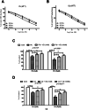
Fig. 1
Fig. 1
MTT data of the cytotoxic effect of lead (Pb) and cadmium (Cd) (0.1, 1, 10, 100, and 1000 µM) on cultured human osteoblasts at 24, 48, and 72 h post-exposure (A, B). C, D The protective effect of VD (1 and 10 nM) to alleviate the cytotoxic effects of Pb and Cd on human osteoblasts 72 h post-exposure. Both metals were cytotoxic to the cultured cells in a concentration- and exposure-duration-dependent manner. Cd was more cytotoxic with lower estimated EC50s in the tested time points. Vitamin D significantly alleviated both metals’ cytotoxic effects
Fig. 2
Fig. 2
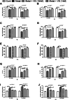
The effect of lead (Pb) and cadmium (Cd) at concentrations of 1 and 10 µM on the cellular bioenergetics of the cultured human osteoblasts and the protective effect of vitamin D (VD) at 1 and 10 nM concentrations. The cells were treated with each metal differently and VD for 72 h. ATP (A, B), mitochondrial complex I (MCI) (C, D), mitochondrial complex III (MCIII) (E, F), mitochondrial membrane potential (MMP) (G, H), and lactate production assays (I, J) were conducted. Both metals significantly inhibited the treated cells’ bioenergetics in a concentration-dependent manner. Cd showed higher inhibitory effects in all assays. Vitamin D led to significant improvements in the metal-treated cells’ ATP production, higher activity of MMP, MCI, and MCIII, and significantly decreased lactate production. Significance was evaluated by one-way ANOVA with Dunnett’s post-test comparing the outcomes in the presence and absence of VD. *p-value < 0.05, **p-value < 0.01, and***p-value < 0.0001
Fig. 3
Fig. 3
The effect of lead (Pb) and cadmium (Cd) at concentrations of 1 and 10 µM on the redox status of cultured human osteoblasts and the protective effect of Vitamin D (VD) at 1 and 10 nM concentrations. Cells were treated with each metal differently and VD for 72 h. Reactive oxygen species (ROS) (A, B), lipid peroxidation thiobarbituric acid (TBARS) product (C, D), antioxidant enzyme catalase (CAT) activities (E, F), and antioxidant enzyme superoxide dismutase (SOD) activities (G, H) were measured. Both metals significantly increased ROS and TBARS production, with a significant decrease in CAT and SOD antioxidant activities in a concentration-dependent pattern. Vitamin D significantly counteracted metal-induced oxidative damage and alleviated the inhibitory effect of the metals on CAT and SOD to varying extents. Significance was evaluated by one-way ANOVA with Dunnett’s post-test comparing the outcomes in the presence or absence of VD. *p-value < 0.05, **p-value < 0.01, and***p-value < 0.0001
Fig. 4
Fig. 4
The effect of lead (Pb) and cadmium (Cd) at 1 and 10 µM concentrations, 72 h post-exposure, on caspase-3, -8, and -9 activity in cultured human osteoblasts and the protective effect of Vitamin D (VD) at concentrations of 1 and 10 nM. Both metals significantly inhibited the secretory functions of the treated cells in the three assays in a concentration-dependent manner. Vitamin D significantly counteracted the metal-induced secretory dysfunction of the treated cells. Significance was evaluated by one-way ANOVA with Dunnett’s post-test comparing the outcomes in the presence and absence of VD. *p-value < 0.05, **p-value < 0.01, and ***p-value < 0.0001
Fig. 5
Fig. 5
The effect of lead (Pb) and cadmium (Cd) at concentrations of 1 and 10 µM, 72 h post-exposure, on osteocalcin (A, B), procollagen type I peptide (C, D) and alkaline phosphatase (ALP) (E, F) production in cultured human osteoblasts. Both metals significantly inhibited the secretory functions of the treated cells in the three assays in a concentration-dependent manner. Vitamin D significantly counteracted the metal-induced secretory dysfunction of the treated cells. Significance was evaluated by one-way ANOVA with Dunnett’s post-test comparing the outcomes in the presence and absence of VD. *p-value < 0.05, **p-value < 0.01 and.***p-vale < 0.0001
Fig. 6
Fig. 6
Potential mechanisms of cadmium (Cd) and lead (Pb) cytotoxicity. (A) Trace metals have prooxidant and mitochondrial disruption effects that lead to the generation of reactive oxygen species (ROS), depending on their ability to lose electrons. Elevated ROS production is frequently associated with DNA damage and can trigger various intracellular signaling pathways, including apoptotic pathways (Adopted with permission from the Kyoto Encyclopedia of Genes and Genomes (KEGG) (https://www.kegg.jp/pathway/map05208) [36]. (B) Predicted protein interaction network of cadmium: CAT: catalase; MT2A: metallothionein 2A; TXN thioredoxin; HMOX1 heme oxygenase; OGG1 8-oxoguanine DNA glycosylase; ESR1: estrogen receptor 1; JUN: jun proto-oncogene; CDH1: cadherin 1; ALDH5A1 aldehyde dehydrogenase 5 family member A1; and IL6: interleukin 6. (C) Predicted protein interaction network of lead: PKC1 protein serine/threonine kinase; KES1 (member of the oxysterol binding protein); CMD1: calmodulin; DCP2 (catalytic subunit of the Dcp1p-Dcp2p decapping enzyme complex); MPS1 (dual-specificity kinase required for spindle pole body duplication and spindle checkpoint); APN2 Class II (a basic endonuclease involved in DNA damage repair); YPK2: protein kinase; HEM2: heme A-farnesyltransferase; and HEM15: ferrochelatase. These interactions explain some of the oxidative stress-, apoptosis-, and mitochondrial disruption-related effects of these metals. Graphical representations of the potential protein partners of Cd and Pb were identified from the STITCH database (http://stitch.embl.de/) [37]

Similar articles

Cited by

References

    1. Bharti R, Sharma R (2022) Effect of heavy metals: an overview. Mater Today: Proc 51:880–885
    1. Zhang S, Sun L, Zhang J, Liu S, Han J, Liu Y (2020) Adverse impact of heavy metals on bone cells and bone metabolism dependently and independently through Anemia. Adv Sci 7(19):2000383. 10.1002/advs.202000383 - PMC - PubMed
    1. Campbell JR, Rosier RN, Novotny L, Puzas JE (2004) The association between environmental lead exposure and bone density in children. Environ Health Perspect 112(11):1200–1203. 10.1289/ehp.6555 - PMC - PubMed
    1. Campbell JR, Auinger P (2007) The association between blood lead levels and osteoporosis among adults–results from the third national health and nutrition examination survey (NHANES III). Environ Health Perspect 115(7):1018–1022. 10.1289/ehp.9716 - PMC - PubMed
    1. Cui A, Xiao P, Hu B, Ma Y, Fan Z, Wang H, Zhou F, Zhuang Y (2022) Blood lead level is negatively associated with bone mineral density in U.S. children and adolescents aged 8–19 years. Front Endocrinol 13:928752. 10.3389/fendo.2022.928752 - PMC - PubMed

LinkOut - more resources