

Potential prognostic biomarker SERPINA12: implications for hepatocellular carcinoma
- PMID: 39235554
- PMCID: PMC12000224
- DOI: 10.1007/s12094-024-03689-w
Potential prognostic biomarker SERPINA12: implications for hepatocellular carcinoma
Abstract
Background: Hepatocellular carcinoma (HCC) remains one of the most prevalent malignant tumors, exhibiting a high morbidity and mortality rate. The mechanism of its occurrence and development requires further study. The objective of this study was to investigate the role of SERPINA12 in the diagnosis, prognosis prediction and biological function within HCC.
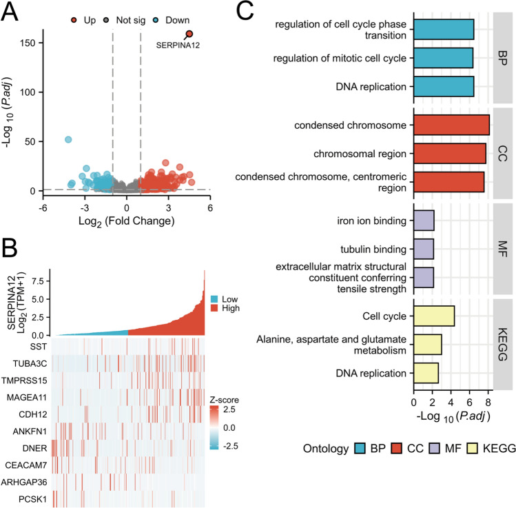
Methods: The Cancer Genome Atlas (TCGA) data were employed to analyze the relationship between clinical features and SERPINA12 expression in HCC. Kaplan-Meier curves were utilized to analyze the correlation between SERPINA12 expression and prognosis in HCC. The function of SERPINA12 was determined by enrichment analysis, and the relationship between SERPINA12 expression and immune cell infiltration was investigated. The expression of SERPINA12 was examined in 75 patients with HCC using RT-qPCR and immunohistochemistry, and survival analysis was performed.
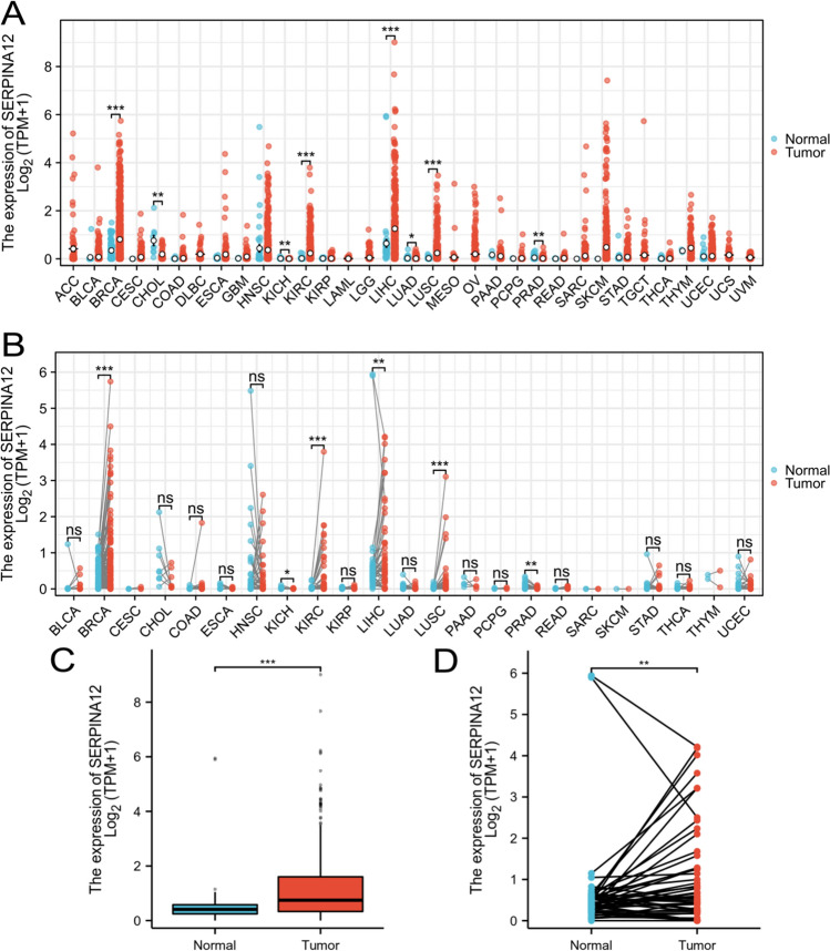
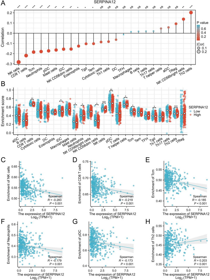
Results: The expression of SERPINA12 from TCGA database was found to be significantly higher in HCC tissues than in normal tissues and carried a poor prognosis. ROC curve demonstrated the diagnostic potential of SERPINA12 for HCC. The multivariate Cox regression analysis showed that pathologic T stage, tumor status, and SERPINA12 expression were independently associated with patient survival. The SERPINA12 expression was found to correlate with immune cell infiltration. Our RT-qPCR and immunohistochemical analysis revealed high expression of SERPINA12 in tumor tissues. Survival analysis indicated its association with poor prognosis.
Conclusion: SERPINA12 is a promising biomarker for diagnosis and prognosis, and it is associated with immune cell infiltration.
Keywords: Bioinformatics; Hepatocellular cancer; Prognosis; SERPINA12.
© 2024. The Author(s).
Conflict of interest statement
Declarations. Conflict of interest: The authors have declared that there are no potential conflicts of interest associated with this article. Ethical approval: Ethical approval was obtained from the Ethics Committee of the Affiliated Tumor Hospital of Xinjiang Medical University. Informed consent: All participants in the sample collection had provided informed consent, personally or through authorized legal representatives.
Figures


Similar articles
-
BUB1 serves as a biomarker for poor prognosis in liver hepatocellular carcinoma.BMC Immunol. 2025 Mar 11;26(1):20. doi: 10.1186/s12865-025-00698-4. BMC Immunol. 2025. PMID: 40069598 Free PMC article.
-
High expression of SMPD4 promotes liver cancer and is associated with poor prognosis.BMC Res Notes. 2025 Apr 10;18(1):159. doi: 10.1186/s13104-025-07212-4. BMC Res Notes. 2025. PMID: 40211349 Free PMC article.
-
Upregulation of FEN1 Is Associated with the Tumor Progression and Prognosis of Hepatocellular Carcinoma.Dis Markers. 2020 Jan 13;2020:2514090. doi: 10.1155/2020/2514090. eCollection 2020. Dis Markers. 2020. PMID: 32399086 Free PMC article.
-
Overexpression of EWSR1 (Ewing sarcoma breakpoint region 1/EWS RNA binding protein 1) predicts poor survival in patients with hepatocellular carcinoma.Bioengineered. 2021 Dec;12(1):7941-7949. doi: 10.1080/21655979.2021.1982844. Bioengineered. 2021. PMID: 34612781 Free PMC article.
-
The diagnostic and prognostic value of H2AFY in hepatocellular carcinoma.BMC Cancer. 2021 Apr 15;21(1):418. doi: 10.1186/s12885-021-08161-4. BMC Cancer. 2021. PMID: 33858382 Free PMC article.
Cited by
-
ADME gene-driven prognostic model for bladder cancer: a breakthrough in predicting survival and personalized treatment.Hereditas. 2025 Mar 19;162(1):42. doi: 10.1186/s41065-025-00409-4. Hereditas. 2025. PMID: 40108724 Free PMC article.
References
-
- Rumgay H, Arnold M, Ferlay J, Lesi O, Cabasag CJ, Vignat J, Laversanne M, McGlynn KA, Soerjomataram I. Global burden of primary liver cancer in 2020 and predictions to 2040. J Hepatol. 2022;77(6):1598–606. 10.1016/j.jhep.2022.08.021. (Epub 2022 Oct 5. PMID: 36208844; PMCID: PMC9670241). - PMC - PubMed
-
- Ferlay J, Colombet M, Soerjomataram I, Parkin DM, Piñeros M, Znaor A, Bray F. Cancer statistics for the year 2020: an overview. Int J Cancer. 2021. 10.1002/ijc.33588. (Epub ahead of print. PMID: 33818764). - PubMed
-
- Wang Y, Deng B. Hepatocellular carcinoma: molecular mechanism, targeted therapy, and biomarkers. Cancer Metastasis Rev. 2023;42(3):629–52. 10.1007/s10555-023-10084-4. (Epub 2023 Feb 2 PMID: 36729264). - PubMed
-
- Booth A, Magnuson A, Fouts J, Foster M. Adipose tissue, obesity and adipokines: role in cancer promotion. Horm Mol Biol Clin Investig. 2015;21(1):57–74. 10.1515/hmbci-2014-0037. (PMID: 25781552). - PubMed
MeSH terms
Substances
LinkOut - more resources
Full Text Sources
Medical
