. 2024 Oct;94(4):599-613.
doi: 10.1007/s00280-024-04695-z. Epub 2024 Sep 5.

Avermectin B1 mediates antitumor activity and induces autophagy in osteosarcoma through the AMPK/ULK1 signaling pathway

Affiliations

Avermectin B1 mediates antitumor activity and induces autophagy in osteosarcoma through the AMPK/ULK1 signaling pathway

Xiang Fei et al. Cancer Chemother Pharmacol. 2024 Oct.

Abstract

Background: Osteosarcoma is the most common malignant bone tumor in children and adolescents. Conventional chemotherapy remains unsatisfactory due to drug toxicity and resistance issues. Therefore, there is an urgent need to develop more effective treatments for advanced osteosarcoma. In the current study, we focused on evaluating the anticancer efficacy of avermectin B1, a novel avermectin analog, against osteosarcoma cells.

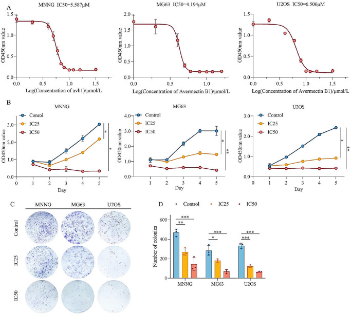
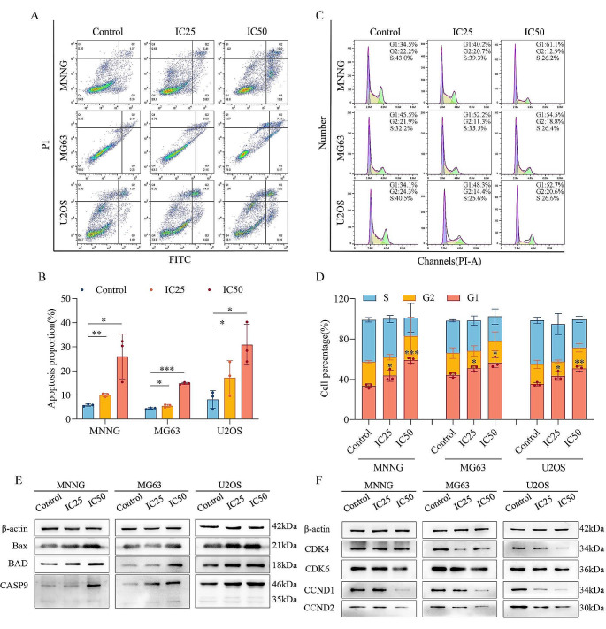
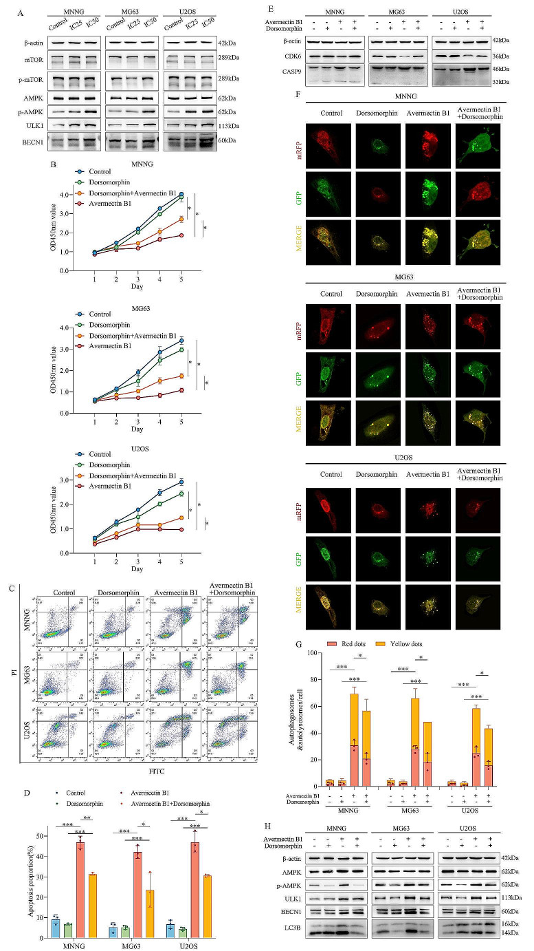
Methods: The half-inhibitory concentration of avermectin B1 was calculated in three osteosarcoma cell lines. Then, functional experiments were conducted to evaluate the effects of avermectin B1 on cell proliferation, the cell cycle, apoptosis and autophagy. Moreover, the AMPK/ULK1 signaling pathway was detected by Western blot assay. Finally, the in vivo effect of avermectin B1 on tumor growth and metastasis was investigated using the xenograft mouse model. To examine the role of the AMPK/ULK1 pathway, an AMPK-specific inhibitor (dorsomorphin) was used in combination with avermectin B1.

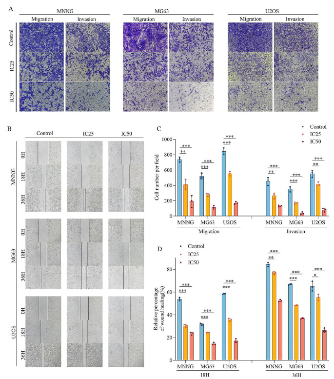
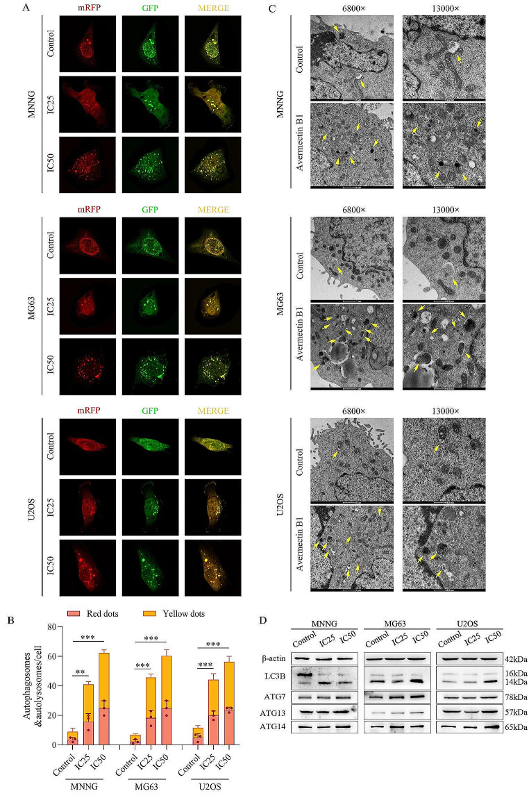
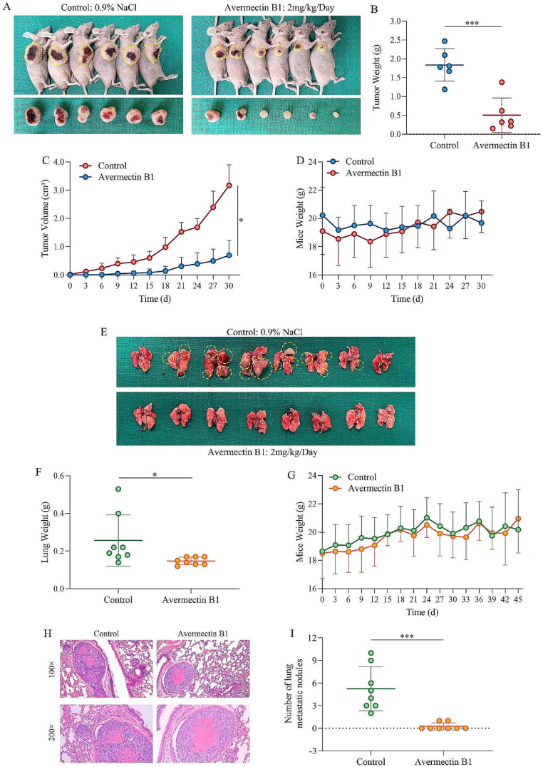
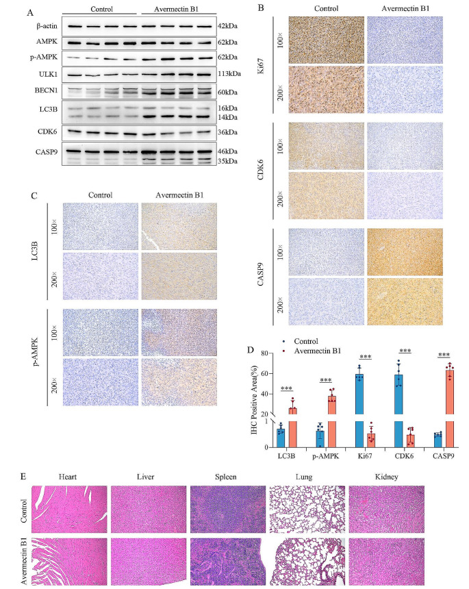
Results: Avermectin B1 inhibited the proliferation of osteosarcoma cells in a dose-dependent manner based on CCK8 and colony formation assays. Then, it was found to inhibit migration and invasion by wound healing assay and cell migration and invasion assay. In addition, avermectin B1 induced osteosarcoma cell apoptosis and autophagy. In vivo, avermectin B1 effectively inhibited osteosarcoma cell growth and pulmonary metastasis. Mechanistically, avermectin B1 activated the AMPK/ULK1 pathway to exert antitumor activity in vitro and in vivo. Dorsomorphin significantly attenuated the Avermectin B1-induced antitumor activities.

Conclusion: Our study suggests that avermectin B1 is a potential agent to treat osteosarcoma cells through the AMPK/ULK1 signaling pathway.

Keywords: AMPK/ULK1 pathway; Apoptosis; Autophagy; Avermectin B1; Osteosarcoma.


Conflict of interest statement

The authors declare no competing interests.

Figures

Fig. 1
Fig. 1
Effects of avermectin B1 on the proliferation of osteosarcoma. (A) OS cells were treated with different concentrations of avermectin B1 for 48 h, and cell viability was detected by CCK-8 assay. (B) Assessment of the proliferative capacity of osteosarcoma cells treated with avermectin B1. (C) Colony formation assays in osteosarcoma cells after treatment with avermectin B1 (magnification, ×40). (D) Quantification of the number of colonies. Data are shown as the means ± SDs with scatter plots from at least three independent experiments. Statistical analysis was performed using one-way ANOVA followed by post-hoc tests. *p < 0.05, **p < 0.01, *** p < 0.001
Fig. 2
Fig. 2
Effects of avermectin B1 on the migration and invasion of osteosarcoma. (A) Transwell assays were conducted to determine the migrative and invasive ability of MNNG, MG63 and U2OS cell lines. (B) Wound healing assay of osteosarcoma cell lines treated with avermectin B1. (C) Quantification of migrated and invaded cells. (D) Quantification of the wound healing rate. Data are shown as the means ± SDs with scatter plots from at least three independent experiments. Statistical analysis was performed using one-way ANOVA followed by post-hoc tests. *p < 0.05, **p < 0.01, *** p < 0.001
Fig. 3
Fig. 3
Avermectin B1 induces cell apoptosis and cell cycle arrest in osteosarcoma. (A) Flow cytometry was conducted to measure the apoptotic rate of three osteosarcoma cell lines after treatment with IC25 and IC50 of avermectin B1. (B) Quantification of the apoptosis proportion. (C) The cell cycle distribution was detected by flow cytometry after treatment with IC25 and IC50 of avermectin B1. (D) Quantification of the cell cycle percentage. (E) Western blotting was performed to detect the expression levels of the apoptosis-related proteins Bax, BAD, and caspase-9 in avermectin B1-treated cells. (F) The expression levels of the cell cycle-related proteins CDK4, CDK6, Cyclin D1 and Cyclin D2. Data are shown as the means ± SDs with scatter plots from at least three independent experiments. Statistical analysis was performed using one-way ANOVA followed by post-hoc tests. *p < 0.05, **p < 0.01, *** p < 0.001
Fig. 4
Fig. 4
Avermectin B1 induced autophagy in osteosarcoma cells. (A) The LC3 puncta were analyzed by mRFP-GFP-LC3 construct. (B) Quantification of the autophagosomes (yellow dots) and autolysosomes (free red dots). (C) Autophagosome-like structures (indicated by the yellow arrows) were assayed by transmission electron microscopy (magnification, ×6800 and ×13,000). (D) Western blot experiments were performed to measure the expression changes in LC3B and the autophagy-associated proteins ATG7, ATG13 and ATG14. Data are shown as the means ± SDs with scatter plots from at least three independent experiments. Statistical analysis was performed using one-way ANOVA followed by post-hoc tests. **p < 0.01, *** p < 0.001
Fig. 5
Fig. 5
Inhibition of autophagy inhibits the avermectin B1-mediated reduction in cell survival and apoptosis induction in osteosarcoma in vitro. (A) Western blot experiments were conducted to measure the expression changes in AMPK/ULK1 signaling pathway proteins after avermectin B1 treatment. (B) MNNG, MG63 and U2OS cells were treated with IC50 of avermectin B1 and 2 mM dorsomorphin alone or in combination as indicated and subjected to CCK8 assay. (C) Flow cytometry was conducted to measure the apoptotic rate of three osteosarcoma cell lines treated with IC50 of avermectin B1 and 2 mM dorsomorphin alone or in combination. (D) Quantification of the apoptosis proportion. (E) MNNG, MG63 and U2OS cells were treated with the IC50 of avermectin B1 and 2 mM dorsomorphin alone or in combination. Then, Western blotting was performed to detect the expression levels of the apoptosis-related protein caspase-9 and the cell cycle protein CDK6 in avermectin B1-treated cells. β-Actin was used as a loading control. (F) The LC3 puncta were analyzed by mRFP-GFP-LC3 construct when MNNG, MG63 and U2OS cells were treated with avermectin B1 and dorsomorphin alone or in combination. (G) Quantification of the autophagosomes (yellow dots) and autolysosomes (free red dots). (H) The protein expression levels of the AMPK/ULK1 pathway proteins BECN1 and LC3B after treatment with avermectin B1, dorsomorphin or avermectin B1 + dorsomorphin. Data are shown as the means ± SDs with scatter plots from at least three independent experiments. Statistical analysis was performed using Student’s t test. *p < 0.05, **p < 0.01, *** p < 0.001
Fig. 6
Fig. 6
Avermectin B1 inhibits osteosarcoma cell growth and metastasis in vivo. (A) The top panel shows a photograph of a tumor-bearing mouse, and the bottom panel shows a photograph of a subcutaneous tumor. (B) Weight of subcutaneous tumors in the control and avermectin B1 treatment groups in the subcutaneous tumor model. (C) Growth curve of the measured tumor volume in the subcutaneous tumor model. (D, G) Mouse weights separately in the subcutaneous tumor model and lung metastasis model. (E) Lung metastases formed in the avermectin B1-treated and control groups. (F) Lung weight of the lung metastasis model in the control and avermectin B1 treatment groups. (H) Lung metastasis nodules were stained with hematoxylin-eosin and observed by microscopy (magnification, ×100 and ×200). (I) Statistical analysis of lung metastases in the avermectin B1 and control groups in the lung metastasis model. Statistical analysis was performed using Student’s t test. Error bars represent the SD. *p < 0.05, *** p < 0.001
Fig. 7
Fig. 7
Avermectin B1 activates AMPK/ULK1 signaling pathway in human osteosarcoma cells in vivo. (A) Western blot analysis of AMPK, p-AMPK, ULK1, BECN1, LC3B, CDK6 and Caspase 9 in subcutaneous tumor tissues. (B, C) Ki67, CDK6, Caspase 9, p-AMPK and LC3B protein expression in tumor tissues was analyzed by immunohistochemistry (×100 and ×200). (D) Percentage of immunohistochemically positive area for Ki67, CDK6, Caspase 9, p-AMPK and LC3B in subcutaneous tumor tissues. (E) H&E staining images of the heart, liver, spleen, lung and kidney in mice. Data are shown as the means ± SDs with scatter plots from at least six independent experiments. Statistical analysis was performed using Student’s t test. *** p < 0.001

Similar articles

References

    1. Strauss SJ, Frezza AM, Abecassis N, Bajpai J, Bauer S, Biagini R et al (2021) Bone sarcomas: ESMO-EURACAN-GENTURIS-ERN PaedCan Clinical Practice Guideline for diagnosis, treatment and follow-up. Ann Oncol 32(12):1520–1536. 10.1016/j.annonc.2021.08.1995 - PubMed
    1. Sadykova LR, Ntekim AI, Muyangwa-Semenova M, Rutland CS, Jeyapalan JN, Blatt N et al (2020) Epidemiology and risk factors of Osteosarcoma. Cancer Invest 38(5):259–269. 10.1080/07357907.2020.1768401 - PubMed
    1. Harrison DJ, Geller DS, Gill JD, Lewis VO, Gorlick R (2018) Current and future therapeutic approaches for osteosarcoma. Expert Rev Anticancer Ther 18(1):39–50. 10.1080/14737140.2018.1413939 - PubMed
    1. Meltzer PS, Helman LJ (2021) New Horizons in the Treatment of Osteosarcoma. N Engl J Med 385(22):2066–2076. 10.1056/NEJMra2103423 - PubMed
    1. Whelan JS, Davis LE (2018) Osteosarcoma, Chondrosarcoma, and Chordoma. J Clin Oncol 36(2):188–193. 10.1200/jco.2017.75.1743 - PubMed

MeSH terms

Substances

LinkOut - more resources