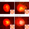
Proximal Location of Optic Disc Hemorrhage and Glaucoma Progression
- PMID: 39235817
- PMCID: PMC11378069
- DOI: 10.1001/jamaophthalmol.2024.3323
Proximal Location of Optic Disc Hemorrhage and Glaucoma Progression
Abstract
Importance: Although optic disc hemorrhage (DH) is widely recognized as a glaucoma risk factor, its clinical relevance in relation to proximity has not been investigated.
Objective: To determine the association of the proximal location of DH with glaucoma progression.
Design, setting, and participants: In this longitudinal observational cohort study, 146 eyes of 146 patients at Seoul National University Hospital who had had 1 or more DH with at least 5 years of follow-up and had at least 5 reliable visual field examinations were included. These data were analyzed January 10, 2010, through June 27, 2017.
Exposures: Laminar, marginal, rim, and parapapillary subtypes of DH were identified based on their respective proximal locations. The laminar and marginal subtypes were classified into the cup-type group, while the rim and parapapillary subtypes were classified into the peripapillary-type group. Kaplan-Meier survival analysis was used to compare survival experiences and multivariate analysis with the Cox proportional hazard model to identify risk factors for glaucoma progression. Regression analyses, both univariate and multivariate, were used to discover significant indicators of mean deviation (MD) loss.
Main outcome and measure: The primary outcome was glaucoma progression. Glaucoma progression was defined either as structural or functional deterioration.
Results: For all of the eyes, the mean follow-up period was 10.9 (3.7) years (range, 5.1-17.8 years), the mean age at which DH was first detected was 55.1 (11.3) years (range, 21-77 years), and 94 participants were female (64.1%). Over the mean follow-up period of 10.9 years, glaucoma progression was detected in 94 eyes (61.4%) with an MD change of -0.48 dB per year. The cup-type group showed a faster rate of MD change relative to the peripapillary-type group (-0.56 vs -0.32 dB per year; difference = -0.24; 95% CI, -0.37 to -0.11; P = .01). The cup group showed a higher cumulative probability of progression of glaucoma (80.4%) relative to the peripapillary group (54.4%; difference = 26.0%; 95% CI, 11.4%-40.6%; P < .001) in a life table analysis. The presence of cup hemorrhage was associated with an increased risk of glaucoma progression (hazard ratio, 3.28; 95% CI, 2.12-5.07; P < .001) in the multivariate Cox proportional hazard model. Cup-type DH was associated to MD loss rate in regression analysis.
Conclusions and relevance: This study showed glaucoma progression was higher in cases of DH classified as the cup type. These findings support the potential utility of assessing the proximal location of DH to predict how glaucoma might progress.
Conflict of interest statement
Figures


Comment in
-
Disc Hemorrhages in Open-Angle Glaucoma-Between a Rock and a Hard Place?JAMA Ophthalmol. 2024 Oct 1;142(10):950-951. doi: 10.1001/jamaophthalmol.2024.3330. JAMA Ophthalmol. 2024. PMID: 39235827 Free PMC article. No abstract available.
References
Publication types
MeSH terms
LinkOut - more resources
Full Text Sources
Medical
Miscellaneous

