. 2024 Dec 10;30(69):e202401667.
doi: 10.1002/chem.202401667. Epub 2024 Oct 31.

Recognition-Encoded Molecules: A Minimal Self-Replicator

Affiliations

Recognition-Encoded Molecules: A Minimal Self-Replicator

Daniele Rosa-Gastaldo et al. Chemistry. .

Abstract

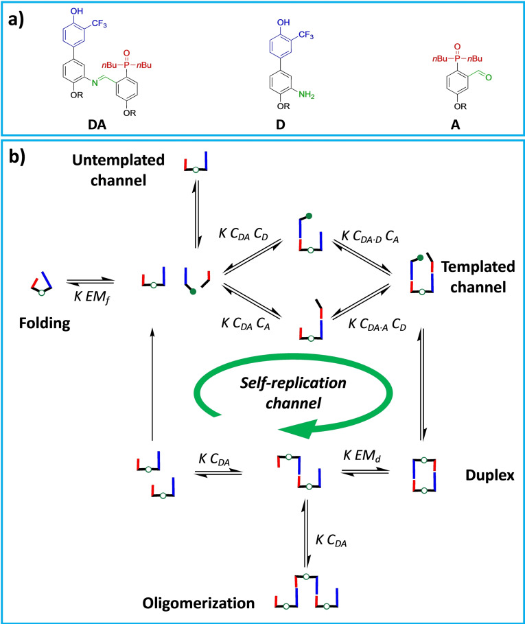
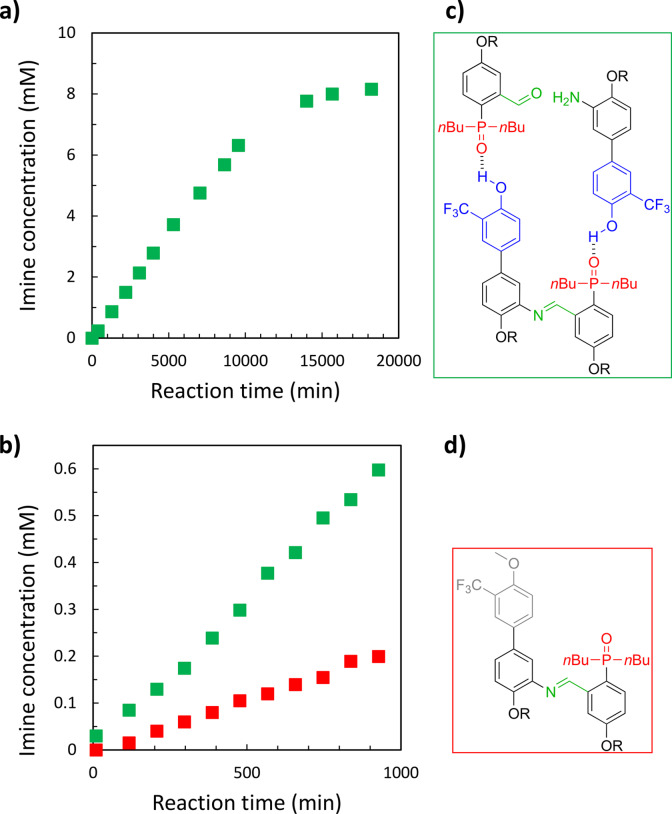
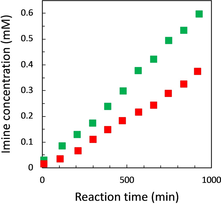
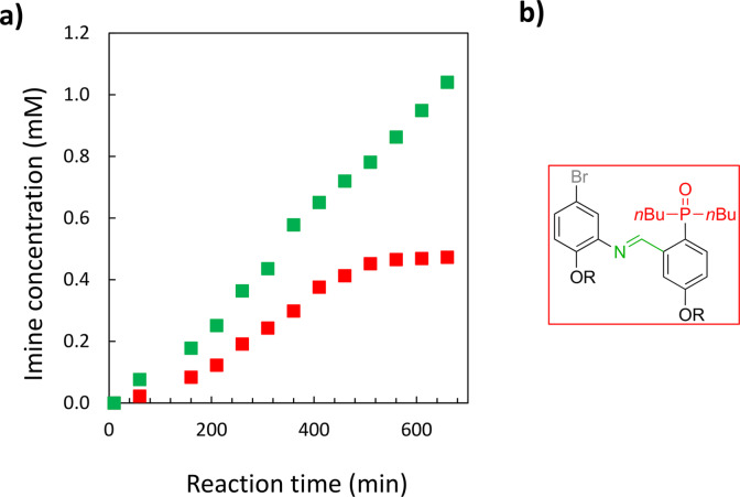
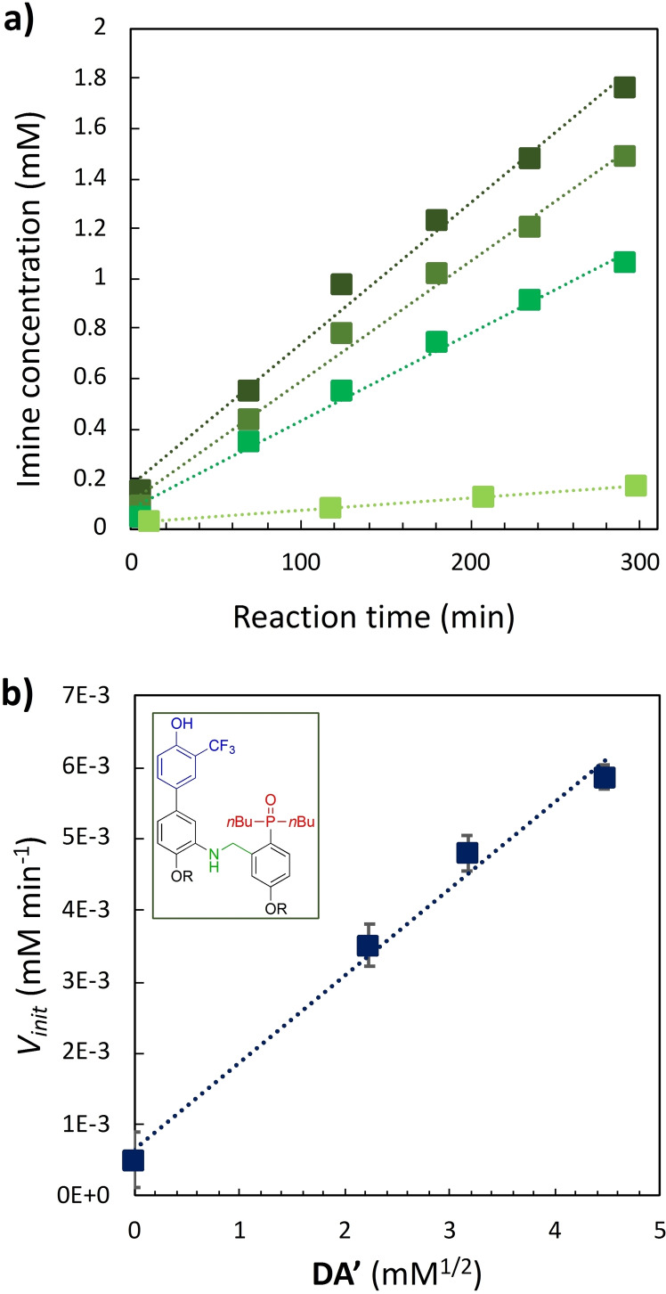
Nucleic acids, with their unique duplex structure, which is key for information replication, have sparked interest in self-replication's role in life's origins. Early template-based replicators, initially built on short oligonucleotides, expanded to include peptides and synthetic molecules. We explore here the potential of a class of synthetic duplex-forming oligoanilines, as self-replicators. We have recently developed oligoanilines equipped with 2-trifluoromethylphenol-phosphine oxide H-bond base pairs and we investigate whether the imine formed between aniline and aldehyde complementary monomers can self-replicate. Despite lacking a clear sigmoidal kinetic profile, control experiments with a methylated donor and a competitive inhibitor support self-replication. Further investigations with the reduced aniline dimer demonstrate templated synthesis, revealing a characteristic parabolic growth. After showing sequence selective duplex formation, templated synthesis and the emergence of catalytic function, the self-replication behaviour further suggests that the unique properties of nucleic acids can be paralleled by synthetic recognition-encoded molecules.

Keywords: Duplex; H-bonding; Oligoaniline; Recognition; Self-replicator.


Conflict of interest statement

The authors declare no conflict of interest.

Figures

Figure 1
Figure 1
a) Chemical structure of the recognition encoded molecules that compose the system: the monomers are equipped with hydrogen bond donor (trifluoromethylphenol, blue, for the aniline D) and acceptor (phosphine oxide, red, for the aldehyde A). Imine heterodimer DA is shown, and the linker chemistry is highlighted in green. R = 2‐ethylhexyl. b) Cartoon showing the pathways involved in this minimal replicating system: the imine template can be formed through a non‐templated channel and via self‐replication. The self‐assembly channels involving the imine dimer DA are highlighted: the first base‐pairing interaction can take place in an intramolecular fashion, leading to the 1,2‐folding path, or intermolecularly with a DA dimer or D/A monomers, leading respectively to the duplex channel or supramolecular oligomerization (template inhibition), or to the trimeric complex (templated channel). The outcome depends on the concentrations of the components C, the association constant for the intermolecular base‐pairing interaction K, and the effective molarities for folding (EMf ) and duplex formation (EMd ).
Figure 2
Figure 2
a) Concentration of self‐assembled DA species for increasing concentration of imine formed in a chloroform solution of aniline D and aldehyde A, 50 mM each. Trimeric complex [DA ⋅ D ⋅ A] is shown in green, duplex [DA ⋅ DA] in red, complexes [DA ⋅ D] and [DA ⋅ A] in dark grey and [free DA] in light grey.
Figure 3
Figure 3
a) Imine formation in chloroform‐d at 298 K between D and A (50 mM each in a well‐sealed tube). b) Imine formation in chloroform‐d at 298 K between D and A (50 mM each, green squares) and DOMe and A (50 mM each, red squares). The best fit of the data is reported in Figure S2.3 and Figure S2.5 in the SI. c) Structures of the DA ⋅ D ⋅ A trimeric complex (green square, top) and d) DOMeA imine (red square, bottom). R = 2‐ethylhexyl.
Figure 4
Figure 4
Percentage of imine formation rate increase caused by the presence of an equimolar amount of phenol. The kinetic experiments were performed as detailed in the section S1.1 of the SI. A detail of the structures of the involved anilines and aldehydes is reported in Scheme S1.1. The formation rate of DA was compared with the one of DOMeA. For the other imines, 50 mM of trioctylphosphine oxide (0 mM when using aldehyde A) and 0 mM (no‐phenol control experiment) or 50 mM of 4‐bromo‐2‐(trifluoromethyl)phenol (phenol experiment) were added into the solutions. See section 3 of the SI. R = 2‐ethylhexyl.
Figure 5
Figure 5
a) Imine formation in chloroform‐d at 298 K between D and A (50 mM each in air‐tight tube) without (green squares) and in the presence of trioctylphosphine oxide (50 mM, red squares). The best fit of the data is reported in Figure S2.7.
Figure 6
Figure 6
a) Imines formation at 298 K in a chloroform‐d solution containing aldehyde A (50 mM) and anilines D and BrNH2 (50 mM each). Imine DA is represented with green squares, imine BrNH2A with red squares. b) Structure of BrNH2A imine (R = 2‐ethylhexyl).
Figure 7
Figure 7
a) Imine formation in chloroform‐d at 298 K between D and A (50 mM each in a well‐sealed tube) in the presence of aniline template DA’ (0, 5, 10, 20 mM, respectively from light to dark green) Vinit data, as result of the best fit line reported, are summarized in Table S2.1 in the SI. b) The initial reaction rate (V init) of imine formation between D and A (50 mM each) is plotted as a function of the square root of DA’ concentration. The line of best fit shown is Vinit =1.22 ⋅ 10−3 [DA’]1/2+6.46 ⋅ 10−4. According to equation (2) a=1.22 ⋅ 10−3 mM1/2 min−1 and b=6.46 ⋅ 10−4 mM min−1 (respectively 6.19 ⋅ 10−7 M1/2 s−1 and 1.16 ⋅ 10−8 M s−1). The structure of aniline DA’ is shown in the insert.

References

    1. Vasas V., Fernando C., Santos M., Kauffman S., Szathmáry E., Biol. Direct 2012, 7, 1. - PMC - PubMed
    1. Kiedrowski G. V., Angew. Chem. 1986, 98, 932–934.
    1. Appel R., Niemann B., Schuhn W., Knoch F., Angew. Chem. Int. Ed. Eng. 1986, 25, 932–932.
    1. Zielinski W. S., Orgel L. E., Nature 1987, 327, 346–347. - PubMed
    1. Lee D. H., Granja J. R., Martinez J. A., Severin K., Ghadiri M. R., Nature 1996, 382, 525–528. - PubMed

LinkOut - more resources