. 2024 Sep 5;19(9):e0309807.
doi: 10.1371/journal.pone.0309807. eCollection 2024.

Curcumin alleviates osteoarthritis in mice by suppressing osteoclastogenesis in subchondral bone via inhibiting NF-κB/JNK signaling pathway

Affiliations

Curcumin alleviates osteoarthritis in mice by suppressing osteoclastogenesis in subchondral bone via inhibiting NF-κB/JNK signaling pathway

Dong Ding et al. PLoS One. .

Abstract

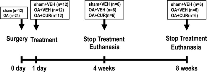
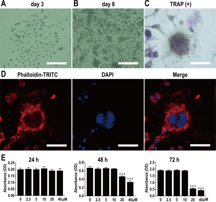
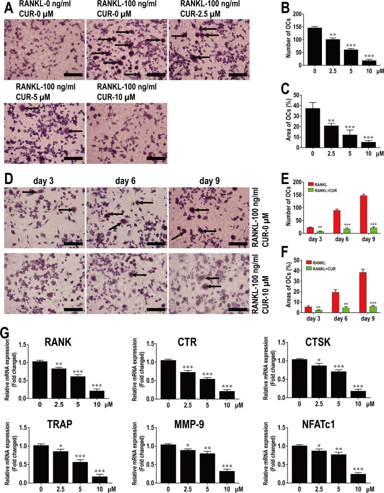
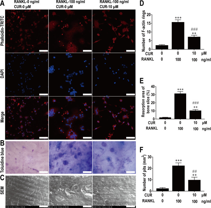
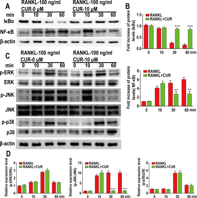
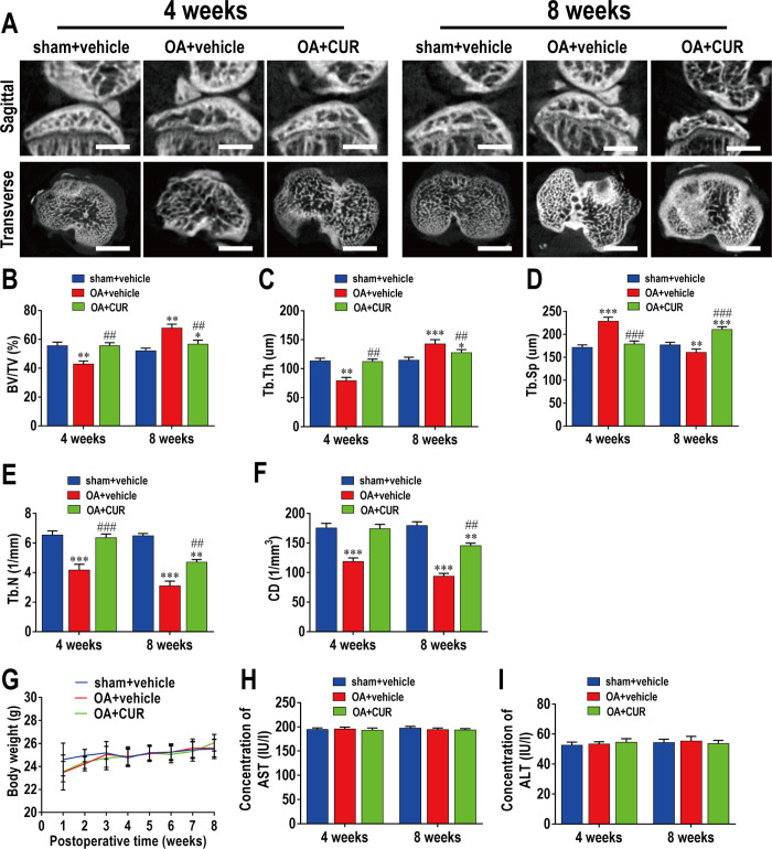
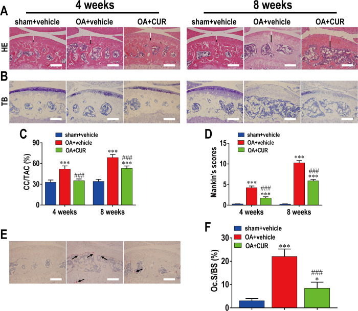
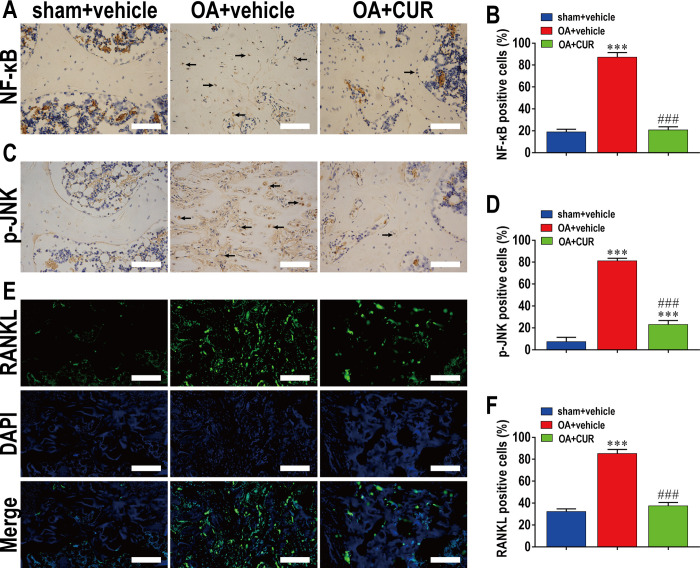
This study explored the mechanism of curcumin (CUR) suppressing osteoclastogenesis and evaluated its effects on osteoarthritis (OA) mouse. Bone marrow-derived macrophages were isolated as osteoclast precursors. In the presence or absence of CUR, cell proliferation was detected by CCK-8, osteoclastogenesis was detected by tartrate-resistant acid phosphatase (TRAP) staining, F-actin rings formation was detected by immunofluorescence, bone resorption was detected by bone slices, IκBα, nuclear factor kappa-B (NF-κB) and mitogen-activated protein kinase (MAPK) signaling pathways were detected using western blot, osteoclastogenesis-related gens were measured using quantitative polymerase chain reaction. A knee OA mouse model was designed by destabilizing the medial meniscus (DMM). Thirty-six male mice were divided into sham+vehicle, OA+vehicle, and OA+CUR groups. Mice were administered with or without CUR at 25 mg/kg/d from the first post-operative day until sacrifice. After 4 and 8 weeks of OA induction, micro-computed tomography was performed to analyze microstructure changes in subchondral bone, hematoxylin and eosin staining was performed to calculate the thickness of the calcified and hyaline cartilage layers, toluidine blue O staining was performed to assess the degenerated cartilage, TRAP-stained osteoclasts were counted, and NF-κB, phosphorylated Jun N-terminal Kinases (p-JNK), and receptor activator of nuclear factor κB ligand (RANKL) were detected using immunohistochemistry. CUR suppressed osteoclastogenesis and bone resorption without cytotoxicity. CUR restrained RANKL-induced activation of NF-κB, p-JNK and up-regulation of osteoclastogenesis-related genes. CUR delayed cartilage degeneration by suppressing osteoclastogenesis and bone resorption in early OA. The mechanism of CUR inhibiting osteoclastogenesis might be associated with NF-κB/JNK signaling pathway, indicating a novel strategy for OA treatment.


Conflict of interest statement

The authors have declared that no competing interests exist.

Figures

Fig 1
Fig 1. Schematic diagram of animal experiment design.
A total of 36 male C57BL/6 mice were randomly assigned into 3 groups (n = 6 per group): sham+vehicle group, OA+vehicle group, and OA+CUR group. Mice in each group respectively received VEH, VEH, and CUR (25 mg/kg/d) for 4 and 8 weeks postoperatively.
Fig 2
Fig 2. Identification of osteoclast and viability of BMMs.
Observation of BMMs on 3rd (A) and 8th d (B) under light microscopy. Scale bar, 200 μm. (C) Observation of osteoclasts with multiple nuclei using TRAP staining under light microscopy. (D) Observation of the structure of F-actin ring stained with phalloidin-TRITC and DAPI under LSCM. Scale bar, 40 μm. (E) Effects of CUR on BMMs viability. n = 3 per group. ***P < 0.001, compared with control group.
Fig 3
Fig 3. CUR inhibits the differentiation of BMMs into osteoclasts in vitro.
(A) TRAP staining of osteoclasts induced with 30 ng/ml M-CSF and 100 ng/ml RANKL in the presence of various concentrations of CUR for 9 days. Scale bar, 200 μm. Quantitative analysis of the numbers (B) and areas (C) of osteoclasts from panel A. (D) TRAP staining of osteoclasts induced with 30 ng/ml M-CSF and 100 ng/ml RANKL in the presence or absence of 10 μM CUR for different days. Scale bar, 200 μm. Quantitative analysis of the numbers (E) and areas (F) of osteoclasts from panel D. (G) BMMs were induced with 30 ng/ml M-CSF and 100 ng/ml RANKL in the presence of various concentrations of CUR for 9 days and then osteoclastogenesis-related genes were detected using RT-qPCR. n = 3 per group. *P < 0.05, **P < 0.01 and ***P < 0.001, compared with control group.
Fig 4
Fig 4. CUR attenuates F-actin rings formation and bone resorption of osteoclast in vitro.
(A) BMMs were induced with 30 ng/ml M-CSF and 100 ng/ml RANKL in the presence or absence of 10 μM CUR for 9 days and then stained with TRITC-conjugated phalloidin and DAPI to show F-actin rings and nucleus. Scale bar, 50 μm. (B) BMMs were plated on bone slices and induced with 30 ng/ml M-CSF and 100 ng/ml RANKL in the presence or absence of 10 μM CUR for 9 days, bone resorption areas were examined by staining with toluidine blue. Scale bar, 50 μm. (C) Bone resorption pits were observed by SEM. Scale bar, 20 μm. Quantitative analysis of (D) F-actin rings, (E) resorption areas and (F) resorption pits. n = 3 per group. **P < 0.01 and ***P < 0.001, compared with control group; ##P < 0.01 and ###P < 0.001, compared with RANKL-induced group.
Fig 5
Fig 5. CUR inhibits RANKL-induced NF-κB and JNK activation during osteoclastogenesis.
BMMs were pretreated with or without CUR (10 μM) for 4 h and then induced with 30 ng/ml M-CSF and 100 ng/ml RANKL for indicated time period (0, 10, 30, 60 min), and WB was performed for quantitative analysis of NF-κB (A and B) and MAPKs (C and D) signaling pathways. n = 3 per group. **P < 0.01 and ***P < 0.001, compared with control group.
Fig 6
Fig 6. CUR ameliorates bone loss without toxicity in the early stage of DMM-induced OA.
(A) The DMM-induced OA mice were treated with or without CUR for 4 and 8 weeks, and then μCT was performed to examine the microarchitecture in tibial subchondral bone. Scale bar, 1,000 μm. (B, C, D, E, and F) Quantitative μCT analyses of microarchitecture in tibial subchondral bone: (B) BV/TV (%), (C) Tb.Th, (D) Tb.Sp, (E) Tb.N and (F) CD. (G, H and I) Quantitative analysis of body weight and transaminases: (G) body weight, (H) AST, and (H) ALT. n = 6 per group/time point. **P < 0.01 and ***P < 0.001, compared with sham+vehicle group; ##P < 0.01 and ###P < 0.001, compared with OA+vehicle group.
Fig 7
Fig 7. CUR ameliorates cartilage degeneration by inhibiting osteoclastogenesis in the early stage of DMM-induced OA.
The DMM-induced OA mice were treated with or without CUR for 4 and 8 weeks, and histological analysis of cartilage and osteoclasts in subchondral bone were stained with (A) HE, (B) TB and (E) TRAP, respectively. Scale bar, 100 μm. Quantitative analysis of (C) CC/TAC, (D) Mankin’s scores, and (F) Oc.S/BS. n = 6 per group/time point. *P < 0.05 and ***P < 0.001, compared with sham+vehicle group; ###P < 0.001, compared with OA+vehicle group.
Fig 8
Fig 8. CUR inhibits NF-κB, p-JNK and RANKL expression in the early stage of DMM-induced OA.
The DMM-induced OA mice were treated with or without CUR for 4 weeks, and expression of (A) NF-κB, (C) p-JNK, and (E) RANKL was shown by immunohistochemistry and immunofluorescence staining. Scale bar, 100 μm. Quantitative analysis of (B) NF-κB, (D) p-JNK, and (F) RANKL. n = 6 per group/time point. ***P < 0.001, compared with sham+vehicle group; ###P < 0.001, compared with OA+vehicle group.
Fig 9
Fig 9. Potential mechanism by which CUR-mediated attenuation of OA via inhibiting osteoclastogenesis.
RANKL binds to RANK and recruits TRAF6 to activate NF-κB and MAPKs pathways. The signal is then transmitted to NFATc1 and c-Fos. Sequentially the stimulated NFATc1 migrates into nucleus and initiates the expression of osteoclast-related genes, including RANK, CTR, CTSK, TRAP, MMP-9, and NFATc1. While CUR can ameliorate osteoclastogenesis via inhibiting the activation of NF-κB/JNK signal pathway.

Similar articles

Cited by

References

    1. G P, MJ T. Osteoarthritis year in review 2020: epidemiology & therapy. OSTEOARTHR CARTILAGE. 2021;29(2):180–9. - PubMed
    1. KD A, YM G. Epidemiology of osteoarthritis: State of the evidence. CURR OPIN RHEUMATOL. 2015;27(3):276–83. - PMC - PubMed
    1. JR K, JJ Y, HA K. Therapeutics in Osteoarthritis Based on an Understanding of Its Molecular Pathogenesis. INT J MOL SCI. 2018;19(3). - PMC - PubMed
    1. JA K, EC S, A O, J C, LC L, N B, et al.. A comparison of three strategies to reduce the burden of osteoarthritis: A population-based microsimulation study. PLOS ONE. 2021;16(12):e261017. doi: 10.1371/journal.pone.0261017 - DOI - PMC - PubMed
    1. W H, Y C, C D, S D. Microenvironment in subchondral bone: predominant regulator for the treatment of osteoarthritis. ANN RHEUM DIS. 2021;80(4):413–22. doi: 10.1136/annrheumdis-2020-218089 - DOI - PMC - PubMed