

The effect of excessive trabeculation on cardiac rotation-A multimodal imaging study
- PMID: 39236040
- PMCID: PMC11376564
- DOI: 10.1371/journal.pone.0308035
The effect of excessive trabeculation on cardiac rotation-A multimodal imaging study
Abstract
Background: Cardiac rotational parameters in primary symptomatic left ventricular noncompaction (LVNC) with preserved left ventricular ejection fraction (LVEF) are not well understood. We aimed to analyze cardiac rotation measured with cardiac magnetic resonance feature-tracking (CMR-FT) and speckle-tracking echocardiography (Echo-ST) in LVNC morphology subjects with preserved LVEF and different genotypes and healthy controls.
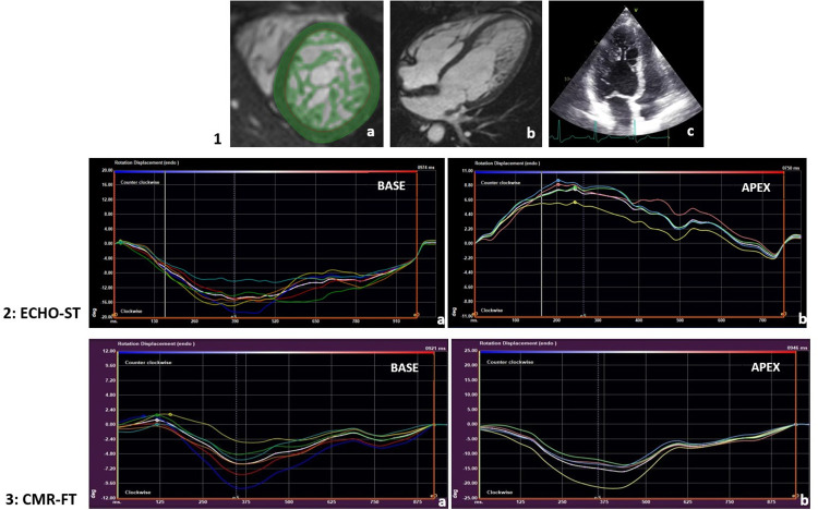
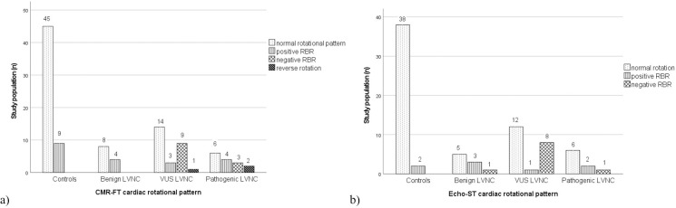
Methods: Our retrospective study included 54 LVNC subjects with preserved LVEF and 54 control individuals. We evaluated functional and rotational parameters with CMR in the total study population and with echocardiography in 39 LVNC and 40 C individuals. All LVNC subjects were genotyped with a 174-gene next-generation sequencing panel and grouped into the subgroups: benign (B), variant of uncertain significance (VUS), and pathogenic (P).
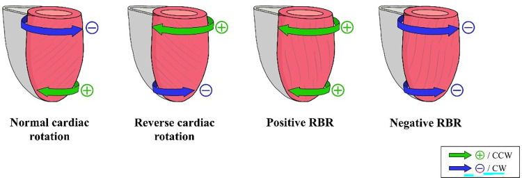
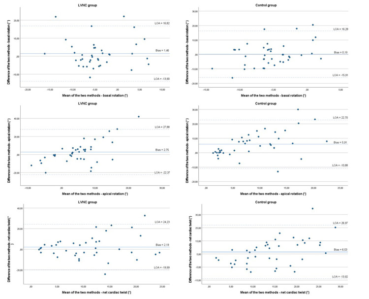
Results: In comparison with controls, LVNC subjects had reduced apical rotational degree (p = 0.004) and one-third had negative apical rotation. While the degree of apical rotation was comparable between the three genetic subgroups, they differed significantly in the direction of apical rotation (p<0.001). In contrast to control and B groups, all four studied cardiac rotational patterns were identified in the P and VUS subgroups, namely normal rotation, positive and negative rigid body rotation, and reverse rotation. When the CMR-FT and Echo-ST methods were compared, the direction and pattern of cardiac rotation had moderate to good association (p<0.001) whereas the rotational degrees showed no reasonable correlation or agreement.
Conclusion: While measuring cardiac rotation using both CMR-FT and Echo-ST methods, subclinical mechanical differences were identified in subjects with LVNC phenotype and preserved LVEF, especially in cases with genetic involvement.
Copyright: © 2024 Grebur et al. This is an open access article distributed under the terms of the Creative Commons Attribution License, which permits unrestricted use, distribution, and reproduction in any medium, provided the original author and source are credited.
Conflict of interest statement
The authors have declared that no competing interests exist.
Figures
References
MeSH terms
LinkOut - more resources
Full Text Sources
