. 2024 Sep 5;19(9):e0308389.
doi: 10.1371/journal.pone.0308389. eCollection 2024.

Inositol trisphosphate and ryanodine receptor signaling distinctly regulate neurite pathfinding in response to engineered micropatterned surfaces

Affiliations

Inositol trisphosphate and ryanodine receptor signaling distinctly regulate neurite pathfinding in response to engineered micropatterned surfaces

Joseph T Vecchi et al. PLoS One. .

Abstract

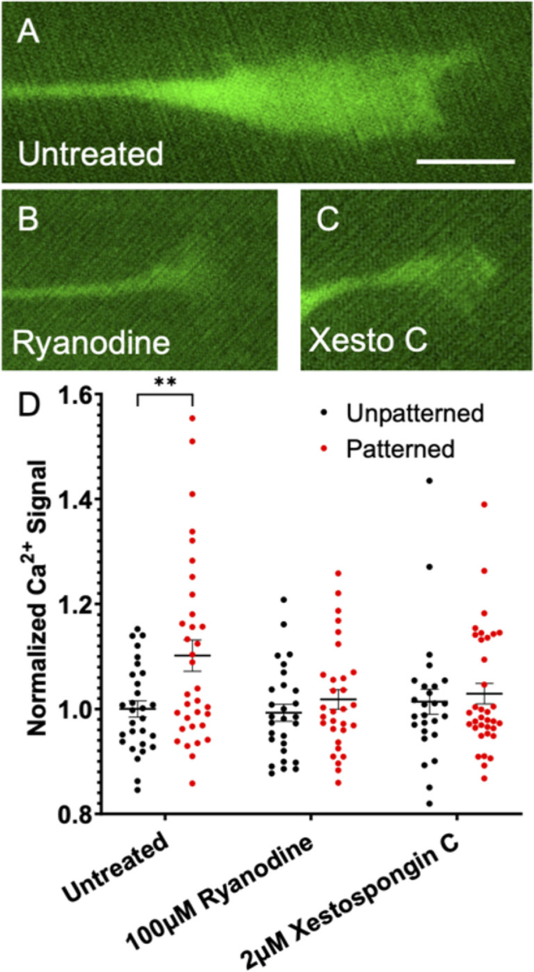
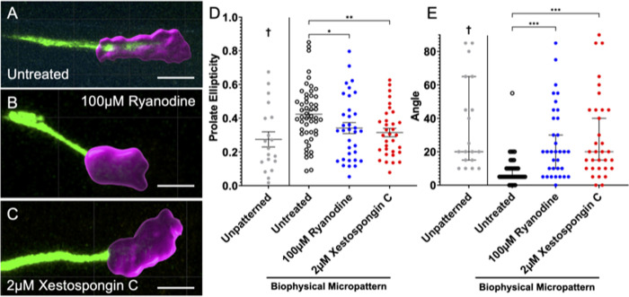
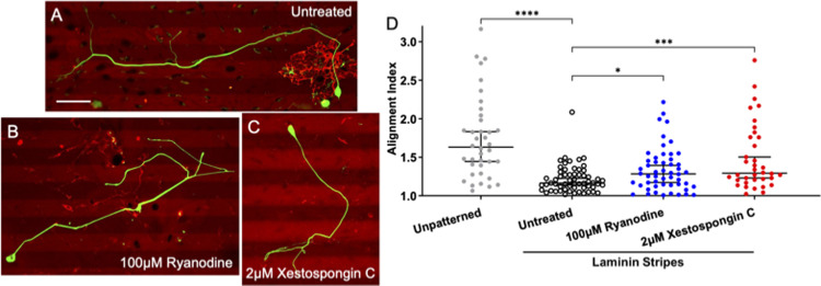
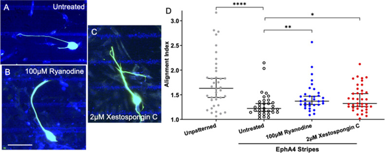
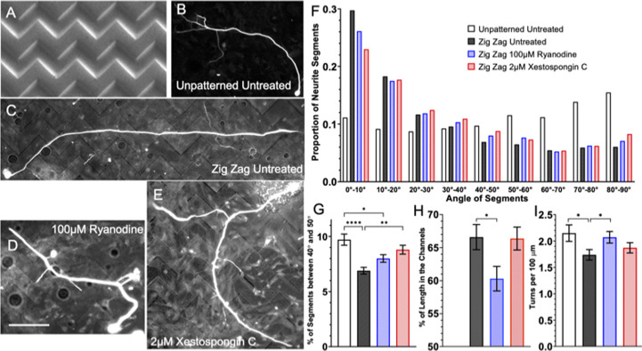
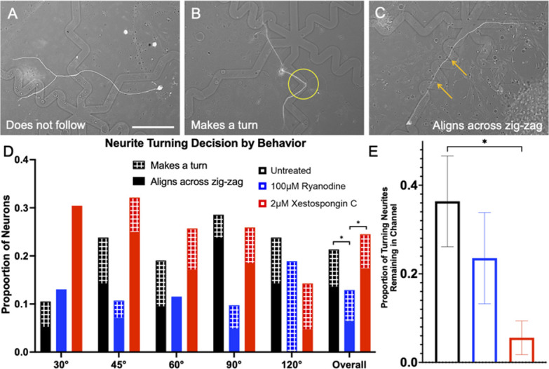
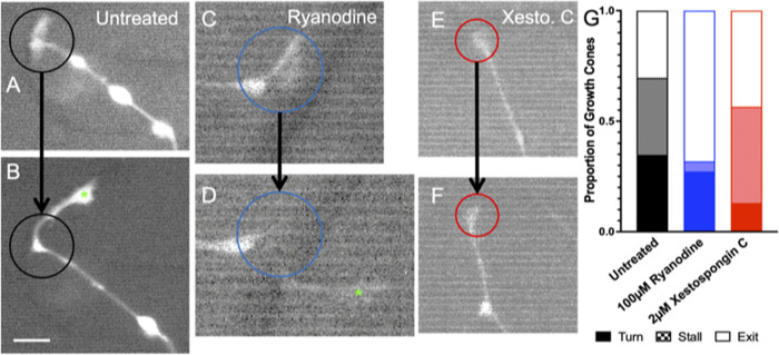
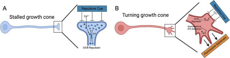
Micro and nanoscale patterning of surface features and biochemical cues have emerged as tools to precisely direct neurite growth into close proximity with next generation neural prosthesis electrodes. Biophysical cues can exert greater influence on neurite pathfinding compared to the more well studied biochemical cues; yet the signaling events underlying the ability of growth cones to respond to these microfeatures remain obscure. Intracellular Ca2+ signaling plays a critical role in how a growth cone senses and grows in response to various cues (biophysical features, repulsive peptides, chemo-attractive gradients). Here, we investigate the role of inositol triphosphate (IP3) and ryanodine-sensitive receptor (RyR) signaling as sensory neurons (spiral ganglion neurons, SGNs, and dorsal root ganglion neurons, DRGNs) pathfind in response to micropatterned substrates of varied geometries. We find that IP3 and RyR signaling act in the growth cone as they navigate biophysical cues and enable proper guidance to biophysical, chemo-permissive, and chemo-repulsive micropatterns. In response to complex micropatterned geometries, RyR signaling appears to halt growth in response to both topographical features and chemo-repulsive cues. IP3 signaling appears to play a more complex role, as growth cones appear to sense the microfeatures in the presence of xestospongin C but are unable to coordinate turning in response to them. Overall, key Ca2+ signaling elements, IP3 and RyR, are found to be essential for SGNs to pathfind in response to engineered biophysical and biochemical cues. These findings inform efforts to precisely guide neurite regeneration for improved neural prosthesis function, including cochlear implants.


Conflict of interest statement

The authors have declared that no competing interests exist.

Figures

Fig 1
Fig 1. Basal Ca2+ level in growth cones increases on patterned substrate via IP3 and RyR signaling.
(A-C) Representative images of rDRGN growth cones expressing Pirt-GcaMP3 growing on topographically patterned substrates either untreated (A), treated with 100μM ryanodine (B), or 2μM xestospongin C (C). (D) Ca2+ signal in the growth cones which is background corrected and normalized to the unpatterned untreated condition. Two-way ANOVA with follow-up Dunnett’s multiple comparison testing suggests that the patterned untreated condition is the only significantly different condition (p < 0.01). n = 26–35 growth cones per condition from three independent culture replicates from one neonatal mouse. Error bars represent +/-SEM. Scale bar = 5μm.
Fig 2
Fig 2. Inhibiting Ca2+ release from internal stores alters growth cone morphology on topographically patterned substrates.
(A-C). Representative images of fixed rDRGN growth cones growing on topographically patterned substrates either untreated (A), treated with 100μM ryanodine (B), or 2μM xestospongin C (C). Growth cone (magenta) was selected and its shape described using Imaris. (D). Growth cone shape was approximated as a spheroid and its prolate ellipticity was calculated. One-way ANOVA with follow-up Dunnett’s multiple comparisons testing shows that untreated growth cones on the patterned substrate had greater prolate ellipticity values. Error bars represent +/-SEM, p < 0.05. E. The major axis of this spheroid was found, and the angle difference between this axis and the neurite shaft was measured to the nearest 5°. Kruskal-Wallis test with follow-up Dunn’s multiple comparisons testing shows that this angle was smaller for the untreated neurons on the patterned substrate. Error bars represent 95% confidence interval. n = 50, 34, 33 growth cones, respectively derived from three independent culture replicates from one neonatal mouse. p < 0.001. Scale bars = 5 μm. † indicates dataset from previously published study.
Fig 3
Fig 3. Inhibiting Ca2+ release from internal stores impairs the ability SGNs to follow chemo-permissive laminin stripes.
(A-C). Representative images of fixed SGNs growing in laminin stripe patterned substrates either untreated (A), treated with 100μM ryanodine (B), or 2μM xestospongin C (C) NF200 is labeled green and laminin red. (D) Alignment Index (Total neurite length divided by length in horizontal direction) of SGNs to the micropatterned laminin substrates. Kruskal-Wallis with follow-up Dunn’s multiple comparisons testing shows that both treatments impair the ability of SGNs to align to the laminin stripes. Graph shows median Alignment Index +/- 95% CI, p < 0.05. n = 38, 59, 52, 36 neurons derived from three independent culture replicates of a pool of spiral ganglia harvested from multiple litter mates. Scale bar = 50 μm.
Fig 4
Fig 4. Inhibiting Ca2+ release from internal stores impairs the ability SGNs to follow chemo-repulsive EphA4 stripes.
(A-C). Representative images of fixed SGNs growing in on EphA4-Fc stripe patterned substrates either untreated (A), treated with 100μM ryanodine (B), or 2μM xestospongin C (C). NF200 is labeled aqua, laminin labeled pink, and EphA4-Fc blue. (D) Alignment Index (Total neurite length divided by length in horizontal direction) of SGNs to the micropatterned EphA4-Fc substrates. Kruskal-Wallis with follow-up Dunn’s multiple comparisons testing shows that both treatments impair the ability of SGNs to align to the EphA4-Fc stripes. Graph shows median Alignment Index +/- 95% CI, p < 0.05. n = 38, 33, 33, 39 neurons derived from three independent culture replicates of a pool of spiral ganglia harvested from multiple litter mates. Scale bar = 50 μm.
Fig 5
Fig 5. Inhibiting RyR or IP3 signaling causes different impairment of neurite guidance on a zigzag micropattern.
(A) SEM image of the topographically micropatterned zigzag substrate. (B) Representative image of fixed SGNs growing on unpatterned substrate.(C-E). Representative images of fixed SGNs growing on topographically micropatterned zigzag substrate either untreated (C), treated with 100μM ryanodine (D), or 2μM xestospongin C (E). (F) Distribution of SGN neurite segment angles relative to the horizontal axis. (G) Proportion of neurite segments which are oriented between 40° and 50° relative to the horizontal, i.e., the angle of each segment of the zigzag micropattern. Error bars are +/- standard error of proportion. Significance indicated for chi-square test with follow-up z-testing. p < 0.01. (H) Average percent of length for each neuron that is in the micropatterned feature. 50% would represent a random tendency for the neuron. (I) Average number of turns measured per neuron normalized by 100 μm of length. For H & I, one-way ANOVA with follow-up Dunnett’s multiple comparisons testing was done to compare groups. Error bars are +/- SEM, p < 0.05. n = 53, 100, 109, 89 neurons derived from three independent culture replicates of a pool of spiral ganglia harvested from multiple litter mates. Scale bar = 50 μm.
Fig 6
Fig 6. Xestospongin C treated SGNs follow the turn challenges, but do not remain in the microfeature.
(A-C). Representative images of fixed SGNs growing in response to topographical microfeature turn challenges. (A) SGN not following the microfeatures (B) SGN making a turn and holding its position in a 90° feature turn, (C) SGN aligning across the 45° turn microfeature. (D) Proportion of neurites which were observed to follow microfeature turns by angle and drug treatment. Patterned segment of a bar is the proportion which made one turn, while solid demonstrated alignment across multiple turns. Multinomial logistic regression shows effect of Ryanodine treatment and microfeature angle on guidance. Measurements in each treatment were compiled and results from Chi-square test with follow up z-tests shown for overall data. Error bars represent +/- standard error for a proportion. n = 103, 155, 155. (E) Proportion of the neurites that made a turn and held its position in the microfeature. Chi-squared test with follow up z-tests indicate a lower proportion of xestospongin C treated remain in the channel during a turn. Error bars +/- SEM, p < 0.05. n = 22, 17, 36 neurite shafts from three independent culture replicates of a pool of spiral ganglia harvested from multiple litter mates. Scale bar = 50 μm.
Fig 7
Fig 7. Ryanodine treated growth cones do not respond to the biophysical microfeatures while xestospongin C treated turn at a decreased frequency.
(A, B) Before and after images of an untreated growth cone navigating a turn. Black circle shows same ROI in each and new growth is marked by green asterisk in B. (C, D) Before and after images of an 100μM ryanodine treated growth cone that exited the microfeatures after encountering a turn. Blue circle shows same ROI in each and new growth is marked by green asterisk in D. (E, F). Before and after images of an 2μM xestospongin C treated growth cone that stalled after encountering a turn. Red circle shows same ROI in each. G. Frequency comparison of turning behaviors of rDRGN growth cones while navigating 4μm amplitude. Chi-squared test with follow up z-tests indicate a lower proportion of ryanodine treated neurite display stalling and xestospongin C treated turn. n = 22, 22, 23 growth cones derived from six independent culture replicates from one neonatal mouse. Scale bar = 10 μm.
Fig 8
Fig 8. Model of RyR and IP3 signaling in neurite guidance to patterned substrate cues.
A. A stalled neurite whereby a repulsive cue initiates Ca2+ influx via yet to be identified plasma membrane sensors and channels, which may vary for each type of repulsive cue. This local increase in Ca2+ leads to RyR mediated CICR and repulsion. B. A turning neurite whereby the phenomenon in Fig 8A is occurring in the region being turning away from. While in the region being turned toward, the growth cone is sensing attractive cues on the permissive substrate, which triggers IP3 signaling and attractive growth.

Similar articles

References

    1. McCormick LE, Gupton SL. Mechanistic advances in axon pathfinding. Current Opinion in Cell Biology. 2020;63:11–9. doi: 10.1016/j.ceb.2019.12.003 - DOI - PMC - PubMed
    1. Domínguez-Romero ME, Slater PG. Unraveling Axon Guidance during Axotomy and Regeneration. International Journal of Molecular Sciences. 2021;22(15):8344. doi: 10.3390/ijms22158344 - DOI - PMC - PubMed
    1. Schmidbauer D, Fink S, Rousset F, Löwenheim H, Senn P, Glueckert R. Closing the Gap between the Auditory Nerve and Cochlear Implant Electrodes: Which Neurotrophin Cocktail Performs Best for Axonal Outgrowth and Is Electrical Stimulation Beneficial? International Journal of Molecular Sciences. 2023;24(3):2013. doi: 10.3390/ijms24032013 - DOI - PMC - PubMed
    1. Aregueta-Robles UA, Woolley AJ, Poole-Warren LA, Lovell NH, Green RA. Organic electrode coatings for next-generation neural interfaces. Frontiers in Neuroengineering. 2014;7. doi: 10.3389/fneng.2014.00015 - DOI - PMC - PubMed
    1. Thompson CH, Zoratti MJ, Langhals NB, Purcell EK. Regenerative Electrode Interfaces for Neural Prostheses. Tissue Eng Part B Rev. 2016;22(2):125–35. doi: 10.1089/ten.TEB.2015.0279 - DOI - PubMed

LinkOut - more resources