. 2024 Sep 5;20(9):e1012493.
doi: 10.1371/journal.ppat.1012493. eCollection 2024 Sep.

Dual-role epitope on SARS-CoV-2 spike enhances and neutralizes viral entry across different variants

Affiliations

Dual-role epitope on SARS-CoV-2 spike enhances and neutralizes viral entry across different variants

Gang Ye et al. PLoS Pathog. .

Abstract

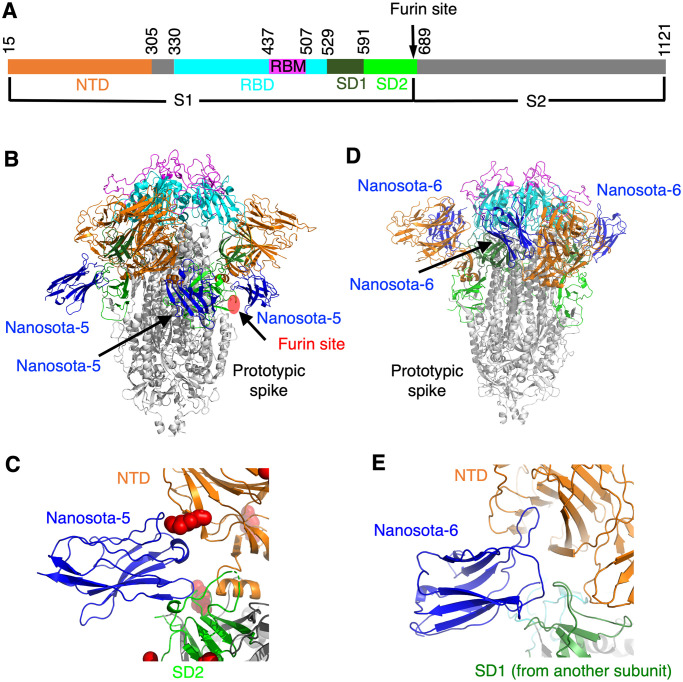
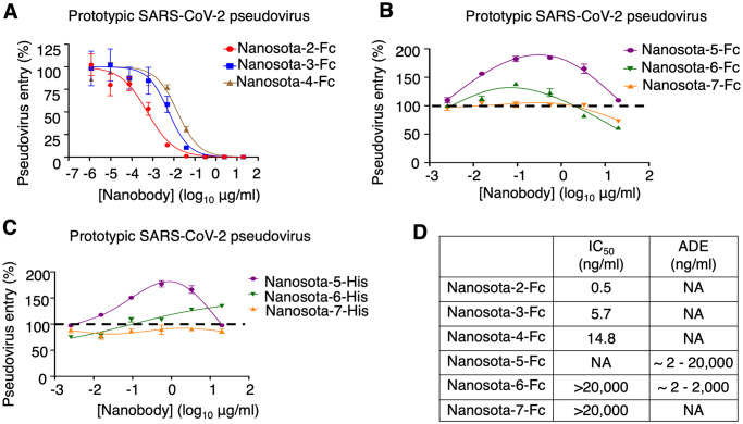
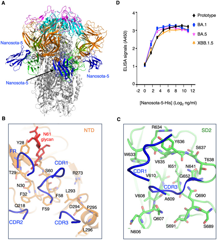
Grasping the roles of epitopes in viral glycoproteins is essential for unraveling the structure and function of these proteins. Up to now, all identified epitopes have been found to either neutralize, have no effect on, or enhance viral entry into cells. Here, we used nanobodies (single-domain antibodies) as probes to investigate a unique epitope on the SARS-CoV-2 spike protein, located outside the protein's receptor-binding domain. Nanobody binding to this epitope enhances the cell entry of prototypic SARS-CoV-2, while neutralizing the cell entry of SARS-CoV-2 Omicron variant. Moreover, nanobody binding to this epitope promotes both receptor binding activity and post-attachment activity of prototypic spike, explaining the enhanced viral entry. The opposite occurs with Omicron spike, explaining the neutralized viral entry. This study reveals a unique epitope that can both enhance and neutralize viral entry across distinct viral variants, suggesting that epitopes may vary their roles depending on the viral context. Consequently, antibody therapies should be assessed across different viral variants to confirm their efficacy and safety.


Conflict of interest statement

The University of Minnesota has filed a patent on Nanosota-5, -6, and -7 with F.L., G.Y., F.B., and B.L. as inventors.

Figures

Fig 1
Fig 1. Structures of prototypic SARS-CoV-2 spike complexed with individual non-RBD-targeting nanobodies.
(A) Schematic drawing of SARS-CoV-2 spike ectodomain (prototypic variant). S1: receptor-binding subunit. S2: membrane-fusion subunit. NTD: N-terminal domain of S1. RBD: receptor-binding domain of S1. RBM: receptor-binding motif of RBD. SD1: subdomain 1 of S1. SD2: subdomain 2 of S1. Furin site: cleavage site for the furin protease. (B) Structure of prototypic SARS-CoV-2 spike complexed with Nanosota-5. Three bound Nanosota-5 molecules are colored in blue. The spike domains are colored in the same way as (A). The furin cleavage site is labeled. (C) The binding site of Nanosota-5 on prototypic SARS-CoV-2 spike. A glycan N-linked to Asn61 on the spike is shown as red balls. NTD and SD2 bound by Nanosota-5 are from the same spike protomer. (D) Structure of prototypic SARS-CoV-2 spike complexed with Nanosota-6. Three bound Nanosota-6 molecules are colored in blue. (E) The binding site of Nanosota-6 on prototypic SARS-CoV-2 spike. NTD and SD1 bound by Nanosota-6 are from two spike protomers.
Fig 2
Fig 2. Non-RBD epitopes enhance the cell entry of prototypic SARS-CoV-2 pseudoviruses.
Retroviruses pseudotyped with prototypic SARS-CoV-2 spike (i.e., prototypic SARS-CoV-2 pseudoviruses) entered ACE2-expressing cells in the presence of one of the nanobodies at various concentrations. Entry efficiency was characterized as the luciferase signal accompanying entry. The efficacy of each nanobody in neutralizing pseudovirus entry was expressed as the concentration capable of neutralizing pseudovirus entry by 50% (i.e., IC50). (A) Activities of three Fc-tagged RBD-targeting nanobodies: they all potently neutralized SARS-CoV-2 pseudovirus entry. Error bars represent SEM (n = 3). (B) Activities of three Fc-tagged non-RBD-targeting nanobodies: Nanosota-5-Fc enhances pseudovirus entry, Nanosota-6-Fc enhances pseudovirus entry at low concentrations and neutralizes pseudovirus entry at high concentrations, and Nanosota-7-Fc neutralizes pseudovirus entry at high concentrations. Error bars represent SEM (n = 4). (C) Activities of three His-tagged non-RBD-targeting nanobodies: Nanosota-5-His and Nanosota-6-His enhance pseudovirus entry using an FcR-independent mechanism. Error bars represent SEM (n = 4). (D) Summary of the activities of the six spike-binding nanobodies. NA: not available.
Fig 3
Fig 3. A non-RBD epitope enhances cell infection by live prototypic SARS-CoV-2.
Recombinant SARS-CoV-2-Venus (prototypic variant) was used to infect Vero E6 cells in the presence of various concentrations of Nanosota-5-Fc. PBS buffer was used as a negative control. (A) Nanosota-5-Fc enhances cell infection by live prototypic SARS-CoV-2 at a high virus titer. (B) Nanosota-5-Fc enhances cell infection by live prototypic SARS-CoV-2 at a low virus titer. (C) Nanosota-3-Fc, an RBD-targeting neutralizing nanobody, was used for comparison to Nanosota-5-Fc. Infection efficiency was characterized as the percentage of infected cells detected by flow cytometry. MOI stands for multiplicity of infection. Comparisons of viral infections between the negative control and various concentrations of Nanosota-5-Fc or Nanosota-3-Fc were performed using an unpaired two-tailed Student’s t-test. Error bars represent SEM (n = 3). ***p<0.001; **p<0.01.
Fig 4
Fig 4. A non-RBD epitope neutralizes the cell entry of SARS-CoV-2 Omicron variant.
(A) Pseudovirus entry assay shows that Nanosota-5-Fc neutralizes the cell entry of three Omicron subvariants. Error bars represent SEM (n = 4). IC50 values were calculated for each of the three Omicron subvariants. (B) Cell-cell fusion assay shows that Nanosota-5-Fc decreases XBB.1.5-spike-mediated cell-cell fusion (left), but increases prototypic-spike-mediated cell-cell fusion (right). Spike-expressing cells and ACE2-expressing cells were incubated together for fusion in the presence of various concentrations of Nanosota-5-Fc. Comparisons of cell-cell fusion between the negative control (PBS buffer) and various concentrations of Nanosota-5-Fc were performed using an unpaired two-tailed Student’s t-test. Error bars represent SEM (n = 4). ***p<0.001; **p<0.01. **p<0.05. NS: not statistically significant. (C) Nanosota-5-Fc neutralizes cell infection by live Omicron subvariants. The efficacy of Nanosota-5-Fc against Omicron subvariants was calculated and expressed as the concentration capable of maintaining the cell viability by 50% (IC50) compared to control virus. Error bars represent SEM (n = 4).
Fig 5
Fig 5. Structure of XBB.1.5 spike complexed with non-RBD-targeting nanobody.
(A) Structure of XBB.1.5 spike complexed with Nanosota-5. Three bound Nanosota-5 molecules are colored in blue. The spike domains are colored as in Fig 1A. (B) Interactions between Nanosota-5 and the NTD of XBB.1.5 spike. Three complementarity-determining regions (CDRs) and part of a framework region (FR) of Nanosota-5 (shown as ribbons) are directly involved in binding the NTD residues (shown as sticks). (C) Interactions between Nanosota-5 and the SD2 of XBB.1.5 spike. CDR1 and CDR3 of Nanosota-5 (shown as ribbons) are directly involved in binding the SD2 residues (shown as sticks). All Nanosota-5-contacting residues in the NTD and SD2 are conserved from the prototypic to Omicron spikes (see S6 Fig). (D) ELISA comparing the binding of Nanosota-5-His to the spike ectodomains from prototypic SARS-CoV-2, Omicron BA.1, Omicron BA.5, and Omicron XBB.1.5.
Fig 6
Fig 6. Mechanism for non-RBD epitopes exhibiting opposite functions across different SARS-CoV-2 variants.
Flow cytometry assay shows that Nanosota-5-Fc increases ACE2 binding by prototypic spike (A) but decreases ACE2 binding by XBB.1.5 spike (B). A mixture of recombinant ACE2 ectodomain and recombinant Nanosota-5-Fc was incubated with spike-expressing cells, and the binding between the recombinant ACE2 ectodomain and the cell-surface spike was measured using flow cytometry. Nanosota-2, which, like ACE2, only binds to the standing up RBD, replaced the ACE2 ectodomain in (C). PBS buffer was used as a negative control. Comparisons between the negative control and Nanosota-5-Fc for their impact on spike/ACE2 binding were performed using an unpaired two-tailed Student’s t-test. Error bars represent SEM (n = 3). ***p<0.001, **p<0.01, *p<0.05. (D) Post-attachment pseudovirus entry. Pseudoviruses were incubated with cells before adding Nanosota-5-Fc and allowing pseudovirus entry to occur.

Similar articles

Cited by

References

    1. Graham BS, Gilman MSA, McLellan JS. Structure-Based Vaccine Antigen Design. Annual Review of Medicine. 2019;70(1):91–104. doi: 10.1146/annurev-med-121217-094234 . - DOI - PMC - PubMed
    1. Shafqat A, Omer MH, Ahmad O, Niaz M, Abdulkader HS, Shafqat S, et al.. SARS-CoV-2 epitopes inform future vaccination strategies. Frontiers in immunology. 2022;13:1041185. Epub 2022/12/13. doi: 10.3389/fimmu.2022.1041185 . - DOI - PMC - PubMed
    1. Liu Y, Soh WT, Kishikawa JI, Hirose M, Nakayama EE, Li S, et al.. An infectivity-enhancing site on the SARS-CoV-2 spike protein targeted by antibodies. Cell. 2021;184(13):3452–66.e18. Epub 20210524. doi: 10.1016/j.cell.2021.05.032 . - DOI - PMC - PubMed
    1. Wan Y, Shang J, Sun S, Tai W, Chen J, Geng Q, et al.. Molecular Mechanism for Antibody-Dependent Enhancement of Coronavirus Entry. Journal of Virology. 2020;94(5):e02015–19. doi: 10.1128/JVI.02015-19 - DOI - PMC - PubMed
    1. Katzelnick LC, Gresh L, Halloran ME, Mercado JC, Kuan G, Gordon A, et al.. Antibody-dependent enhancement of severe dengue disease in humans. Science. 2017;358(6365):929–32. Epub 2017/11/04. doi: 10.1126/science.aan6836 . - DOI - PMC - PubMed

MeSH terms

Supplementary concepts