. 2024 Sep 10;121(37):e2411583121.
doi: 10.1073/pnas.2411583121. Epub 2024 Sep 5.

Gold nanorod-assisted theranostic solution for nonvisible residual disease in bladder cancer

Affiliations

Gold nanorod-assisted theranostic solution for nonvisible residual disease in bladder cancer

Paolo Armanetti et al. Proc Natl Acad Sci U S A. .

Abstract

Residual nonvisible bladder cancer after proper treatment caused by technological and therapeutic limitations is responsible for tumor relapse and progression. This study aimed to demonstrate the feasibility of a solution for simultaneous detection and treatment of bladder cancer lesions smaller than one millimeter. The α5β1 integrin was identified as a specific marker in 81% of human high-grade nonmuscle invasive bladder cancers and used as a target for the delivery of targeted gold nanorods (GNRs). In a preclinical model of orthotopic bladder cancer expressing the α5β1 integrin, the photoacoustic imaging of targeted GNRs visualized lesions smaller than one millimeter, and their irradiation with continuous laser was used to induce GNR-assisted hyperthermia. Necrosis of the tumor mass, improved survival, and computational modeling were applied to demonstrate the efficacy and safety of this solution. Our study highlights the potential of the GNR-assisted theranostic strategy as a complementary solution in clinical practice to reduce the risk of nonvisible residual bladder cancer after current treatment. Further validation through clinical studies will support the findings of the present study.

Keywords: bladder tumor; computational modeling; hyperthermia; nanoparticles; photoacoustic imaging.


Conflict of interest statement

Competing interests statement:F.C., A.C., M.A., I.L., E.A., M. Maturi, E.L., and M.C.F. are inventors of patents regarding GNRs functionalized with peptide Iso4.

Figures
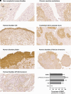
Fig. 1.
Fig. 1.
Expression of the α5 integrin subunit in human NMIBC. Representative immunohistochemistry photomicrographs of human bladder sections of non-neoplastic tissue (A and B), CIS (C and D), HG pTa (pTaG3, E), and HG pT1 (pT1G3) in the urothelial layer (F) and in the lamina propria (G, representative tumor area highlighted by dashed circle); all tissues were obtained by TURB. [Scale bar, (A and B), 50 µm; (CG), 100 µm.] Ur, urothelium, Lp, lamina propria. Quantification of tissues with cells positive for the membranous expression of the α5 subunit and located in the urothelial layer (H); N.n.= non-neoplastic tissue.
Fig. 2.
Fig. 2.
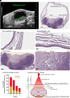
Theranostic application of GNRs@Chit-Iso4 against the murine MB49-Luc orthotopic bladder tumor. (A) Axial frame of the murine bladder with a tumor detected by ultrasound imaging and specific photoacoustic imaging of GNRs@Chit-Iso4 obtained after spectral unmixing using the reference photoacoustic spectra of melanin, deoxygenated and oxygenated blood (after intravesical instillation of 10 nmol Au; 100 µL of 100 µM GNRs@Chit-Iso4): one representative frame taken in the middle of the bladder, with the time gain compensation (TGC) shown at the right side of the image. (B) Hematoxylin–eosin stain of the tissue section from the same animal shown in panel A, after 3 min of irradiation with continuous laser at a laser power density of 0.664 W/cm2. (C) The non-neoplastic tissue near the necrotic tumor mass. (D) The normal bladder wall that was passed through by the laser. (E) The tumor mass with the necrotic area highlighted by the yellow dashed border, followed by a magnification of the region within the red dotted frame showing the coagulative necrosis characterized by the absence of cell nuclei, and the pyknosis characterized by the condensation of chromatin in the nuclei of the cells. (F) Quantification of the thermal damage in the neoplastic tissue, estimated as necrosis and pyknosis from the tumor surface to the inner part of the mass (measurements on 6 hematoxylin/eosin slides each tumor, four tumors from four different animals). (G) Scheme and parameters used to establish the gradient of laser power density at the upper and lower side of the bladder; bladder dimensions and distance from the skin were obtained from US images of the mouse in the supine position. Data shown in panels AF are from one representative animal out of three tested. Data shown in panel F were from eight ex vivo murine bladders.
Fig. 3.
Fig. 3.
The specificity of thermal therapy mediated by GNRs@Chit-Iso4 against bladder cancer cells expressing the integrin α5β1. Intravesical instillation of 1,970 ng of Au (100 μL of 100 μM of GNRs@Chit-Iso4) was performed in five animals with bladder orthotopic tumors of different sizes: after 15 min of incubation followed by three intravesical washes with saline solution, the binding of GNRs@Chit-Iso4 to the tumor area was visualized by PAUS imaging (SI Appendix, Fig. S7). (A) Left: Kinetic of temperature rise during laser irradiation and temperature fall during the laser off period; Right: maximum of temperature rise measured after 180 s of laser irradiation; each symbol in the dot plot represents a single animal. (B) The total temperature and heat calculated during the i) laser on/off period, ii) on period, and iii) off period. (C) Quantification of gold and maximum of the measured temperature rise according to the luminal area of the bladder tumor. (D) Recovery of gold. (E) Density of gold on neoplastic tissue. Each dot represents a single animal, with raw data reported in SI Appendix, Table S2. Lines and bars in panels B and C show means and SEM. (F) Tumor bioluminescence before and after laser irradiation (3 and 5 min) in the absence and presence of GNRs@Chit-Iso4 (intravesical instillation of 10 nmol Au, 100 μL of 100 μM of GNRs@Chit-Iso4); each symbol represents one animal before and after laser irradiation of the bladder, tested 9 to 12 d after the intravesical instillation of the MB49-Luc cells (statistical significance by the paired t test is shown by asterisks). (G) Cell viability of MB49-Luc cells cultivated in vitro was established 16 h after treatment with GNRs@Chit or GNRs@Chit-Iso4 and left not irradiated or irradiated with a 808 nm continuous laser. The experiment was performed in quadruplicate, and data are expressed as mean ± SEM.
Fig. 4.
Fig. 4.
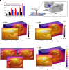
Computational model for GNRs@Chit-iso4-assisted PTT of ex vivo bladder and small bladder cancer lesions. (A) Comparison of temperature rise across the top and bottom surfaces of the tumor estimated from the model of ex vivo bladder. (B) Schematic of the computational model representing the region of interest during GNR-assisted PTT of bladder cancer in the mouse. The blue region represents the tumor, while the thin region in red represents the layer of the tumor with GNR attachment. The position of the laser probe in contact with the skin is also shown. (C) Contours of temperature after 180 s of laser irradiation at a laser power of 0.66 W in the model that considers an Au density of 7.5 ng/mm2 at the tumor surface (the Inset shows an enlarged view of the temperature contours across the tumor). (D) As in panel B, but considering 300 s of irradiation, with the temperature scale capped at 48.9 °C for better visualization (the Inset shows enlarged views of the temperature contours across the tumor). (E) Spread of the 42 °C isotherm (green) from the tumor boundary for cases with tumor radii of 1, 1.5, and 2 mm. (F) As in panel B but considering a 10-fold higher Au density (75 ng/mm2) of the tumor surface (the Inset shows an enlarged view of the temperature contours across the tumor).
Fig. 5.
Fig. 5.
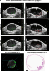
PA imaging of GNRs@Chit-Iso4 allows the identification of small neoplastic lesions, which rapidly merge to give rise to neoplastic mass with big volume and irregular shape. (A) Three representative axial frames of US imaging of a murine bladder 9 d after the intravesical instillation of murine bladder cancer cells MB49-Luc from the scanning of the entire bladder volume, with neoplastic lesions detected only by the PA imaging of GNRs@Chit-Iso4 (intravesical instillation of 10 nmol Au; 100 μL of 100 μM of GNRs@Chit-Iso4) indicated by the green arrows, followed by the 3D reconstruction of PA and US imaging. The PA imaging of GNRs@Chit-Iso4 is after unmixing the photoacoustic signal of melanin, deoxy- and oxy-genated blood. (B) US imaging of the bladder from the same animal reported in panel A, 3 d after the detection of the neoplastic lesions by the PA imaging of GNRs@Chit-Iso4: three axial frames showing the presence of two distinct tumor masses (frame 26, red dashed contours) that get in contact (frame 28, red dashed contours) and merge in a single tumor mass with irregular shape (frame 30, red dashed contour). Time gain compensation is shown on the right side of each PAUS and US image. (C) Histology of the orthotopic bladder cancer 12 d after the intravesical instillation of the murine bladder cancer cell line MB49-Luc. Data are representative of the follow-up of one animal out of five.
Fig. 6.
Fig. 6.
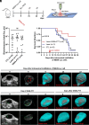
GNR-assisted PTT of orthotopic bladder cancer improves the survival of the preclinical model. (A) Scheme of the experimental procedure, starting from the intravesical instillation of MB49-Luc cells at day 0, bioluminescence (BLI) quantification, and PAUS imaging at day 9 and then intravesical instillation of GNRs@Chit-Iso4 (100 μL of 100 μM of GNRs@Chit-Iso4) followed by GNR-assisted PTT; the tumor growth was evaluated in the two following time points by US imaging, and the mice were euthanized according to the approved procedure by the Institutional Animal Care and Use Committee of San Raffaele Scientific Institute and were performed according to the prescribed guidelines. The cartoon shows the GNR-assisted PTT performed with the laser probe placed 11 mm from the skin. (B) Quantification of bioluminescence of the bladder tumor 9 d after the implantation of the MB49-Luc cells (n.s. = not significant). (C) Survival analysis of animals instilled with the GNRs@Chit-Iso4 (intravesical instillation of 10 nmol Au) and irradiated with a laser power density 0.664 W/cm2 for 3 min vs. laser irradiated animals without GNRs vs. control animals (CTL, no GNRs, no laser irradiation); survival analysis was truncated at day 37 after the instillation of murine bladder cancer cells MB49-Luc according to ethical guidelines. Statistical analysis was performed by using the log-rank (Mantel–Cox) test. (D) Axial frame and 3D visualization of US imaging of the murine bladder with tumor (red arrows showing multiple neoplastic mass), followed by the 3D reconstruction of the bladder volume (light blue) and quantification of the tumor volume (red) during the follow up; tumor volume is reported at the bottom of each image. One animal representative of 11. (E) Axial frame and 3D visualization of the murine bladder with tumor recognized by US and PA imaging of GNRs@Chit-Iso4, followed by the 3D reconstruction of the bladder volume and quantification of the tumor volume after GNR-assisted PTT. One animal representative of five. (F) Axial frame and 3D visualization of the murine bladder in which the tumor was not detected by US imaging but in which several neoplastic lesions were identified by the PA imaging of GNRs@Chit-Iso4, followed by the 3D reconstruction of the bladder volume and quantification of the tumor volume after GNR-assisted PTT. One animal representative of four (n.a.; not available).

References

    1. Ferlay J., et al. , Cancer incidence and mortality worldwide: Sources, methods and major patterns in GLOBOCAN 2012. Int. J. Cancer 136, E359–386 (2015). - PubMed
    1. Comperat E., et al. , Clinicopathological characteristics of urothelial bladder cancer in patients less than 40 years old. Virchows Arch. 466, 589–594 (2015). - PubMed
    1. European Association of Urology, Non-muscle-invasive Bladder Cancer, Chapter 5: Diagnosis. https://uroweb.org/guidelines/non-muscle-invasive-bladder-cancer/chapter.... Accessed 1 June 2014.
    1. Zapala P., et al. , Clinical rationale and safety of restaging transurethral resection in indication-stratified patients with high-risk non-muscle-invasive bladder cancer. World J. Surg Oncol. 16, 6 (2018). - PMC - PubMed
    1. Rakesh Heer R. L., et al. , A randomized trial of PHOTOdynamic surgery in non–muscle-invasive bladder cancer. NEJM Evid. 1, EVIDoa2200092 (2022). - PubMed

LinkOut - more resources