. 2024 Oct;300(10):107746.
doi: 10.1016/j.jbc.2024.107746. Epub 2024 Sep 3.

Perturbations in mitochondrial metabolism associated with defective cardiolipin biosynthesis: An in-organello real-time NMR study

Affiliations

Perturbations in mitochondrial metabolism associated with defective cardiolipin biosynthesis: An in-organello real-time NMR study

Antonio J Rua et al. J Biol Chem. 2024 Oct.

Abstract

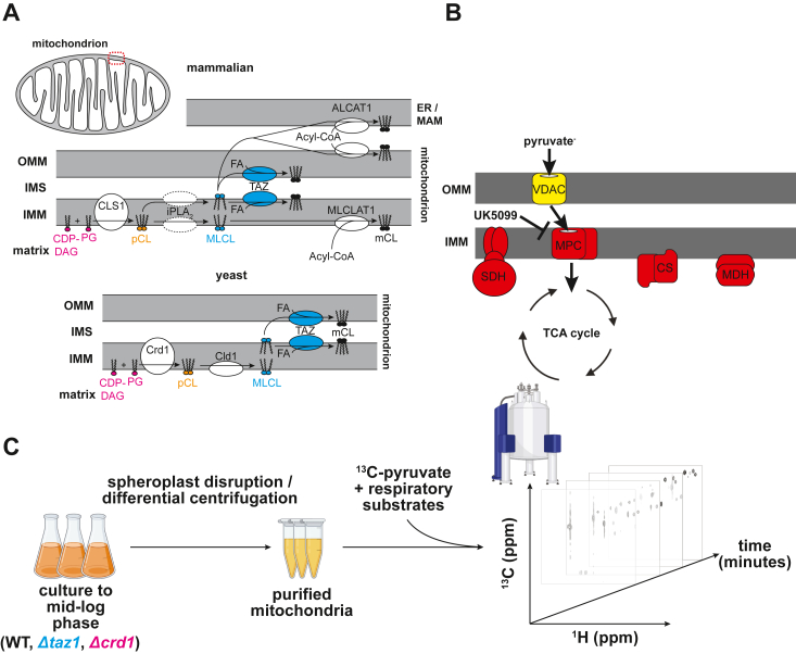
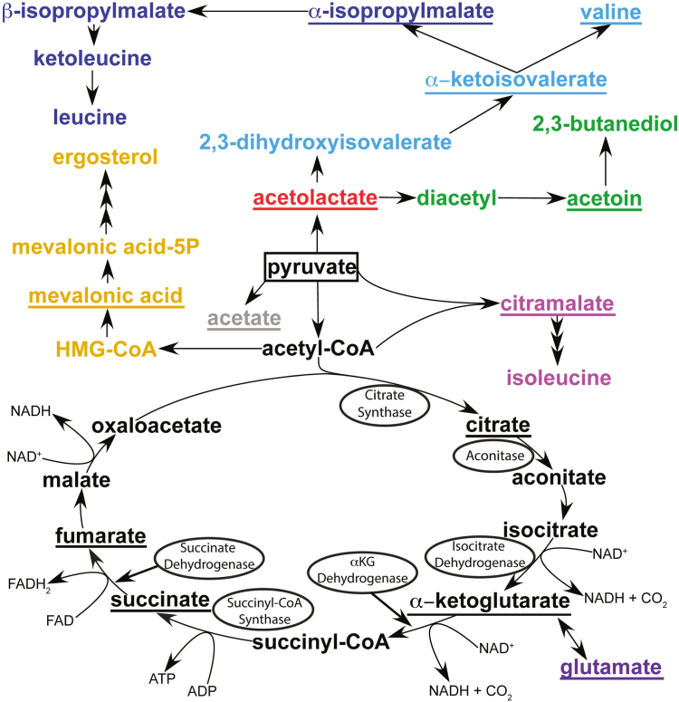
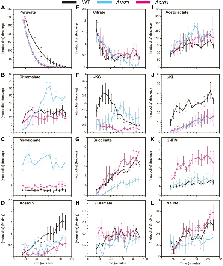
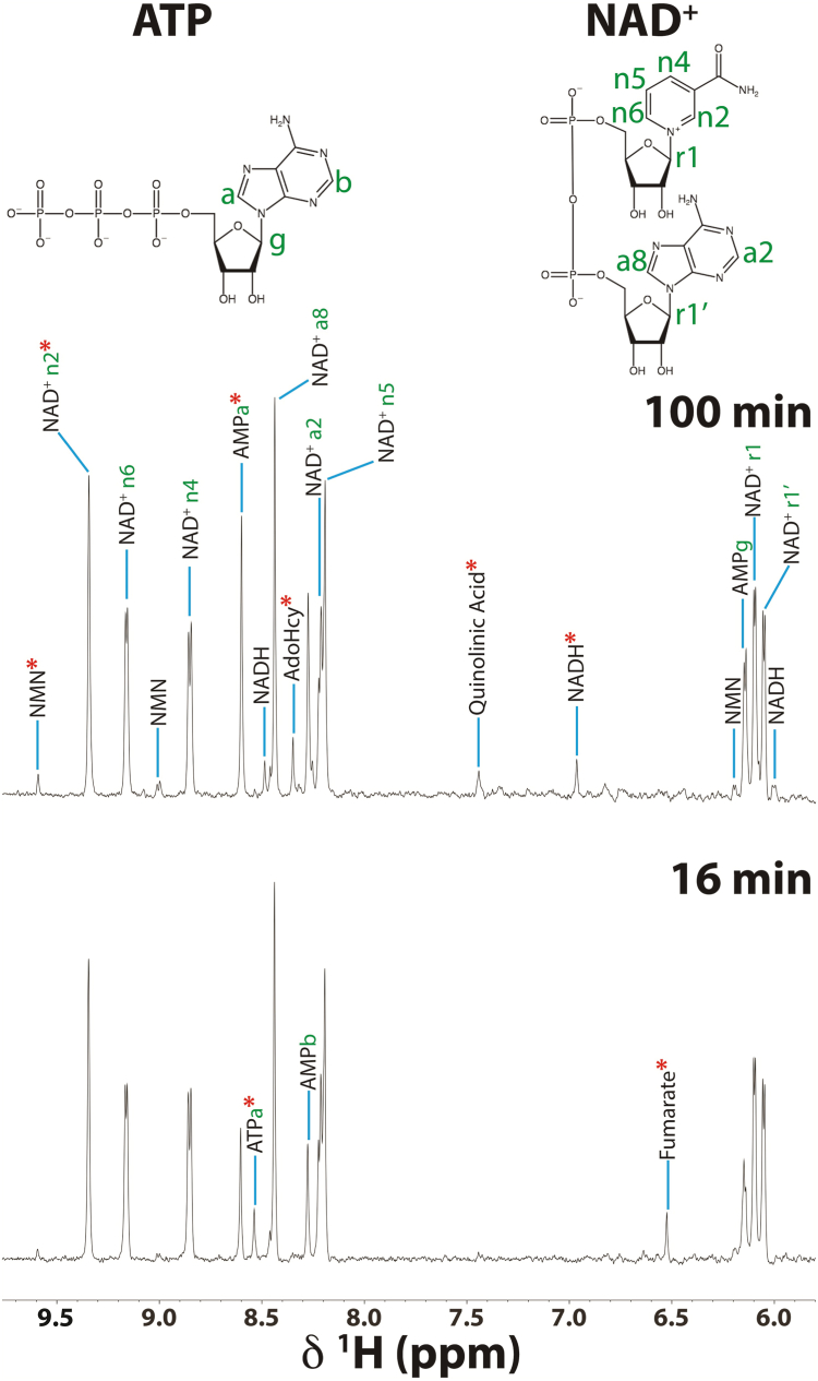
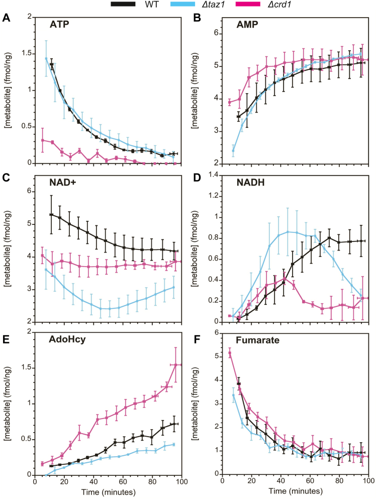
Mitochondria are central to cellular metabolism; hence, their dysfunction contributes to a wide array of human diseases. Cardiolipin, the signature phospholipid of the mitochondrion, affects proper cristae morphology, bioenergetic functions, and metabolic reactions carried out in mitochondrial membranes. To match tissue-specific metabolic demands, cardiolipin typically undergoes an acyl tail remodeling process with the final step carried out by the phospholipid-lysophospholipid transacylase tafazzin. Mutations in tafazzin are the primary cause of Barth syndrome. Here, we investigated how defects in cardiolipin biosynthesis and remodeling impacts metabolic flux through the TCA cycle and associated yeast pathways. Nuclear magnetic resonance was used to monitor in real-time the metabolic fate of 13C3-pyruvate in isolated mitochondria from three isogenic yeast strains. We compared mitochondria from a WT strain to mitochondria from a Δtaz1 strain that lacks tafazzin and contains lower amounts of unremodeled cardiolipin and mitochondria from a Δcrd1 strain that lacks cardiolipin synthase and cannot synthesize cardiolipin. We found that the 13C-label from the pyruvate substrate was distributed through twelve metabolites. Several of the metabolites were specific to yeast pathways including branched chain amino acids and fusel alcohol synthesis. While most metabolites showed similar kinetics among the different strains, mevalonate concentrations were significantly increased in Δtaz1 mitochondria. Additionally, the kinetic profiles of α-ketoglutarate, as well as NAD+ and NADH measured in separate experiments, displayed significantly lower concentrations for Δtaz1 and Δcrd1 mitochondria at most time points. Taken together, the results show how cardiolipin remodeling influences pyruvate metabolism, tricarboxylic acid cycle flux, and the levels of mitochondrial nucleotides.

Keywords: 3-methylglutaconic acid (3MGA); Barth syndrome (BTHS); Krebs cycle; adenosine triphosphate (ATP); metabolic disease; mitochondrial respiration; nuclear magnetic resonance (NMR); tricarboxylic acid (TCA) cycle.


Conflict of interest statement

Conflicts of interest The author declares that they have no conflicts of interest with the contents of this article.

Figures

Figure 1
Figure 1
Cardiolipin remodeling and pyruvate uptake in mitochondria.A, steps for CL biosynthesis and remodeling in mammals (upper) and yeast (lower). Mitochondria consist of an outer (OMM) and inner (IMM) membrane, which delineates the intermembrane space (IMS) from the matrix. Precursor CL (pCL) with variable acyl chain composition is synthesized de novo on the IMM by the joining of cytidine diphosphate diacylglycerol (CDP-DAG) to phosphatidylglycerol (PG) by cardiolipin synthase (CLS1/Crd1). CL then undergoes acyl chain remodeling first by the action of a deacylase (presumed to be a member of the iPLA2 family in mammals, Cld1 in yeast) to form monolyso-cardiolipin (MLCL). Then, a donor fatty acid (FA) is catalytically joined to MLCL by the transacylase tafazzin (TAZ) to form mature CL (mCL). Alternatively, in mammals, ALCAT1 can remodel mCL in the OMM or mitochondrial-associated ER membrane (ER/MAM) via utilization of a donor acyl tail from acyl-CoA. MLCLAT1 can also form mCL from acyl-CoA in the IMM. B, mitochondrial pyruvate uptake and metabolism. Cytosolic pyruvate is first taken up by mitochondria via the nonselective voltage-dependent anion carrier (VDAC) protein and subsequently imported directly into the matrix by the selective mitochondrial pyruvate carrier (MPC). Pyruvate is then metabolized by enzymes of the TCA cycle, which includes the integral IMM succinate dehydrogenase (SDH) complex, as well as the peripherally bound citrate synthase (CS) and malate dehydrogenase (MDH) proteins. The covalent inhibitor UK5099 blocks pyruvate uptake into the mitochondrial matrix by binding MPC. C, schematic of in-organello real-time NMR approach to monitor mitochondrial metabolic flux. First, mitochondria are isolated from WT, Δtaz1, and Δcrd1 yeast strains by spheroplast disruption and differential centrifugation. Then, mitochondria are incubated with 13C3-pyruvate and respiratory substrates. Finally, a series of 2D 1H-13C HSQC NMR experiments are recorded over time to track the flux of the 13C-label.
Figure 2
Figure 2
Purity and activity of mitochondrial isolated from yeast strains defective in CL biosynthesis.A, representative Western blots against mitochondrial markers (Tom40 and Tom70). Uncropped blots for Tom70 are included in Fig. S1. B, quantification of Tom70 abundance. Tom70 band intensity was quantified in ImageJ from n = 3 independent replicates originating from three independent mitochondrial isolations for each strain. Values were normalized to signal obtained from loading 15 μg protein from WT mitochondria. Error bars represent means ± SDs. C, respirometry measurements prior to (pre-) and following (post-) incubation at 30 °C for 65 min. Shown here are representative traces. D, quantification of state 2 respiration rate. State 2 respiration pre- and post-65 min of incubation at 30 °C was measured following addition of malate, pyruvate, and NADH for n = 3 independent biological replicates. p-values were obtained from two-way ANOVA and Tukey’s post hoc test. E, 1H-13C HSQC spectra obtained following t = 15 (cyan) and t = 100 (black) minutes of incubation with 13C3-pyruvate. Left panels were acquired in the presence of UK5099, which blocks mitochondrial pyruvate uptake through the MPC.
Figure 3
Figure 3
Representative13C3-pyruvate flux experiment in WT mitochondria. Each panel represents an individual 2D 1H-13C HSQC spectrum collected at the specified time over the course of the experiment. Numbers listed next to each peak correspond to NMR signals from the metabolites listed in Table 1 and are shown in different colors depending on the time of their first appearance in the spectra (green, initial; cyan, 28 min; orange, 60 min; red, 117 min). “1t” corresponds to the enol tautomer of pyruvate.
Figure 4
Figure 4
Pathways of pyruvate metabolism in yeast mitochondria. Metabolites derived from pyruvate that are observed in the NMR experiments are underlined. The starting substrate (pyruvate) is boxed. Tricarboxylic acid (TCA) cycle enzymes that are known to require CL for activity are circled. Color legend: black, TCA cycle; green, butanediol pathway; light/dark blue, valine and leucine arms of the branched chain amino acid (BCAA) pathway, respectively; pink, citramalate pathway leading to isoleucine; violet, glutamine oxoglutarate aminotransferase (GOGAT)/glutamine dehydrogenase (GDH) pathway; yellow, sterol pathway; gray, acetate formation through pyruvate decarboxylation (PD). Refer to Table 1 for references.
Figure 5
Figure 5
Kinetic traces of mitochondrial metabolite flux observed by 2D1H-13C HSQC experiments. Data points are colored according to yeast strain: black, WT; cyan, Δtaz1; pink, Δcrd1. Y values (fmol metabolite per ng of mitochondrial protein) represent means ± SEM from n ≥ 3 individual experiments. X-error bars represent variation (±SEM) in timing of the n ≥ 3 experiments. T = 0 corresponds to the time of 13C3-pyruvate addition. Panels are arranged so that (AD) correspond to pathways for which a single metabolite is observed (A: pyruvate, B: citramalate, C: mevalonate, D: acetoin), (EH) correspond to the TCA cycle and glutamate pathways (E: citrate, F: αKG, G: succinate, H: glutamate), and (IL) corresponds to the BCAA pathways (I: acetolactate, J: αKI, K: 2-IPM, L: valine). For metabolites with multiple peaks (Table 1), the peaks used to determine concentrations were 1, pyruvate; an average of 16 and 17, acetolactate; 15, αKI; an average of 4 and 7, 2-IPM; and an average of 6 and 8, valine.
Figure 6
Figure 6
Downfield region of the 1D1H NMR spectrum following pyruvate addition. Representative 1D 1H NOESY spectra of WT mitochondria at the start (bottom) and end (top) of the NMR timecourses using 3 mM unlabeled 12C3-pyruvate in place of 3 mM 13C3-pyruvate. Peaks are labeled with their indicated NMR assignments (120). Only peaks not appearing in the spectra at 100 min are labeled in the spectra at 16 min. The positions of protons corresponding to the NMR assignments of ATP and NAD+ are labeled in their structures. The red ∗ symbols indicate NMR signals that were sufficiently isolated in the spectra to allow quantification of the respective metabolite concentration (see Fig. 7).
Figure 7
Figure 7
Mitochondrial metabolites observed by 1D1H NOESY experiments. Kinetic profiles of selected metabolites obtained from the 1D 1H NMR data illustrated in Figure 6. (A) ATP, (B) AMP, (C) NAD+, (D) NADH, (E) S-adenosyl homocysteine (AdoHcy), (F) fumarate.

Update of

Similar articles

Cited by

References

    1. Aversa R., Petrescu R.V.V., Apicella A., Petrescu F.I.T. Mitochondria are naturally micro robots - a review. Am. J. Eng. Appl. Sci. 2016;9:991–1002.
    1. Giacomello M., Pyakurel A., Glytsou C., Scorrano L. The cell biology of mitochondrial membrane dynamics. Nat. Rev. Mol. Cell Biol. 2020;21:204–224. - PubMed
    1. Spinelli J.B., Haigis M.C. The multifaceted contributions of mitochondria to cellular metabolism. Nat. Cell Biol. 2018;20:745–754. - PMC - PubMed
    1. Bhatti J.S., Bhatti G.K., Reddy P.H. Mitochondrial dysfunction and oxidative stress in metabolic disorders - a step towards mitochondria based therapeutic strategies. Biochim. Biophys. Acta Mol. Basis Dis. 2017;1863:1066–1077. - PMC - PubMed
    1. Murphy M.P., O’Neill L.A.J. Krebs cycle reimagined: the emerging roles of succinate and itaconate as signal transducers. Cell. 2018;174:780–784. - PubMed

MeSH terms

LinkOut - more resources