. 2024 Sep 5;14(1):20699.
doi: 10.1038/s41598-024-71739-y.

IGF-I concentration determines cell fate by converting signaling dynamics as a bifurcation parameter in L6 myoblasts

Affiliations

IGF-I concentration determines cell fate by converting signaling dynamics as a bifurcation parameter in L6 myoblasts

Ryosuke Okino et al. Sci Rep. .

Abstract

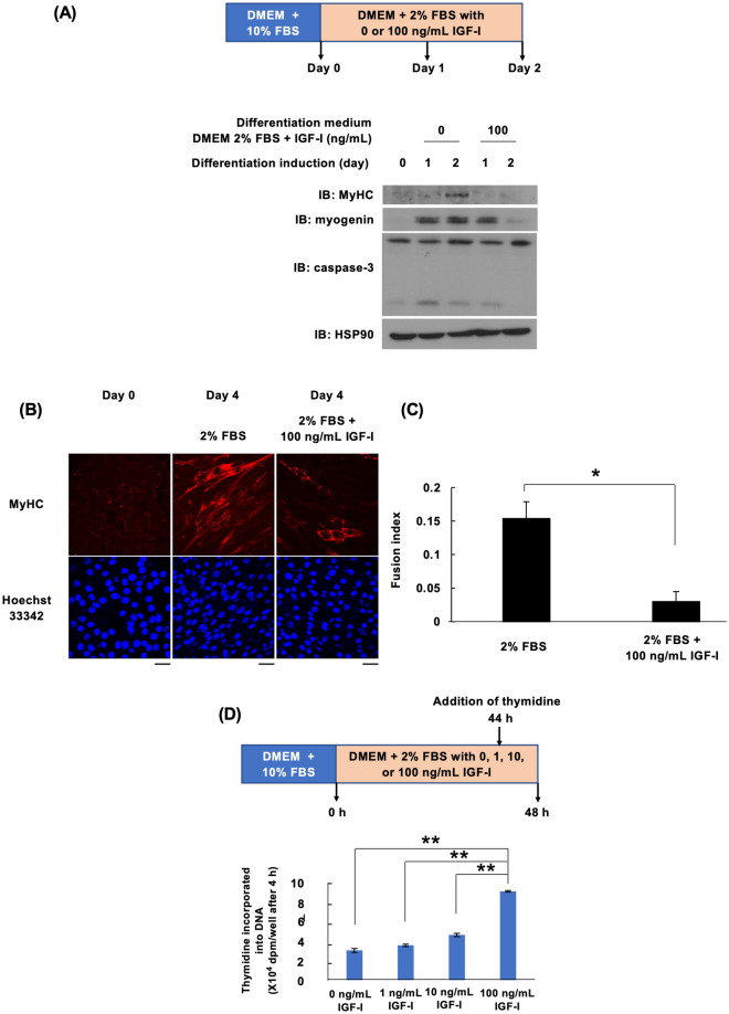
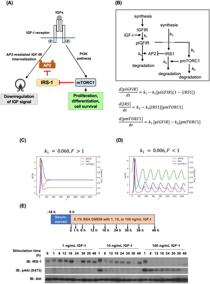
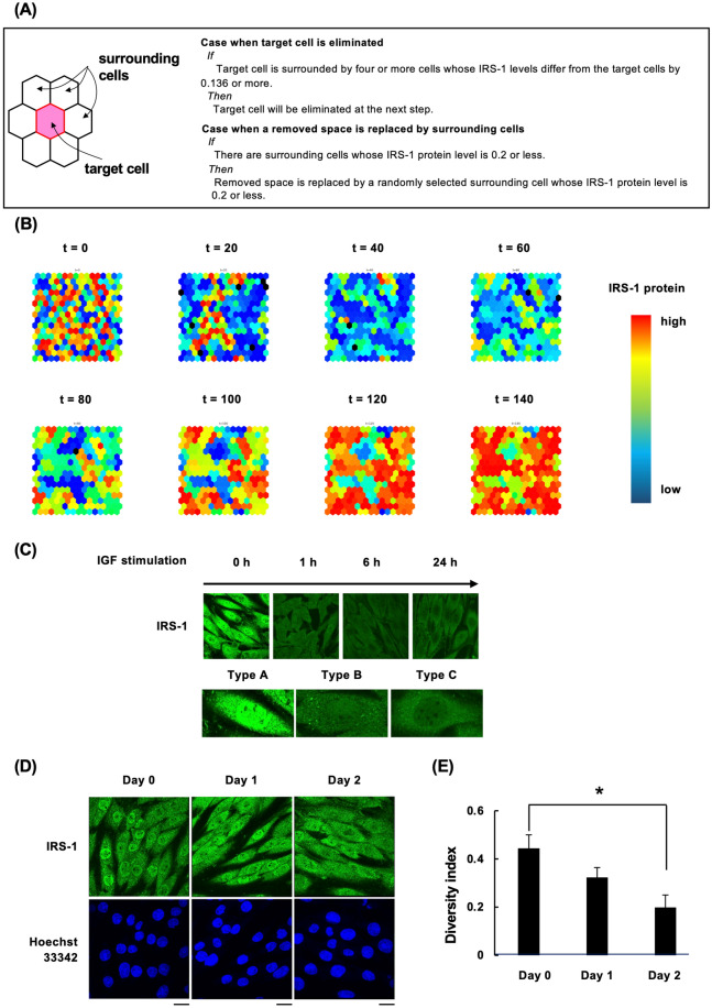
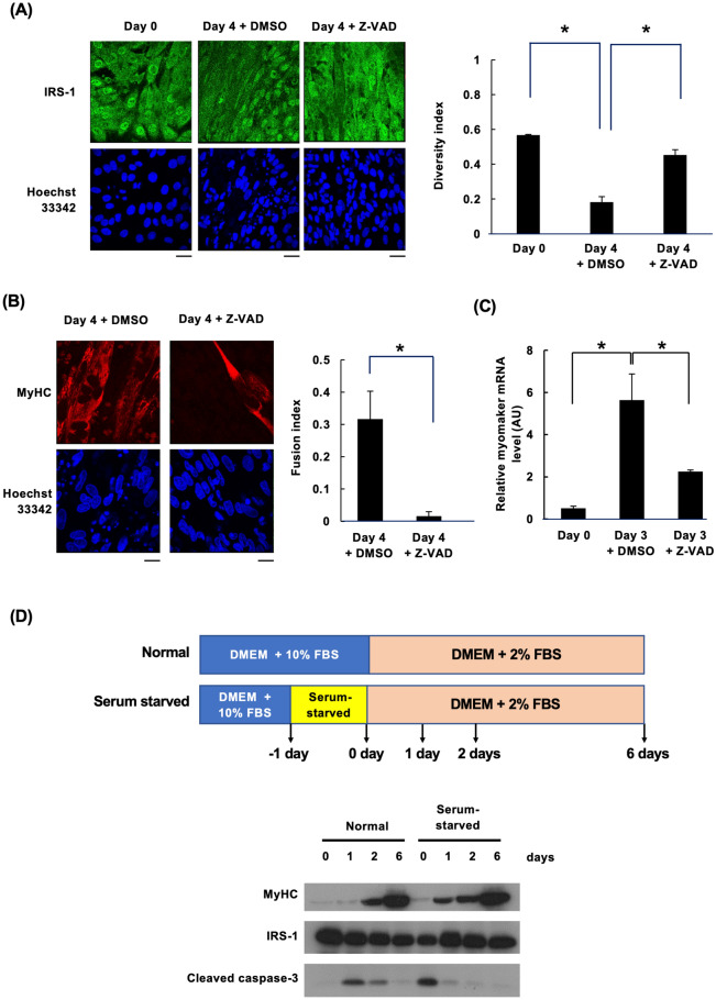
Insulin-like growth factor (IGF)-I mediates long-term activities that determine cell fate, including cell proliferation and differentiation. This study aimed to characterize the mechanisms by which IGF-I determines cell fate from the aspect of IGF-I signaling dynamics. In L6 myoblasts, myogenic differentiation proceeded under low IGF-I levels, whereas proliferation was enhanced under high levels. Mathematical and experimental analyses revealed that IGF-I signaling oscillated at low IGF-I levels but remained constant at high levels, suggesting that differences in IGF-I signaling dynamics determine cell fate. We previously reported that differential insulin receptor substrate (IRS)-1 levels generate a driving force for cell competition. Computational simulations and immunofluorescence analyses revealed that asynchronous IRS-1 protein oscillations were synchronized during myogenic processes through cell competition. Disturbances of cell competition impaired signaling synchronization and cell fusion, indicating that synchronization of IGF-I signaling oscillation is critical for myoblast cell fusion to form multinucleate myotubes.

Keywords: Bifurcation analysis; Cell competition; Insulin-like growth factor-I; L6 myoblast; Myogenesis; Signaling dynamics; Signaling oscillation.


Conflict of interest statement

The authors declare no competing interests.

Figures

Fig. 1
Fig. 1
Effects of IGF-I concentration on IGF-I signaling pathway dynamics and bioactivity expression. (A) L6 myoblasts were incubated in a differentiation medium (DMEM with 2% fetal bovine serum (FBS)) supplemented with either 0 or 100 ng/mL insulin-like growth factor-I (IGF-I). Cell lysates were collected at 0, 1, or 2 days post-differentiation induction for immunoblotting analysis using the specified antibody. (B) L6 myoblasts were cultured in a differentiation medium. Cells were fixed with 4% paraformaldehyde at 0 and 4 days post-differentiation induction and subjected to immunostaining analysis using the MyHC antibody. Scale bar = 50 µm. (C) The fusion index was determined (n = 3). Data are presented as mean ± SEM. *P < 0.05, determined by Student’s t-test. (D) L6 myoblasts were cultured in a differentiation medium (DMEM with 2% FBS) supplemented with 0, 1, 10, or 100 ng/mL IGF-I. Forty-four hours later, [methyl-3H] thymidine was added, and after a 4-h incubation, the amount of [3H] incorporated into DNA was measured as an indicator of proliferation ability (n = 6). Data are presented as mean ± SEM. **P < 0.01, analyzed using one-way ANOVA followed by Tukey’s post hoc test.
Fig. 2
Fig. 2
Mathematical model of the IGF-I signaling pathway. (A) Schematic illustration of the IGF-I signaling pathway model. Insulin receptor substrate-1 (IRS-1) inhibits AP2 function, thereby suppressing internalization of the active IGF-I receptor from the plasma membrane. Downstream signaling pathway activation induces the degradation of IRS-1. In this model, IRS-1 is not a mediator but rather a regulator of the IGF signaling pathway. (B) A mathematical model based on the IGF-I signaling pathway. (C,D) Simulation results of the IGF-I signaling pathway. C: k1 = 0.006. D: k1 = 0.06. (E) L6 myoblasts were serum-starved for 18 h, followed by stimulation with DMEM + 2% FBS containing 1 or 100 ng/mL IGF-I. Cell lysates were prepared from each sample, and the total cell lysate was used for immunoblotting analysis with anti-IRS-1, anti-pAkt, or anti-Akt antibodies. BSA bovine serum albumin, mTORC1 mechanistic target of rapamycin complex 1, PI3K phosphatidylinositol 3-kinase.
Fig. 3
Fig. 3
Synchronization of IGF-I signaling oscillations. (A) Cellular automaton-based algorithm considering cell competition. When the target cell is surrounded by low-level IRS-1 cells, target cells are eliminated, and the empty space is replaced by the proliferation of neighboring cells. (B) Cellular automaton simulation results. The IRS-1 level in each cell is shown as a heatmap, and the dead space is in black. At time 0, each cell had different IRS-1 protein levels because IRS-1 oscillation was not synchronized. Over time, IRS-1 protein levels gradually synchronized between neighboring cells. (C) L6 myoblasts were serum-starved for 18 h, followed by stimulation with 100 ng/mL IGF-I for the indicated time. Afterward, cells were fixed and immunostained with anti-IRS-1 antibody. (D) L6 myoblasts were differentiated into myotubes in a differentiation medium. On days 0, 1, or 2 after induction of differentiation, cells were fixed and immunostained with anti-IRS-1 antibody. The green color shows IRS-1, and the blue color indicates the nuclei stained with Hoechst 33342. Scale bar = 20 µm; applicable to all images in each panel. (E) The diversity index was calculated from the data of photographs immunostained with IRS-1 and shown as a graph (n = 4). Results are presented as mean ± SEM. *P < 0.05. One-way ANOVA and Tukey’s post hoc test were performed for assessment.
Fig. 4
Fig. 4
Synchronization of IRS-1 oscillation is critical for cell fusion. (A) L6 myoblast differentiation was induced in the differentiation medium containing Dimethyl sulfoxide (DMSO) or Z-VAD-FMK. At 0 or 4 days after induction of differentiation, cells were fixed and immunostained with an anti-IRS-1 antibody. The green color shows IRS-1, and the blue color indicates the nuclei stained with Hoechst 33342. Scale bar = 50 µm; applicable to all images in each panel. The diversity index was calculated and shown in the graph (n = 4). Results are presented as mean ± SEM. *P < 0.05. One-way ANOVA and Tukey’s post hoc test were performed for assessment. (B) L6 myoblasts were differentiated in the differentiation medium for 4 days. Cells were fixed and immunostained with an anti-MyHC antibody. Scale bar = 20. The fusion index was calculated and shown in the graphs (n = 3). Results are presented as mean ± SEM. *P < 0.05. Student’s t-test was performed for assessment. (C) L6 myoblasts were differentiated in the differentiation medium for 3 days. Myomaker mRNA levels in L6 myoblasts were measured via qPCR (n = 3). Results are presented as mean ± SEM. *P < 0.05. One-way ANOVA and Tukey’s post hoc test were performed for assessment. (D) L6 myoblasts were directly differentiated by exchanging the culture medium from DMEM + 10% FBS to DMEM + 2% FBS (normal). L6 myoblasts were serum-starved for 1 day, followed by differentiation induction in DMEM + 2% FBS. Total cell lysates were prepared for immunoblotting analysis using the indicated antibodies.

Similar articles

References

    1. Nakae, J., Kido, Y. & Accili, D. Distinct and overlapping functions of insulin and IGF-I receptors. Endocr. Rev.22, 818–835. 10.1210/EDRV.22.6.0452 (2001). 10.1210/EDRV.22.6.0452 - DOI - PubMed
    1. Fukushima, T. et al. Phosphatidylinositol 3-kinase (PI3K) activity bound to insulin-like growth factor-I (IGF-I) receptor, which is continuously sustained by IGF-I stimulation, is required for IGF-I-induced cell proliferation. J. Biol. Chem.287, 29713–29721. 10.1074/jbc.M112.393074 (2012). 10.1074/jbc.M112.393074 - DOI - PMC - PubMed
    1. Dumont, N. A., Bentzinger, C. F., Sincennes, M. C. & Rudnicki, M. A. Satellite cells and skeletal muscle regeneration. Compr. Physiol.5, 1027–1059. 10.1002/CPHY.C140068 (2015). 10.1002/CPHY.C140068 - DOI - PubMed
    1. Clemmons, D. R. Role of IGF-I in skeletal muscle mass maintenance. Trends Endocrinol. Metab.20, 349–356. 10.1016/J.TEM.2009.04.002 (2009). 10.1016/J.TEM.2009.04.002 - DOI - PubMed
    1. Barton-Davis, E. R., Shoturma, D. I. & Sweeney, H. L. Contribution of satellite cells to IGF-I induced hypertrophy of skeletal muscle. Acta Physiol. Scand.167, 301–305. 10.1046/J.1365-201X.1999.00618.X (1999). 10.1046/J.1365-201X.1999.00618.X - DOI - PubMed

Substances

LinkOut - more resources