. 2024 Sep 5;14(1):20697.
doi: 10.1038/s41598-024-71305-6.

A Gaussia luciferase reporter assay for the evaluation of coronavirus Nsp5/3CLpro activity

Affiliations

A Gaussia luciferase reporter assay for the evaluation of coronavirus Nsp5/3CLpro activity

Asimenia Vlachou et al. Sci Rep. .

Abstract

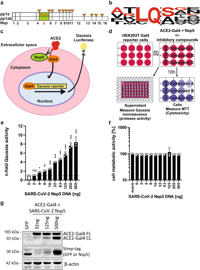
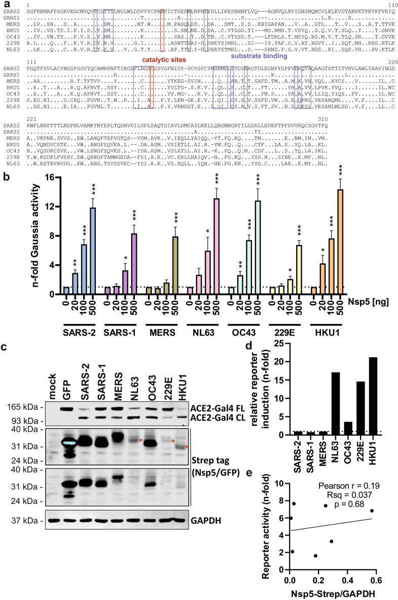
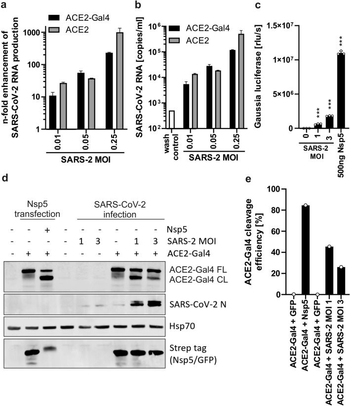
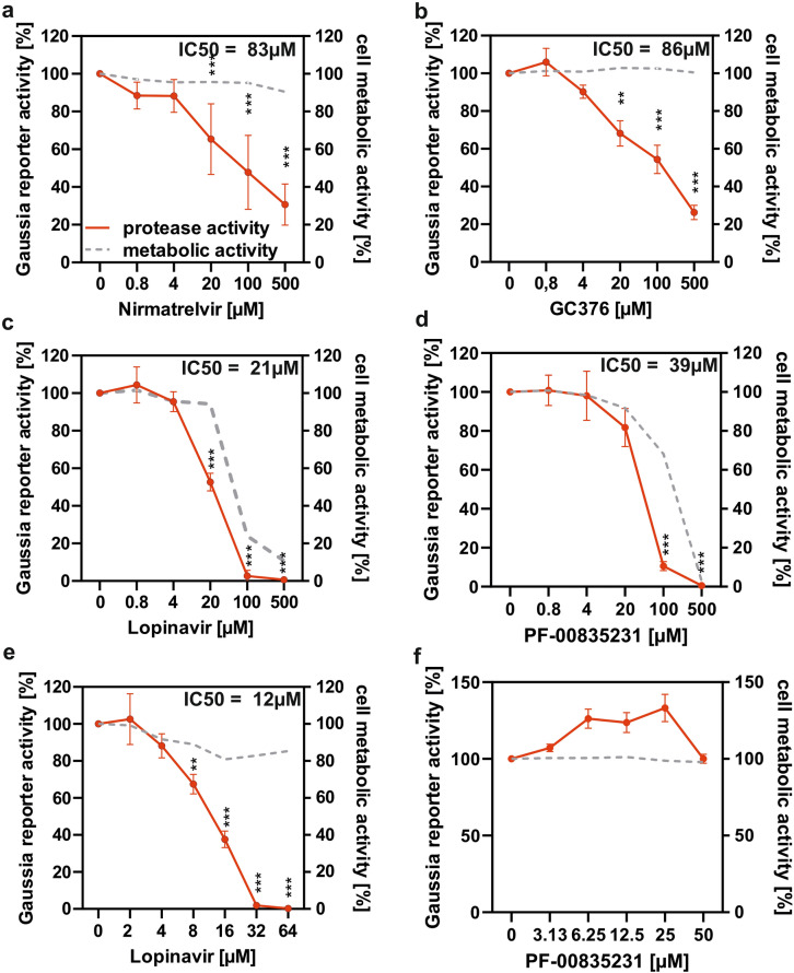
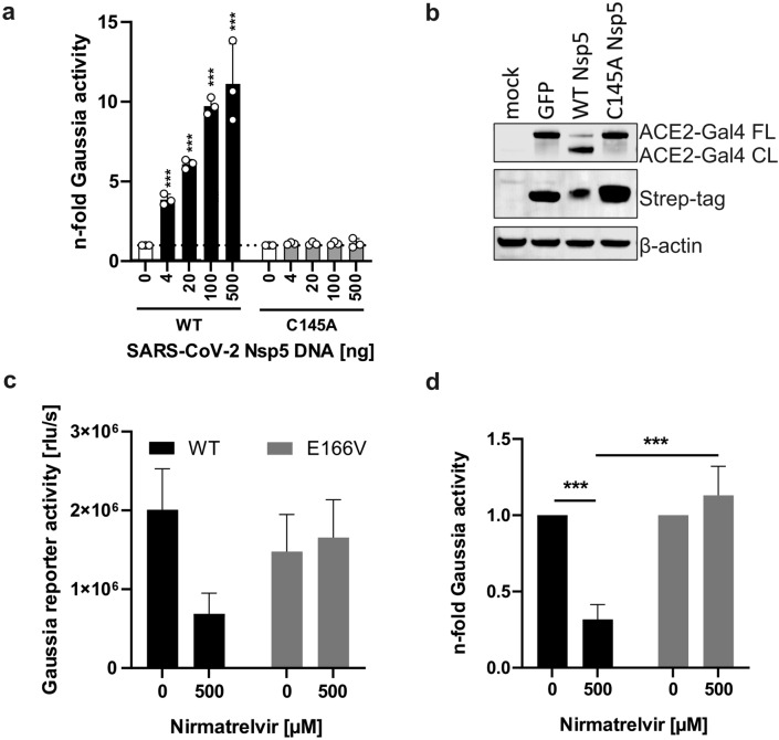
Human coronaviruses (hCoVs) infect millions of people every year. Among these, MERS, SARS-CoV-1, and SARS-CoV-2 caused significant morbidity and mortality and their emergence highlights the risk of possible future coronavirus outbreaks. Therefore, broadly-active anti-coronavirus drugs are needed. Pharmacological inhibition of the hCoV protease Nsp5 (3CLpro) is clinically beneficial as shown by the wide and effective use of Paxlovid (nirmatrelvir, ritonavir). However, further treatment options are required due to the risk of drug resistance. To facilitate the assessment of coronavirus protease function and its pharmacological inhibition, we developed an assay allowing rapid and reliable quantification of Nsp5 activity under biosafety level 1 conditions. It is based on an ACE2-Gal4 transcription factor fusion protein separated by a Nsp5 recognition site. Cleavage by Nsp5 releases the Gal4 transcription factor, which then induces the expression of Gaussia luciferase. Our assay is compatible with Nsp5 proteases from all hCoVs and allows simultaneous measurement of inhibitory and cytotoxic effects of the tested compounds. Proof-of-concept measurements confirmed that nirmatrelvir, GC376 and lopinavir inhibit SARS-CoV-2 Nsp5 function. Furthermore, the assay accurately predicted the impact of Nsp5 mutations on catalytic activity and inhibitor sensitivity. Overall, the reporter assay is suitable for evaluating viral protease activity.

Keywords: 3CLpro; Coronaviruses; Gaussia reporter assay; Nsp5; Protease inhibitor; SARS-CoV-2.


Conflict of interest statement

The authors declare no competing interests.

Figures

Fig. 1
Fig. 1
Design and performance of the Nsp5 reporter assay. (a) Location of Nsp5 cleavage sites in coronavirus polyprotein 1ab. (b) Sequence logo of the Nsp5 recognition site based on SARS-CoV-2 1ab polyprotein. The sequence of representative cleavage site selected for testing is indicated in red. (c) Principle of the reporter assay. Human ACE2 fused to transcription factor Gal4 is expressed in HEK293T cells containing a stable Gaussia luciferase reporter downstream of 5 × Gal4 binding sites. The fusion protein contains Nsp5 recognition site in the flexible glycine linker region. Upon cleavage, Gal4 fused to amino acids 364–550 of mouse NF-kB translocates into the nucleus where it activates the transcription of Gaussia luciferase, which is subsequently secreted into cell supernatant. (d) Experimental outline of the Nsp5 reporter assay. HEK293T cells containing the stable Gaussia luciferase reporter downstream of the 5 × Gal4 binding sites are co-transfected with construct encoding ACE2-Gal4 fusion protein, and varying amounts of Strep-tagged Nsp5 or GFP (negative control). At 18-20 h post transfection supernatants are harvested for luminescence measurement of Gaussia luciferase activity (relative light units per second; rlu/s) using coelenterazine substrate, while the cells are subjected to 2,5-diphenyl-2H-tetrazolium bromide (MTT) assay measuring cell metabolic activity. (e) Normalized n-fold increase in Gaussia luciferase activity over background (GFP + reporter only) by transfected SARS-CoV-2 Nsp5, and (f) corresponding MTT assay results measured at 18 h post transfection. (g) Western blot of transfected cell lysates showing dose-dependent ACE2-Gal4 reporter cleavage (FL full length, CL cleaved protein detected by ACE2 staining). Lane 1 contained GFP only control. Mean of 3 independent experiments + SD. *P < 0.05; **P < 0.01; ***P < 0.001, ns, P > 0.05. Unpaired Student’s t-test.
Fig. 2
Fig. 2
Activation of the reporter assay by hCoV Nsp5. (a) Amino acid sequence alignment of hCoV Nsp5 proteins. Catalytic residues are highlighted in red and substrate binding sites are indicated by blue brackets. Domain mapping was based on similarity to SARS-CoV-2 sequence. (b) Dose-dependent activation of the reporter assay by overexpressed hCoV Nsp5 proteins shown as n-fold enhancement over negative control (ACE2-Gal4 + GFP). Mean of 4 independent experiments + SD. *P < 0.05; **P < 0.01; ***P < 0.001, unpaired Student’s t-test. (c) Western blot of cellular lysates following co-expression of the ACE2-Gal4 reporter and indicated Strep-tagged hCoV Nsp5 proteins or GFP. GAPDH served as protein loading control. Red stars indicate the relative gel position of low expressed proteins. (d) Levels of Gaussia luciferase activity normalized to protein expression levels measured by a Western blot. (e) Lack of significant correlation between reporter activity measured in (b) at the 100 ng dose and Nsp5 protein expression levels observed in western blot (c).
Fig. 3
Fig. 3
Activation of the Nsp5 reporter assay during hCoV infection. (a) Relative levels and (b) absolute copy numbers of SARS-CoV-2 RNA present in the supernatant of infected HEK293T reporter cells transfected with plasmid encoding human ACE2 or ACE2-Gal4 fusion protein 48 h after infection. MOI multiplicity of infection of input virus. Mean of one independent experiment measured by qRT PCR in technical triplicates, + SD. (c) Activation of the Nsp5 Gaussia luciferase reporter assay 18 h after SARS-CoV-2 infection with indicated MOI or by transfection of 500 ng of Nsp5 expression construct. Mean of 3 infections + SD. ***P < 0.001, unpaired Student’s t-test. (d) Western blot of reporter transfected and infected cell lysates showing cleavage of the ACE2-Gal4 reporter 3 days after SARS-CoV-2 infection (indicated by N staining). Protein concentrations were adjusted prior to loading to account for infection-induced cytotoxicity. Hsp70 served as a protein loading control. (e) Quantification of ACE2-Gal4 cleavage efficiency detected by western blot shown in panel (d).
Fig. 4
Fig. 4
Evaluation of protease inhibitors using the Nsp5 reporter assay. HEK293T reporter cells were co-transfected with equal amounts of plasmid encoding the ACE2-Gal4 fusion protein and SARS-CoV-2 Nsp5 or GFP (negative control). After 6 h cells were treated with indicated molar concentration of inhibitors (a) nirmatrelvir, (b) GC376, (c) Lopinavir and (d) PF-00835231 or DMSO control for 20 h. The activity of secreted Gaussia luciferase was measured using luminometer and cellular metabolic activity was evaluated by the MTT assay. The activity of Lopinavir and PF-00835231 the was re-evaluated at lower, nontoxic concentrations (e and f). Red line indicates relative Nsp5 activity as percentage of no inhibitor control measured by luciferase assay (left y-axis) and grey dotted line indicates metabolic activity as percentage of inhibitor untreated cells as determined by MTT assay (right y-axis). Mean of 3–5 independent experiments + SD. *P < 0.05; **P < 0.01; ***P < 0.001, paired Student’s t-test. IC50 values were calculated using Prism GraphPad.
Fig. 5
Fig. 5
(a) Activation of the assay by WT Nsp5 and the C145A catalytically inactive mutant 20 h post transfection. Mean of 3 independent experiments + SD. (b) Representative Western blot showing the expression levels of Nsp5 catalytic mutant and its inability to cleave ACE2 reporter. (c) Gaussia activity induced by WT Nsp5 protein and E166V Nsp mutant in the presence of nirmatrelvir and (d) relative sensitivity of both proteins to the inhibitor. Mean of 3 independent experiments + SD. ***P < 0.001, unpaired Student’s t-test.

Similar articles

References

    1. Coronavirus disease (COVID-19). https://www.who.int/news-room/fact-sheets/detail/coronavirus-disease-(co... (Accessed 18 April 2024).
    1. Hoenigsperger, H., Sivarajan, R. & Sparrer, K. M. Differences and similarities between innate immune evasion strategies of human coronaviruses. Curr. Opin. Microbiol.79, 102466. 10.1016/j.mib.2024.102466 (2024). 10.1016/j.mib.2024.102466 - DOI - PubMed
    1. V’kovski, P., Kratzel, A., Steiner, S., Stalder, H. & Thiel, V. Coronavirus biology and replication: Implications for SARS-CoV-2. Nat. Rev. Microbiol.10.1038/s41579-020-00468-6 (2021). 10.1038/s41579-020-00468-6 - DOI - PMC - PubMed
    1. Peiris, J. S. M., Yuen, K. Y., Osterhaus, A. D. M. E. & Stöhr, K. The severe acute respiratory syndrome. N. Engl. J. Med.349(25), 2431–2441. 10.1056/NEJMra032498 (2003). 10.1056/NEJMra032498 - DOI - PubMed
    1. Lampl, B. M. J., Edenharter, B., Leitzmann, M. F. & Salzberger, B. COVID-19-related deaths: A 2-year inter-wave comparison of mortality data from Germany. Infection51(4), 1147–1152. 10.1007/s15010-023-01982-4 (2023). 10.1007/s15010-023-01982-4 - DOI - PMC - PubMed

LinkOut - more resources