. 2024 Sep 5;7(1):1091.
doi: 10.1038/s42003-024-06792-4.

Small GTP-binding protein GDP dissociation stimulator influences cisplatin-induced acute kidney injury via PERK-dependent ER stress

Affiliations

Small GTP-binding protein GDP dissociation stimulator influences cisplatin-induced acute kidney injury via PERK-dependent ER stress

Yuxue Yang et al. Commun Biol. .

Abstract

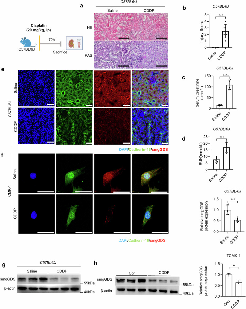
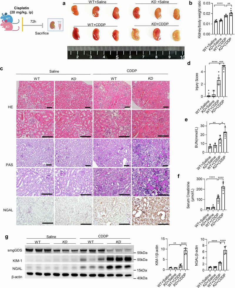
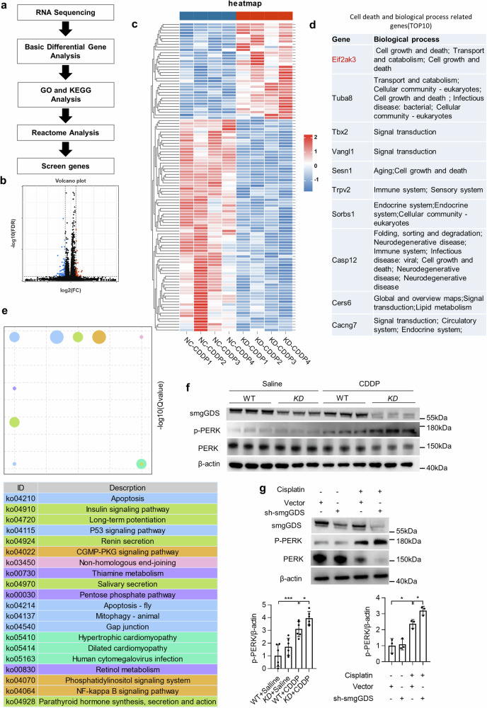
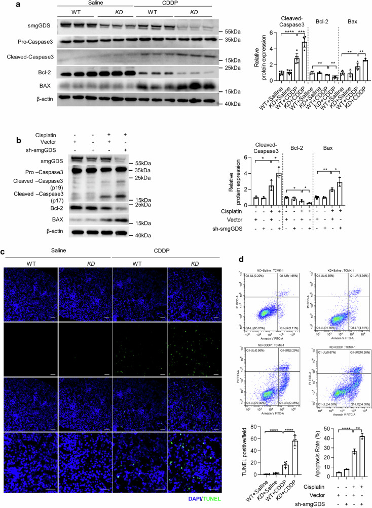
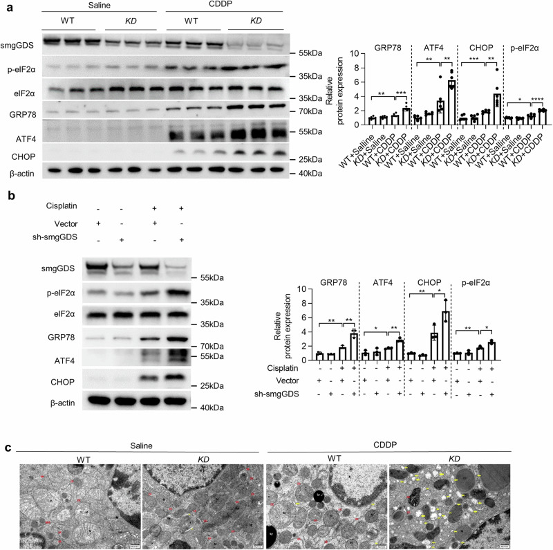
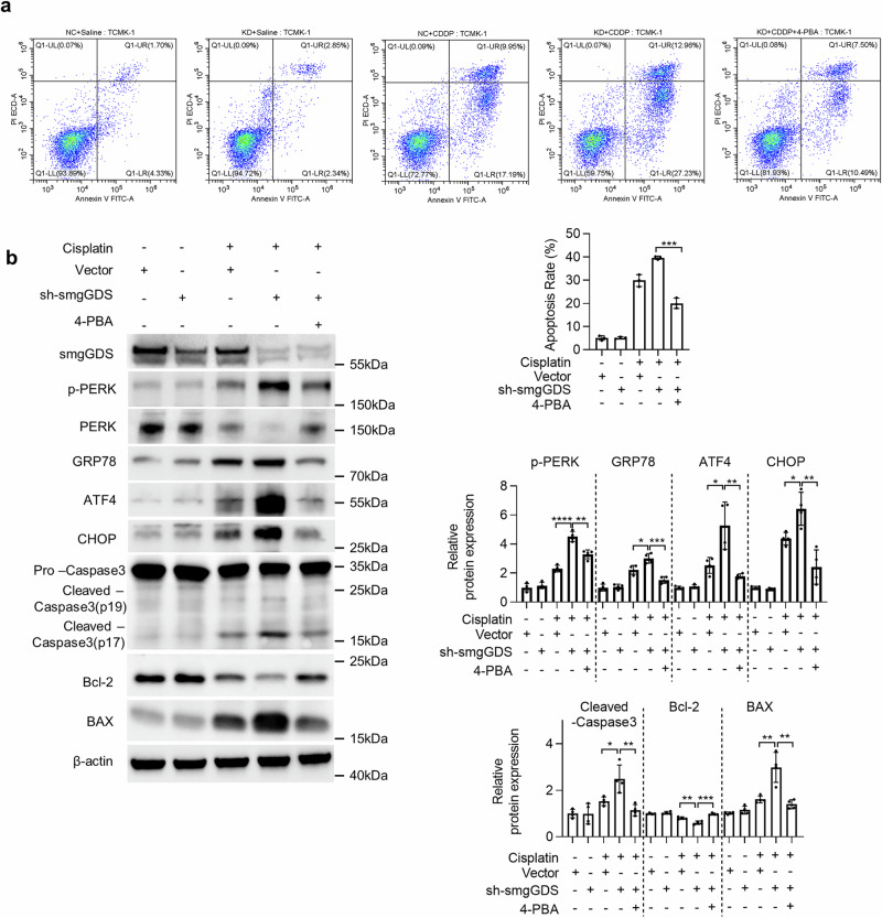
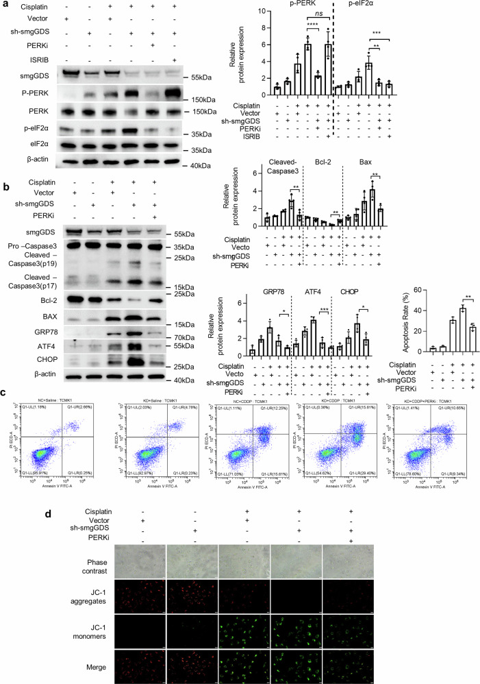
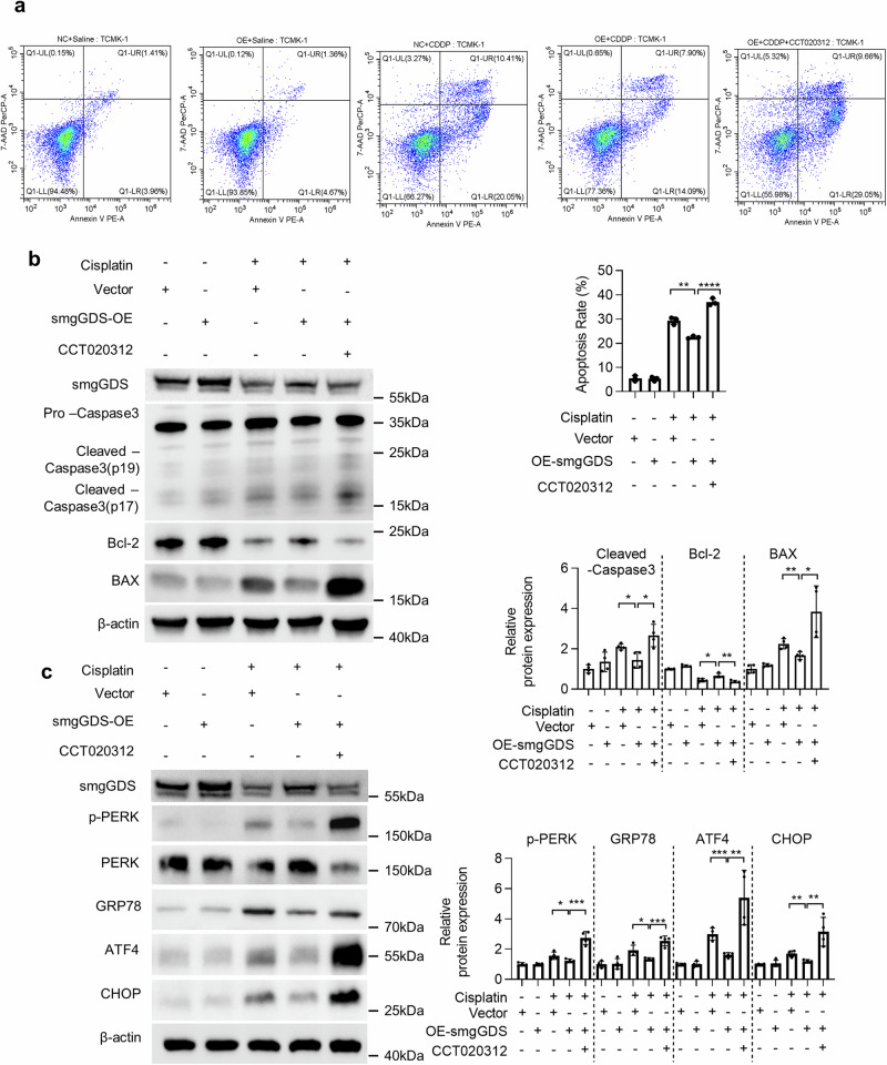
Cisplatin is a common anticancer drug, but its frequent nephrotoxicity limits its clinical use. Small GTP-binding protein GDP dissociation stimulator (smgGDS), a small GTPase chaperone protein, was considerably downregulated during cisplatin-induced acute kidney injury (CDDP-AKI), especially in renal tubular epithelial cells. SmgGDS-knockdown mice was established and found that smgGDS knockdown promoted CDDP-AKI, as demonstrated by an increase in serum creatine, blood urea nitrogen levels and the appearance of tubular patterns. RNA sequencing suggested that protein kinase RNA-like ER kinase (PERK), which bridges mitochondria-associated ER membranes, was involved in smgGDS knockdown following CDDP-AKI, and then identified that smgGDS knockdown increased phosphorylated-PERK in vivo and in vitro. Furthermore, we confirmed that smgGDS deficiency aggravated apoptosis and ER stress in vivo and in vitro. And the ER stress inhibitor 4-Phenylbutyric acid and the inhibition of PERK phosphorylation mitigated smgGDS deficiency-induced ER stress related apoptosis following cisplatin treatment, while the eIF2α phosphorylation inhibitor could not reverse the smgGDS deficiency accelerated cell death. Furthermore, the over-expression of smgGDS could reverse the ER stress and apoptosis caused by CDDP. Overall, smgGDS regulated PERK-dependent ER stress and apoptosis, thereby influencing renal damage. This study identified a target for diagnosing and treating cisplatin-induced acute kidney injury.


Conflict of interest statement

The authors declare no competing interests. The corresponding author, Daxin Wang, had full access to all data in the study and had the final responsibility for the decision to submit for publication.

Figures

Fig. 1
Fig. 1. SmgGDS decreased in CDDP-AKI.
a The HE and PAS staining of C57BL/6 J mice kidney treated with saline or cisplatin (scale bar: 50 μm); (b) the kidney injury score of C57BL/6 J mice kidney treated with saline or cisplatin; (c, d) serum creatinine and blood urea nitrogen determined at 72 h after CDDP or saline injection in C57BL6/J mice; (e) immunofluorescence staining of smgGDS (red) and cadherin-16 (green) in the kidney cortex of C57BL/6 J mice treated with saline or CDDP for 72 h (magnification: ×400, scale bar: 50 μm); (f) immunofluorescence staining of smgGDS (red) and cadherin-16 (green) in TCMK-1 treated with CDDP or saline (control group) (magnification: ×1000, scale bar: 50 μm); (g, h) western blot of smgGDS in kidney (n = 6) and TCMK-1 (n = 3) protein expression treated with saline or CDDP and quantified by ImageJ. CDDP, cisplatin; TCMK-1, mouse renal tubular epithelial cells; DAPI, nuclei; smgGDS, small GTP-binding protein GDP dissociation stimulator. All data presented are the means ± SD. *P < 0.05, **P < 0.01, ***P < 0.001, ****P < 0.0001.
Fig. 2
Fig. 2. SmgGDS insufficiency promoted the cisplatin-induced AKI.
a Coronal sections of the kidney from WT and KD mice injected with CDDP or saline intraperitoneally for 72 h; (b) the ratio of kidney and body weight (n = 6); (c) representative images of HE, PAS, and immunohistochemistry staining of NGAL of kidneys collected from WT and KD mice injected with CDDP or saline intraperitoneally for 72 h (magnification: ×200 HE and PAS upper panel, scale bar: 50 μm; ×400 HE, PAS and immunohistochemistry staining of NGAL lower panel, scale bar: 50 μm); (d) the injury score was evaluated as described in “Methods” (n = 6); (e, f) serum creatinine and blood urea nitrogen determined at 72 h after CDDP or saline injection (n = 6); (g) western blot KIM-1 and NGAL protein expression in the kidney collected from WT and KD mice injected with CDDP or saline and quantified by ImageJ (n = 6). WT wildtype, KD smgGDS+/−, CDDP cisplatin, HE hematoxylin–eosin staining, PAS periodic acid–Schiff staining, DAPI nuclei, smgGDS small GTP-binding protein GDP dissociation stimulator, NGAL neutrophil gelatinase-associated lipocalin, KIM-1 kidney injury molecule 1. All data presented are the means ± SD. *P < 0.05, **P < 0.01, ***P < 0.001, ****P < 0.0001.
Fig. 3
Fig. 3. PERK involved in smgGDS-mediated CDDP-AKI process.
Transcriptome sequencing analysis of vector and smgGDS-knockdown TCMK-1 treated with CDDP (NC + CDDP vs. KD + CDDP). a Scheme of RNA-seq and bioinformatics analysis to screen the effector molecule of smgGDS knockdown; a volcano plot (b) and heat map (c) of different genes compared with NC + CDDP with KD + CDDP (n = 4, Log2(fold change) ≥ 1.5, P < 0.05); (d) TOP10 genes related with the cell death and biological process of smgGDS knockdown affected in the CDDP-AKI; (e) KEGG analysis of top 20 pathways; (f) protein expression of PERK and p-PERK in vivo quantified by ImageJ (n = 6); (g) protein expression of PERK and p-PERK in vitro quantified by ImageJ (n = 3). WT wildtype, KD smgGDS+/−, CDDP cisplatin, smgGDS small GTP-binding protein GDP dissociation stimulator, KEGG Kyoto Encyclopedia of Genes and Genomes. All data presented are the means ± SD. *P < 0.05, **P < 0.01, ***P < 0.001, ****P < 0.0001.
Fig. 4
Fig. 4. SmgGDS insufficiency enhanced mitochondria-dependent apoptosis in CDDP-AKI.
a Western blot to detect the expression and activation of Caspase-3, PARP, Bcl-2, and BAX in the kidneys of WT and KD mice injected with CDDP or saline intraperitoneally for 72 h was quantified by ImageJ (n = 6); (b) western blot to detect the protein expression and activation of Caspase-3, Bcl-2, and BAX in TCMK-1 treated with CDDP or saline was quantified by ImageJ (n = 3); (c) representative images of apoptotic cells labeled by TUNEL in the kidney cortex of WT and KD mice injected with CDDP or saline intraperitoneally for 72 h, and the TUNEL-positive cells were counted as described in the “Methods” Section (scale bar: 50 μm). d Apoptosis detection by flow cytometry labeled with PI and Annexin V FITC in NC and KD TCMK-1 treated with CDDP or saline; CDDP cisplatin, NC control group of TCMK-1, KD knockdown of smgGDS in TCMK-1, WT wildtype, KD smgGDS+/−, TUNEL terminal deoxynucleotidyl transferase-mediated dUTP nick-end labeling; smgGDS, small GTP-binding protein GDP dissociation stimulator. All data presented are the means ± SD. *P < 0.05, **P < 0.01, ***P < 0.001, ****P < 0.0001.
Fig. 5
Fig. 5. SmgGDS insufficiency aggravated endoplasmic reticulum stress in CDDP-AKI.
a Western blot to detect the protein expression of eIF2α, p- eIF2α, GRP78, ATF4 and CHOP in the kidneys of WT and KD mice injected with CDDP or saline intraperitoneally for 72 h; (b) western blot to detect the protein expression of eIF2α, p- eIF2α, GRP78, ATF4 and CHOP in TCMK-1 treated with CDDP or saline; (c) transmission electron microscopy to observe the morphology of subcellular organelles in the kidneys of WT and KD mice injected with CDDP or saline (magnification: ×25.0 k, scale bar: 500 nm); WT wildtype, KD smgGDS+/−, DAPI nuclei, CDDP cisplatin, smgGDS, small GTP-binding protein GDP dissociation stimulator. All data presented are the means ± SD. *P < 0.05, **P < 0.01, ***P < 0.001, ****P < 0.0001.
Fig. 6
Fig. 6. Suppressed of endoplasmic reticulum stress reduced apoptosis in CDDP-induced AKI.
a Apoptosis detected by flow cytometry labeled with PI and Annexin V FITC in NC and KD TCMK-1 treated with CDDP or saline and/or 4-PBA; (b) western blot to detect the influence of 4-PBA to the protein expression of smgGDS, p-PERK, PERK, GRP78, ATF4, CHOP, Cleaved-Caspase3, Pro-Caspase3, Bcl-2, and BAX in NC and KD TCMK-1 treated with CDDP or saline; 4-PBA 4-phenylbutyric acid, CDDP cisplatin, smgGDS small GTP-binding protein GDP dissociation stimulator. All data presented are the means ± SD. *P < 0.05, **P < 0.01, ***P < 0.001, ****P < 0.0001.
Fig. 7
Fig. 7. SmgGDS insufficiency increased apoptosis via PERK-dependent ER stress.
a The protein expression and phosphorylation of PERK and eIF2α in CDDP or saline-treated TCMK-1 treated with PERKi (5 nM) or ISRIB (25 nM); (b) the protein expression and activation related to apoptosis including Caspase-3, Bcl-2, BAX, GRP78, ATF4, and CHOP normalized by β-actin in CDDP or saline-treated TCMK-1 treated with PERKi (5 nM); (c) apoptosis detected by flow cytometry labeled by PI and Annexin V FITC in CDDP or saline-treated NC and KD TCMK-1 treated with PERKi (5 nM); (d) the mitochondria membrane potential detection in CDDP or saline-treated NC and KD TCMK-1 treated with PERKi (5 nM, scale bar: 20 μm). CDDP cisplatin, PERKi selective PERK phosphorylation inhibitor, smgGDS small GTP-binding protein GDP dissociation stimulator, JC-1 mitochondrial membrane potential probe. All data presented are the means ± SD. *P < 0.05, **P < 0.01, ***P < 0.001, ****P < 0.0001.
Fig. 8
Fig. 8. SmgGDS overexpression alleviated CDDP-induced apoptosis via PERK-dependent ER stress.
a Apoptosis detected by flow cytometry labeled by 7-AAD and Annexin V PE in CDDP or saline-treated OE-NC and OE TCMK-1 treated with CCT020312 (10 μM); (b) the protein expression and activation related to apoptosis including smgGDS, Caspase-3, Bcl-2, and BAX normalized by β-actin in CDDP or saline-treated OE-NC and OE TCMK-1 treated with CCT020312 (10 μM); (c) the protein expression including smgGDS, GRP78, ATF4, and CHOP normalized by β-actin in CDDP or saline-treated OE-NC and OE TCMK-1 treated with CCT020312 (10 μM). CDDP, cisplatin; CCT020312, PERK phosphorylation agonist; OE overexpression of smgGDS in TCMK-1, smgGDS small GTP-binding protein GDP dissociation stimulator. All data presented are the means ± SD. *P < 0.05, **P < 0.01, ***P < 0.001, ****P < 0.0001.

Similar articles

Cited by

References

    1. Levey, A. S. & James, M. T. Acute kidney injury. Ann. Intern Med. 167, ITC66–ITC80 (2017). 10.7326/AITC201711070 - DOI - PubMed
    1. Bellomo, R., Kellum, J. A. & Ronco, C. Acute kidney injury. Lancet380, 756–766 (2012). 10.1016/S0140-6736(11)61454-2 - DOI - PubMed
    1. Ronco, C., Bellomo, R. & Kellum, J. A. Acute kidney injury. Lancet394, 1949–1964 (2019). 10.1016/S0140-6736(19)32563-2 - DOI - PubMed
    1. Wen, L. et al. Selective EZH2 inhibitor zld1039 alleviates inflammation in cisplatin-induced acute kidney injury partially by enhancing RKIP and suppressing NF-κB p65 pathway. Acta Pharm. Sin.43, 2067–2080 (2022). 10.1038/s41401-021-00837-8 - DOI - PMC - PubMed
    1. Pabla, N. & Dong, Z. Cisplatin nephrotoxicity: mechanisms and renoprotective strategies. Kidney Int73, 994–1007 (2008). 10.1038/sj.ki.5002786 - DOI - PubMed

Publication types