. 2024 Dec;50(2):378-387.
doi: 10.1038/s41386-024-01971-7. Epub 2024 Sep 5.

Prenatal exposure to the mineralocorticoid receptor antagonist spironolactone disrupts hippocampal area CA2 connectivity and alters behavior in mice

Affiliations

Prenatal exposure to the mineralocorticoid receptor antagonist spironolactone disrupts hippocampal area CA2 connectivity and alters behavior in mice

Stephanie M Jones et al. Neuropsychopharmacology. 2024 Dec.

Abstract

In the brain, the hippocampus is enriched with mineralocorticoid receptors (MR; Nr3c2), a ligand-dependent transcription factor stimulated by the stress hormone corticosterone in rodents. Recently, we discovered that MR is required for the acquisition and maintenance of many features of mouse area CA2 neurons. Notably, we observed that immunofluorescence for the vesicular glutamate transporter 2 (vGluT2), likely representing afferents from the supramammillary nucleus (SuM), was disrupted in the embryonic, but not postnatal, MR knockout mouse CA2. To test whether pharmacological perturbation of MR activity in utero similarly disrupts CA2 connectivity, we implanted slow-release pellets containing the MR antagonist spironolactone in mouse dams during mid-gestation. After confirming that at least one likely active metabolite crossed from the dams' serum into the embryonic brains, we found that spironolactone treatment caused a significant reduction of CA2 axon fluorescence intensity in the CA1 stratum oriens, where CA2 axons preferentially project, and that vGluT2 staining was significantly decreased in both CA2 and dentate gyrus in spironolactone-treated animals. We also found that spironolactone-treated animals exhibited increased reactivity to novel objects, an effect similar to what is seen with embryonic or postnatal CA2-targeted MR knockout. However, we found no difference in preference for social novelty between the treatment groups. We infer these results to suggest that persistent or more severe disruptions in MR function may be required to interfere with this type of social behavior. These findings do indicate, though, that developmental disruption in MR signaling can have persistent effects on hippocampal circuitry and behavior.


Conflict of interest statement

Competing interests: The authors declare no competing interests.

Figures

Fig. 1
Fig. 1. Spiro metabolites are detected in treated dams and pups.
a On the first day of pregnancy (gestational day 0; GD0), each Amigo2-GFP dam was paired with a male C57BL/6J mouse. Pregnancy was confirmed by the presence of a copulation plug after pairing. Spiro pellets (15 mg/pellet) were implanted in pregnant dams at gestational day 12 (+/−1; GD12). High-performance liquid chromatography-mass spectrometry (HPLCMS) was used to confirm the presence of spiro and metabolites in a subset of dams and pups collected on the day of birth (first postnatal day; P0). Other litters were allowed to age until P60 for behavioral assays prior to immunofluorescent studies. b RNA in situ hybridization of the gene encoding MR, Nr3c2 (magenta), in a murine hippocampus at P0. Scale bar is 200 µm. c Log2(x + 1) transformed peak integrated values (arbitrary units; a.u.) of spiro and metabolites in serum and brain homogenates of treated (red circles) and control (black circles) subjects. Displayed analytes include spiro and metabolites canrenone (can), 7α-Thiomethylspironolactone (7-alpha), and 6-beta-7-alpha-thiomethylspironolactone (6-beta). Two-way ANOVA analysis revealed that there was a main effect of treatment on transformed integrated peak values (two-way, ANOVA, F(1,6) = 16363, p < 0.0001). Post hoc analysis revealed that integrated areas differed between control and treatment groups for can, 7-alpha, and 6-beta (Šídák multiple comparisons test, p < 0.0001 for all except for spiro, which was not detected). One metabolite of spiro, 6-beta, was detected in pup brains of a spiro-treated litter on the day of birth (one-tailed, unpaired t-test, t(4) = 22.94, p < 0.0001). Data are presented in box and whisker (min to max, line at median) and floating bar (min to max, line at mean) plots in Prism for serum and brain homogenate data, respectively. d Table showing mass spectral measurements of spiro and metabolites measured by LC-MS.
Fig. 2
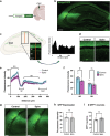
Fig. 2. CA2 innervation of dorsal CA1 is decreased following prenatal spiro.
a Schematic of green fluorescent protein (GFP) expression being driven by the Amigo2 promoter. Amigo2 and GFP are selectively expressed in CA2 of this mouse strain. Image created in Biorender. b Representative image of GFP expression in the murine hippocampus. c To assay CA2 axonal profile, coronal hippocampal sections were immunostained for GFP to enhance endogenous fluorescence. A 10-pixel width linear region-of-interest (ROI) was created through CA1 to capture CA2-axonal GFP signal through the stratum oriens (SO), stratum pyramidale [65], stratum radiatum (SR), and stratum lacunosum-moleculare (SLM) cell layers. Fluorescence intensity values along the line were plotted for the creation of line intensity plot for each animal. Of note, CA2 axonal signal is most dense in SO and SR layers. d Representative images of CA2 axonal signal in CA1 of adult animals from control (left) and spiro-treated litters (right). e Averaged line intensity profile plots for control and spiro-exposed animals. Lines are displayed with cyan and magenta SEM error bars for control and spiro litters, respectively. Control and spiro groups seemed to differ in GFP expression in SO and SR. f The maximum fluorescence intensity value from the SO and SR linear regions were extracted from each animal. We found no main effect for treatment (two-way, rmANOVA, F(1,14) = 3.239, p = 0.0935), but there was a significant interaction between layer and treatment (two-way, rmANOVA, F(1,14) = 8.842, p = 0.0101). Post hoc analysis revealed that maximum fluorescence intensity value differed in SO between control and spiro (Šídák multiple comparisons test, t(28) = 2.555, p = 0.0324), but not in SR (Šídák multiple comparisons test, t(28) = 0.9032, p = 0.6083). g Representative images of GFP immunofluorescence in CA2 of adult animals from control (left panel) and spiro-treated litters (right panel). h Adult, background-subtracted GFP immunofluorescence also did not differ (two-tailed, unpaired t-test t(14) = 0.4979, p = 0.6263). i Similarly, the number of GFP+ cells did not differ across groups (two-tailed, unpaired t-test t(14) = 2.075, p = 0.0569). Scale bars are: b 200 µm; d 50 µm; g 100 µm. Image created with BioRender.com.
Fig. 3
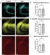
Fig. 3. CA2 marker expression is unaffected by prenatal spiro exposure.
a Representative images of PCP4 immunofluorescence in CA2 of adult animals from control (left panel) and spiro-treated litters (right panel). Scale bars are 100 μm. b Adult PCP4 expression in CA2 did not differ in offspring of control and treated litters (two-tailed, unpaired t-test, t(14) = 0.3903, p = 0.7022). c Representative images of NECAB2 immunofluorescence in CA2 of adult animals from control (left panel) and spiro-treated litters (right panel). Scale bars are 100 μm. d Adult NECAB2 expression in CA2 did not differ in offspring of control and treated litters (two-tailed, unpaired t-test, t(14) = 0.2339, p = 0.8185). e Representative images of MR immunofluorescence in CA2 of adult animals. f MR expression levels did not differ between control (left panel) and spiro-treated litters (right panel) (two-tailed, unpaired t-tests, t(14) = 0.5362, p = 0.6002). All scale bars are 100 µm.
Fig. 4
Fig. 4. Prenatal spiro alters presumed SuM input into CA2 and the dentate gyrus.
a Schematic representation of known SuM projections to CA2 and the DG of the hippocampus. SuM afferents are approximated by vGluT2 immunostaining in these regions, shown in an example in b. c Representative images of vGluT2 staining (red) in CA2 of control (top) and spiro-treated (bottom) mice. d Prenatal exposure to spiro led to decreased  vGluT2 immunostaining in CA2, suggestive of decreased SuM input into this region (two-tailed, unpaired t-test, t(14)  =  3.040, p  =  0.0088). e Representative images of vGluT2 (red) staining in the DG of control (top) and spiro-treated mice. f Spiro exposure led to decreased vGluT2 immunostaining in the DG, also suggestive of decreased SuM input into this region (two-tailed, unpaired t-test with Welch’s correction, t(10.78)  =  4.721, p  =  0.0007). Scale bars are: b 200 µm; c and e 50 µm.
Fig. 5
Fig. 5. Prenatal spiro exposure leads to alterations in some CA2-dependent behaviors.
a Mice were allowed to freely explore a novel open arena (Open Field) for a 10-minute period. Anxiety-like behavior was measured by determining the cumulative time spent in the center of the arena (less time in the center is interpreted as greater anxiety). b The total duration of time spent in center did not differ between control and exposed groups (two-tailed, unpaired t-test, t(50) = 1.274, p = 0.2085). c Animals were tested for novel object reactivity in a familiarized testing arena. d Animals exposed to spiro prenatally spent more time in the interaction zones about novel objects than controls (two-tailed, unpaired t-test, t(50) = 2.295, p = 0.0260). e For a 10-minute period, subject mice were allowed to explore a familiarized three-chamber arena containing one novel conspecific within a cup in one chamber and an empty cup in the opposing chamber, which served as a test for sociability (1). Mice were then introduced to a second novel conspecific contained in the opposing chamber (2). The time spent in the interaction zones over a 10-min duration about the stimuli mice was measured. f Mice in both treatment groups showed a significant preference for the cup with the novel animal over the empty cup. (main effect of chamber: F(1,76) = 59.72, p < 0.0001; each treatment group, p < 0.0001; two-way ANOVA with Sidak’s multiple comparisons test. g The ratio of time spent about the novel mouse to the time spent about the familiar mouse did not differ between control and spiro-treated groups (two-tailed, unpaired t-test, t(35) = 1.216, p = 0.2322).

References

    1. Sapolsky RM, Krey LC, McEwen BS. Glucocorticoid-sensitive hippocampal neurons are involved in terminating the adrenocortical stress response. Proc Natl Acad Sci USA. 1984;81:6174–7. - PMC - PubMed
    1. Diamond DM, Bennett MC, Fleshner M, Rose GM. Inverted-U relationship between the level of peripheral corticosterone and the magnitude of hippocampal primed burst potentiation. Hippocampus. 1992;2:421–30. - PubMed
    1. Oitzl MS, de Kloet ER. Selective corticosteroid antagonists modulate specific aspects of spatial orientation learning. Behav Neurosci. 1992;106:62–71. - PubMed
    1. ter Horst JP, van der Mark MH, Arp M, Berger S, de Kloet ER, Oitzl MS. Stress or no stress: mineralocorticoid receptors in the forebrain regulate behavioral adaptation. Neurobiol Learn Mem. 2012;98:33–40. - PubMed
    1. McEwen BS. Steroid hormones: effect on brain development and function. Horm Res. 1992;37:1–10. - PubMed

MeSH terms

Substances

LinkOut - more resources