. 2024 Sep 5;14(1):20664.
doi: 10.1038/s41598-024-71248-y.

Conditional protein splicing of the Mycobacterium tuberculosis RecA intein in its native host

Affiliations

Conditional protein splicing of the Mycobacterium tuberculosis RecA intein in its native host

Ryan F Schneider et al. Sci Rep. .

Abstract

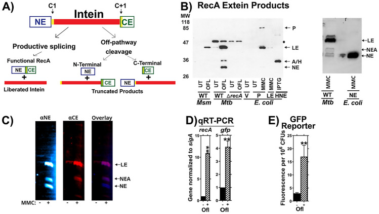
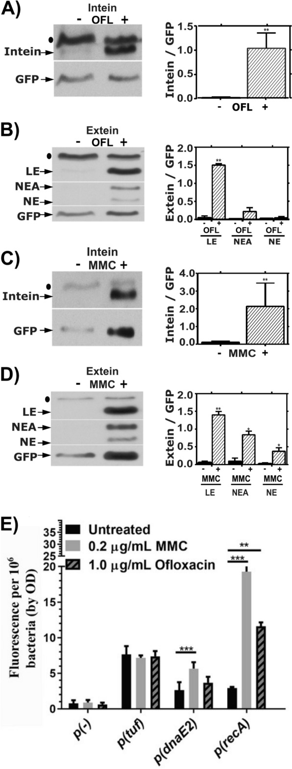
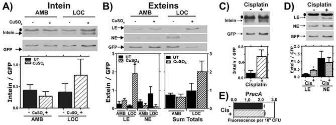
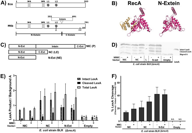
The recA gene, encoding Recombinase A (RecA) is one of three Mycobacterium tuberculosis (Mtb) genes encoding an in-frame intervening protein sequence (intein) that must splice out of precursor host protein to produce functional protein. Ongoing debate about whether inteins function solely as selfish genetic elements or benefit their host cells requires understanding of interplay between inteins and their hosts. We measured environmental effects on native RecA intein splicing within Mtb using a combination of western blots and promoter reporter assays. RecA splicing was stimulated in bacteria exposed to DNA damaging agents or by treatment with copper in hypoxic, but not normoxic, conditions. Spliced RecA was processed by the Mtb proteasome, while free intein was degraded efficiently by other unknown mechanisms. Unspliced precursor protein was not observed within Mtb despite its accumulation during ectopic expression of Mtb recA within E. coli. Surprisingly, Mtb produced free N-extein in some conditions, and ectopic expression of Mtb N-extein activated LexA in E. coli. These results demonstrate that the bacterial environment greatly impacts RecA splicing in Mtb, underscoring the importance of studying intein splicing in native host environments and raising the exciting possibility of intein splicing as a novel regulatory mechanism in Mtb.

Keywords: DNA damage repair; Exaptation; Gene expression; Mobile elements; Post-translational gene regulation; SOS response.


Conflict of interest statement

The authors declare no competing interests.

Figures

Fig. 1
Fig. 1
Validation of the splicing reporter system. (A) Schematic of RecA splicing products. Mtb RecA is translated as a precursor containing an internal 48 kDa RecA intein. The intein is flanked by an N-terminal extein (NE) of 26.5 kDa and a C-terminal extein (CE) of 11 kDa. Splicing depends on the first residues of the intein (cysteine, C1), and the C-extein (cysteine, C + 1). Productive splicing results in precise liberation of the intein from the precursor and allows the N- and C-terminal exteins to be ligated into functional RecA protein (ligated exteins, LE). Alternatively, off-pathway products result in liberation of either the C-extein (C-terminal cleavage) or of the N-extein (N-terminal cleavage). (B) Left: Western blot of whole cell lysates from M. smegmatis (Msm), Mtb H37Rv mc26230 (Mtb) or recombinant E. coli probed with anti- N-extein antibody. RecA splicing was evaluated in response to ofloxacin (OFL), Mitomycin C (MMC), or in untreated cultures (UT) to determine which bands contained splicing or aberrant cleavage products. Msm, which lacks the recA intein sequences, was used as a size marker for LE. ‘WT’ refers to presence of wild-type recA locus in Msm mc2155. E. coli expressing precursor (P), LE, or NE were included for additional reference bands, as marked, and to compare RecA intein splicing activity in a non-native bacterial host with that in Mtb. RecA was examined in Mtb with a wild-type recA locus (WT) or recA deletion (ΔrecA) to identify recA-specific bands. Black dot ● denotes a non-specific background band in Mtb cultures. A/H marks position of RecA-specific band of unknown identity (NEA) and a 6xHis-tagged N-extein protein (HNE) expressed in E. coli BL21 that migrated to similar positions in the gel. Right: E. coli expressing tagless version of NE was used to confirm Mtb NE band. Other abbreviations: Vehicle control (V), Isopropyl β-D-1-thiogalactopyranoside treatment (IPTG). (C) Lysates of MMC-treated Mtb cultures were dual-probed for the (left) NE and (center) CE using fluorescent secondary antibodies. Fluorescent images were overlayed (right) to identify bands cross-reacting bands. LE and NEA bands were visualized with both antibodies. (D) Expression of native recA and recA promoter-driven gfp in mc26230 cultures containing the PrecA:GFP reporter was examined via RT-PCR and (E) GFP fluorescence in response to ofloxacin. **indicates p < 0.01 Panel C: n = 2. Panel D: n = 3.
Fig. 2
Fig. 2
RecA Intein splicing in response to DNA damaging agents. Presence of native excised intein and ligated exteins in Mtb were measured via western blot following treatment with DNA-damaging drugs ofloxacin (Panels A, B) or mitomycin C (Panels C, D). (A) Left: Representative western blot of the intein in response to OFL. Right: quantification of 3 biological repeats, normalized to PrecA:GFP. (B) Left: Representative western blot of LE, NEA and NE in response to OFL. Right: quantification of 3 biological repeats, normalized to PrecA:GFP. (C) Left: Representative western blot of the intein in response to MMC. Right: quantification of 3 biological repeats, normalized to PrecA:GFP. (D) Left: Representative western blot of LE, NEA and NE in response to MMC. Right: quantification of 3 biological repeats, normalized to PrecA:GFP. (E) recA, dnaE2 and tuf transcription in shaking ambient conditions in response to 24 h of treatment with OFL or MMC as read-out by our GFP based reporter assay. ● indicates background bands in the extein or intein western blot. * indicates p < 0.05, **p < 0.01, ***p < 0.001, n = 3 biological replicates for A-E.
Fig. 3
Fig. 3
RecA splicing in response to copper and cisplatin. (A, B) Mtb cultures were grown shaking in ambient (AMB) or hypoxic (LOC) conditions in the presence ( +) or absence (−) of 50 µM copper sulfate (CuSO4) for 7 days. Top: Representative western of intein (A) or LE, and NE products in response to copper treatment in AMB or LOC (B). Bars represent mean and standard deviation. Bottom left: Densitometric quantification of LE and NE from 3 biological western blot replicates. Bottom right: Sum of LE and NE bands from the 3 replicates. Bars represent mean and standard error of the mean. (C, D) Mtb cultures were treated with cisplatin (CIS), and levels of excised intein (C) and LE and NE (D) were examined by western blot and normalized to GFP (top). Densitometric quantification of 3 biological western blot replicates. Bars represent mean and standard deviation (bottom). (E) recA expression in response to cisplatin as read out by our GFP transcriptional reporter assay. ● indicates background bands in the extein or intein western blot, n = 3. * indicates p < 0.05.
Fig. 4
Fig. 4
Stability and degradation of RecA splicing products. (A, B) Mid-log Mtb cultures were treated with Mitomycin C (MMC) for 24 h, then treated with chloramphenicol (CAM) for the indicated times to block new protein synthesis. (A) Western blot analysis of intein levels over time. Top: Representative blot. Bottom: Quantification of 3 biological repeats normalized to GFP. (B) Western blot analysis of ligated extein levels over time. Top: Representative blot. Bottom: Quantification of 3 biological repeats normalized to GFP. (C, D) Mtb cultures were treated ( +) with bortezomib (BTZ) to block proteasome activity or left untreated (−). (C) Western blot analysis of intein levels over time. Top: Representative blot. Bottom: Quantification of 3 biological repeats normalized to GFP. (D) Western blot analysis of ligated extein, LE, NEA, and NE levels after BTZ treatment. Top: Representative blot. Bottom: Quantification of 3 biological repeats normalized to GFP; ** indicates p < 0.01.
Fig. 5
Fig. 5
The N-extein product can activate the SOS response in E. coli. (A) Domain architecture map of (top) E. coli RecA protein and (bottom) Mtb RecA protein. WA and WB indicate the Walker A and B motifs. L1 and L2 (swirls) indicate loop 1 and loop 2. The residue in Mtb RecA that can be phosphorylated is in L2. The pupylation site is marked as pup, and the cyclic-di-AMP binding area is labeled CdA. (B) (left) Previously crystallized Mtb RecA structure from Datta et al. (PDB: 1mo3). The N-extein is colored red, and the C-extein is shaded gold. (right) I-Tasser derived model of folded N-extein colored in red. (C–F) Production of LexA and LexA cleavage in E. coli ∆recA BLR strains complemented with Mtb RecA constructs driven by the native Mtb promoters: NIC—recA including intein; NC—inteinless recA; N-Ext—N-extein producing only. (C) Schematic of the three constructs. (D) Representative western blot probing for LexA. (E) Densitometric analysis of LexA western blots normalized to an unchanging background band (Bkgrnd A). (F) Quantified cleavage of LexA from western blot analysis. N.D. – Not detected. Error bars indicate SEM of 3 biological replicates.

Update of

Similar articles

References

    1. Martin, C. J., Carey, A. F. & Fortune, S. M. A bug’s life in the granuloma. Semin. Immunopathol.38, 213–220. 10.1007/s00281-015-0533-1 (2016). 10.1007/s00281-015-0533-1 - DOI - PMC - PubMed
    1. Singh, A. Guardians of the mycobacterial genome: A review on DNA repair systems in Mycobacterium tuberculosis. Microbiology (Reading)163, 1740–1758. 10.1099/mic.0.000578 (2017). 10.1099/mic.0.000578 - DOI - PubMed
    1. Boshoff, H. I., Reed, M. B., Barry, C. E. 3rd. & Mizrahi, V. DnaE2 polymerase contributes to in vivo survival and the emergence of drug resistance in Mycobacterium tuberculosis. Cell113, 183–193. 10.1016/s0092-8674(03)00270-8 (2003). 10.1016/s0092-8674(03)00270-8 - DOI - PubMed
    1. Stephanou, N. C. et al. Mycobacterial nonhomologous end joining mediates mutagenic repair of chromosomal double-strand DNA breaks. J. Bacteriol.189, 5237–5246. 10.1128/JB.00332-07 (2007). 10.1128/JB.00332-07 - DOI - PMC - PubMed
    1. Sander, P. et al. Mycobacterium bovis BCG recA deletion mutant shows increased susceptibility to DNA-damaging agents but wild-type survival in a mouse infection model. Infect. Immun.69, 3562–3568. 10.1128/IAI.69.6.3562-3568.2001 (2001). 10.1128/IAI.69.6.3562-3568.2001 - DOI - PMC - PubMed

MeSH terms

LinkOut - more resources