. 2024 Sep 6;10(1):207.
doi: 10.1186/s40792-024-02005-6.

An actinomycosis infection resembling peritoneal dissemination of rectal cancer: a case report

Affiliations

An actinomycosis infection resembling peritoneal dissemination of rectal cancer: a case report

Yukiko Fukunaga et al. Surg Case Rep. .

Abstract

Background: Actinomycosis is a suppurative and granulomatous inflammation commonly caused by Actinomyces israelii. Due to its rarity and the paucity of characteristic clinical features, diagnosis of intra-abdominal actinomycosis is challenging, especially when the patient has a treatment history of abdominal cancer.

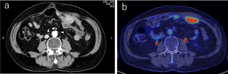
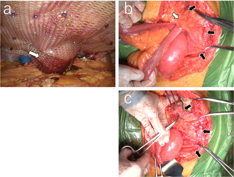
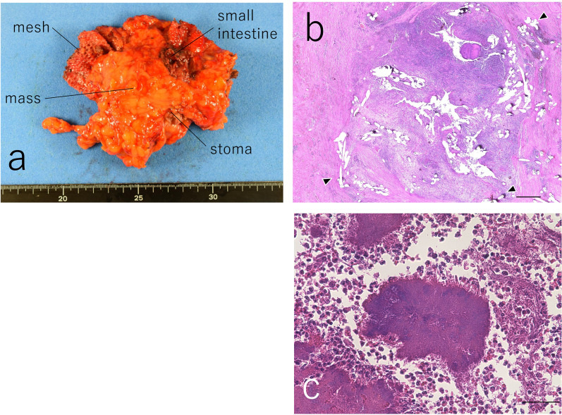
Case presentation: The patient is a 72-year-old man who has a history of multiple abdominal surgeries for rectal cancer, including low anterior resection for primary rectal cancer, partial hepatic resection for metachronous liver metastasis, and Hartmann surgery for local recurrence. The patient has also undergone parastomal hernia repair using the Sugarbaker method. One year after hernia repair, computed tomography (CT) identified a mass lesion between the abdominal wall and the mesh, suggesting the possibility of peritoneal recurrence of rectal cancer. The accumulation of fluorodeoxyglucose (FDG) was evident via positron emission tomography-CT (PET-CT), while tumor marker levels were within the normal range. On laparotomy, the small intestine, abdominal wall, mesh, colon, and stoma were observed to be associated with the mass lesion, and en bloc resection was carried out. However, postoperative histopathological examination revealed an actinomyces infection without any cancerous cells.

Conclusions: This case highlights the challenges faced by surgeons regarding preoperative diagnosis of actinomycosis, especially when it occurs after the resection of abdominal cancer. Also, this case reminds us of the importance of a histopathological examination for abdominal masses or nodules before starting chemotherapy.

Keywords: Abdominal actinomycosis; Hernia; Mesh; Peritoneal dissemination.


Conflict of interest statement

The authors declare that they have no competing interests.

Figures

Fig. 1
Fig. 1
Radiological features of the case. a Abdominal computed tomography (CT) identified a mass near the stoma just below the abdominal wall. b There was no other nodule suggestive of peritoneal dissemination. The abdominal mass was removed along with the stoma, abdominal wall, mesh, and the associated portion of the small intestine
Fig. 2
Fig. 2
Surgical features. a Hernia repair using the Sugarbaker method was performed by placing a mesh on the dorsal side of the sigmoid and fixing it to the abdominal wall around the colostomy (The white arrow indicates the colon behind the mesh). b The small intestine is also involved in the development of lesions. The white arrow indicates the mass lesion covered by adipose tissue (omentum), while the black arrows indicate the mesh. a and b were rotated 180° from the original version). c The mesh (the black arrows) was removed from the abdominal cavity so that the mass lesion could be resected en bloc
Fig. 3
Fig. 3
Macroscopic and microscopic features of the mass lesion. a The removed mass lesion is surrounded by the mesh, small intestine, colostomy, and adipose tissue. b Within the resected granulomatous tissue, fibrous structure of the mesh and multiple small holes (the black arrowheads) created by the mesh were observed. c Amorphous bacterial lesions consisting of hematoxylinophilic hyphae, markedly surrounded by neutrophils, were sparsely identified, occupying approximately 30% of the resected mass. Scale bar equals 50 μm. Images (b) and (c) were taken from different sections

References

    1. Valour F, Sénéchal A, Dupieux C, Karsenty J, Lustig S, Breton P, et al. Actinomycosis: etiology, clinical features, diagnosis, treatment, and management. Infect Drug Resist. 2014;7:183–97. 10.2147/IDR.S39601. 10.2147/IDR.S39601 - DOI - PMC - PubMed
    1. Wong VK, Turmezei TD, Weston VC. Actinomycosis. BMJ. 2011;343: d6099. 10.1136/bmj.d6099. 10.1136/bmj.d6099 - DOI - PubMed
    1. Karateke F, Özyazici S, Menekşe E, Koray DA, Özdoğan M. Unusual presentations of actinomycosis; anterior abdominal wall and appendix: report of three cases. Balk Med J. 2013;30:315–7. 10.5152/balkanmedj.2012.377.10.5152/balkanmedj.2012.377 - DOI - PMC - PubMed
    1. Acquaro P, Tagliabue F, Confalonieri G, Faccioli P, Costa M. Abdominal wall actinomycosis simulating a malignant neoplasm: case report and review of the literature. World J Gastrointest Surg. 2010;2:247–50. 10.4240/wjgs.v2.i7.247. 10.4240/wjgs.v2.i7.247 - DOI - PMC - PubMed
    1. Jabi R, Ramdani H, Elmir S, Elmejjati F, Serji B, El Harroudi T, et al. Pseudotumoral actinomycosis mimicking malignant colic disease: a case report and literature review. Visc Med. 2020;36:333–7. 10.1159/000502895. 10.1159/000502895 - DOI - PMC - PubMed

LinkOut - more resources