. 2024 Sep 5;31(1):87.
doi: 10.1186/s12929-024-01072-z.

Neuroprotective effects of intranasal extracellular vesicles from human platelet concentrates supernatants in traumatic brain injury and Parkinson's disease models

Affiliations

Neuroprotective effects of intranasal extracellular vesicles from human platelet concentrates supernatants in traumatic brain injury and Parkinson's disease models

Liling Delila et al. J Biomed Sci. .

Abstract

Background: The burgeoning field of regenerative medicine has significantly advanced with recent findings on biotherapies using human platelet lysates (HPLs), derived from clinical-grade platelet concentrates (PCs), for treating brain disorders. These developments have opened new translational research avenues to explore the neuroprotective effects of platelet-extracellular vesicles (PEVs). Their potential in managing neurodegenerative conditions like traumatic brain injury (TBI) and Parkinson's disease (PD) warrants further exploration. We aimed here to characterize the composition of a PEV preparation isolated from platelet concentrate (PC) supernatant, and determine its neuroprotective potential and neurorestorative effects in cellular and animal models of TBI and PD.

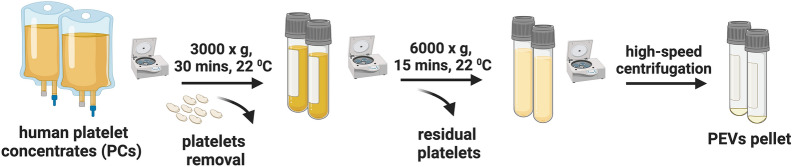
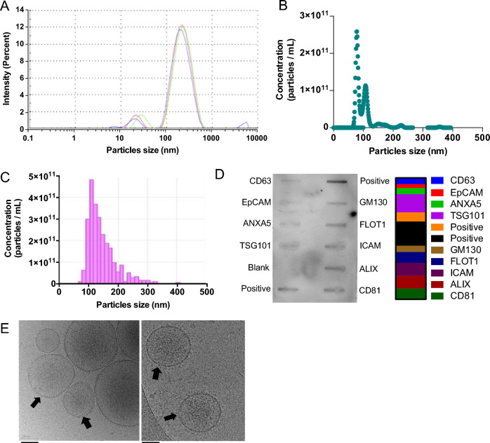
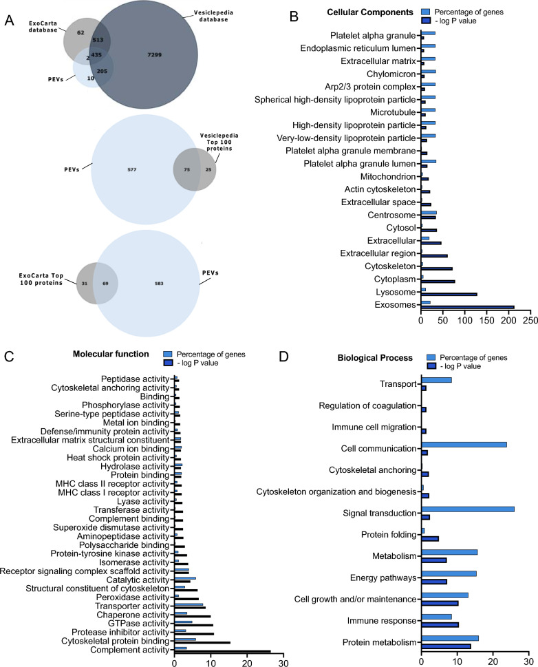
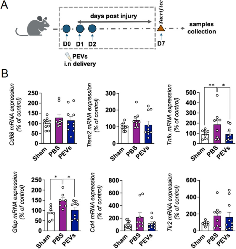
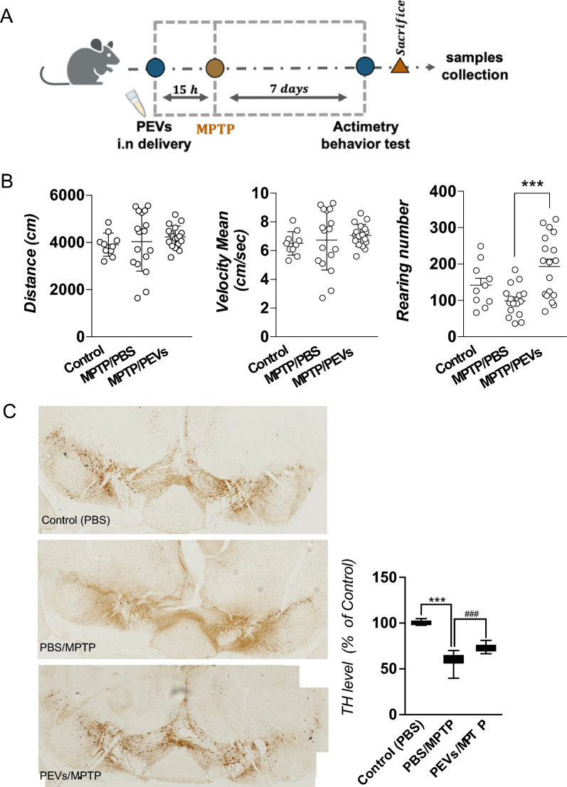
Methods: We isolated PEVs from the supernatant of clinical-grade PC collected from healthy blood donors utilizing high-speed centrifugation. PEVs were characterized by biophysical, biochemical, microscopic, and LC-MS/MS proteomics methods to unveil biological functions. Their functionality was assessed in vitro using SH-SY5Y neuronal cells, LUHMES dopaminergic neurons, and BV-2 microglial cells, and in vivo by intranasal administration in a controlled cortical impact (CCI)-TBI model using 8-weeks-old male C57/BL6 mice, and in a PD model induced by MPTP in 5-month-old male C57/BL6 mice.

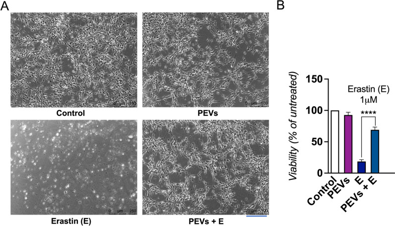
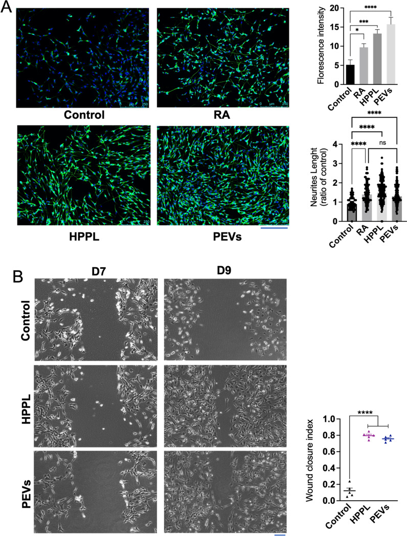
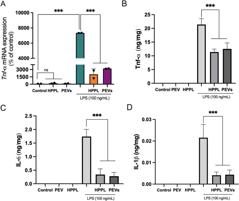
Results: PEVs varied in size from 50 to 350 nm, predominantly around 200 nm, with concentrations ranging between 1010 and 1011/mL. They expressed specific platelet membrane markers, exhibited a lipid bilayer by cryo-electron microscopy and, importantly, showed low expression of pro-coagulant phosphatidylserine. LC-MS/MS indicated a rich composition of trophic factors, including neurotrophins, anti-inflammatory agents, neurotransmitters, and antioxidants, unveiling their multifaceted biological functions. PEVs aided in the restoration of neuronal functions in SH-SY5Y cells and demonstrated remarkable neuroprotective capabilities against erastin-induced ferroptosis in dopaminergic neurons. In microglial cells, they promoted anti-inflammatory responses, particularly under inflammatory conditions. In vivo, intranasally delivered PEVs showed strong anti-inflammatory effects in a TBI mouse model and conserved tyrosine hydroxylase expression of dopaminergic neurons of the substantia nigra in a PD model, leading to improved motor function.

Conclusions: The potential of PEV-based therapies in neuroprotection opens new therapeutic avenues for neurodegenerative disorders. The study advocates for clinical trials to establish the efficacy of PEV-based biotherapies in neuroregenerative medicine.

Keywords: Blood; Central Nervous System; Exosomes; Neurological disorders; Neuroprotection.


Conflict of interest statement

The authors of this manuscript have no conflicts of interest.

Figures

Fig. 1
Fig. 1
Preparation of PEVs. Diagram illustrating the process of preparing PEVs. PC was centrifuged at 3000×g for 30 min to pelletize the platelets and obtain PC supernatant followed by 6000×g for 30 min to remove the residual platelets. PEVs were isolated by high-speed centrifugation at 25,000×g for 90 min and resuspended in PBS. PCs: Platelet concentrates; PEVs: Platelet-extracellular vesicles
Fig. 2
Fig. 2
The bio-physical characterization of PEVs. A The size population profile of PEVs is determined by DLS. B PEVs number and size distribution determined by TRPS. The sample was diluted 10-fold in 0.1 μm-filtered PBS prior to analysis. C PEVs number and size distribution determined by NTA. The sample was diluted 100-fold in 0.1 μm-filtered PBS prior to the analysis. D Images displaying human-specific antibody arrays after incubation with PEVs. 50 μg proteins were loaded into an array. The quantification of the intensity of emitted light corresponding to the presence of bound antibodies to the membrane was displayed (right panel) assessing EVs markers TSG101, ALIX, CD81, FLOT1, CD63, and ANXA5. Cell adhesion protein markers ICAM, EpCAM, and Golgi marker GM130. E Representative CryoEM image displaying a lipid bilayer membrane in PEVs, indicated by a black arrow. Scale bar = 100 nm. PC: Platelet concentrate; PEVs: Platelet-extracellular vesicles
Fig. 3
Fig. 3
Proteomic analysis of PEVs. A Venn diagram showing the number of PEVs proteins that are found in ExoCarta and Vesiclepedia databases and the presence of 72 and 69 PEVs proteins among the top 100 detected in Vesiclepedia and Exo-carta, respectively. B The visualized results of representative functional enrichment used FunRich analytical tool to of the 652 PEVs proteins on the cellular component C Molecular function and D Biological processes. Each bar is proportional to the percentage of genes. The significance cutoff was p < 0.5. PEVs: Platelet-extracellular vesicles
Fig. 4
Fig. 4
The effect of PEVs treatment on the viability of LUHMES cells exposed to erastin. A LUHMES were treated with PEVs and subsequently, a dose of 1 µM neurotoxin erastin was added. Images showing the cell morphology after 24 h. The scale bar is 250 μm. B LUHMES cell viability after 24 h was quantified by a CCK-8 assay. LUHMES were exposed or not to erastin. N = 3, ****p < 0.0001 as compared to the control with erastin, a ferroptosis promoter. PEVs: Platelet-extracellular vesicles; E: Erastin
Fig. 5
Fig. 5
Functional activity of PEVs to promote neuronal growth in vitro on SH-SY5Y neuroblastoma cell line. A Capacity of PEVs to stimulate the neuronal maturation of SH-SY5Y cells. Cells were immuno-stained with β-III tubulin and counterstained with DAPI. Images showing extension of SH-SY5Y neurites under the treatment of PEVs and HPPL. HPPL and RA were used as positive controls to stimulate cell differentiation. Scale bar = 250 μm. Quantitative measurement of the fluorescence intensity (right) showed the capacity of PEVs to induce SH-SY5Y neuronal maturation. N = 3, *p < 0.05; ***p < 0.001 compared to the untreated negative control. From the captured images of β-III tubulin fluorescence, the length of each extension was measured individually to estimate total neurite outgrowth. The ratio of neurite length in treated cells to that in untreated cells was then calculated. N = 3, significant difference (ns), ****p < 0.001 compared to the untreated negative control. B Neuro-restoration effect of PEVs on the differentiated SH-5YSY. A scratch assay was performed using differentiated SH-SY5Y cells. Cells without any treatment (negative control), HPPL (positive control), and PEVs were used. The neuro-restoration effect was monitored by microscopy two days after the treatments on Day 9 (D9). Scale bar = 100 μm. The results are expressed as a wound-healing index, determined by the formula: (initial wound area − final wound area)/initial wound area). N = 3, ****p < 0.001 compared to untreated negative control. RA: Retinoic acid; PEVs: Platelet-extracellular vesicles; HPPL: heat-treated platelet pellet lysate
Fig. 6
Fig. 6
Anti-inflammatory activity of PEVs on activated BV-2 microglia cells. LPS was added to induce BV-2 cell activation 1 h before PEVs and HPPL (used as non-inflammatory control) treatments. The Tnf-α level was measured from RNA isolated from the cell lysate (pooled N = 2). TNF-α, IL-6 and IL-1β protein expression were quantified in the cell supernatant by ELISA (N = 3). The results showed that Tnf-α gene expression, TNF-α, IL-6, and IL-1β proteins were increased in BV-2 microglia cells exposed to 100 ng/ml LPS treatment, and a significantly lower expression was observed after 24 h of PEVs or HPPL treatments. ***p < 0.001 as compared to the control with LPS. ns: no significant difference; LPS: Escherichia coli lipopolysaccharide; PEVs: Platelet-extracellular vesicles; HPPL: heat-treated platelet pellet lysate
Fig. 7
Fig. 7
Anti-inflammatory activity of PEVs in CCI in vivo model of TBI. A Diagram illustrating the CCI-TBI mice to evaluate the anti-inflammatory effect of PEVs treatment. Mild CCI-TBI was applied on the left hemisphere of the mice brain followed by PEVs treatment administered intranasally (concentration 60 µL or 1.2 × 108 on 3 consecutive days), and PBS was used as a control. On day 7, mice were sacrificed and the cortex injury part was collected for further gene expression analysis. B Effect of PEVs treatment on the expressions of inflammatory markers post injury. Changes in cytokines and glial markers expression in the cortex at day 7 post-injury by CCI. The inflammatory markers such as Cd68, Trem-2, Tnf-α, Gfap, Ccl4, and Tlr2 were assessed. The results showed the upregulation of Tnf-α, Cd68, Gfap, and Trem2 induced by the cortical impact and a significant suppression of Gfap and Tnf-α expression following PEVs treatment. Data were presented as means ± SEM (n = 7–10 in each group). *p < 0.05, **p < 0.01. CCI: Controlled cortical impact; TBI: Traumatic brain injury; PBS: Phosphate-buffered saline; PEVs: Platelet-extracellular vesicles
Fig. 8
Fig. 8
Neuroprotective effect of PEVs in MPTP mice model of PD. A Diagram illustrating MPTP mice to evaluate the neuroprotective effect of PEVs treatment. Approximately 15 h prior to the induction of MPTP intoxication (acute dose, 20 mg/kg MPTP injected intraperitoneally), intranasal (i.n) delivery of PEVs was administered at a dose of 4 × 1010. Some mice received saline as a control (sham group). On the seventh day, the open-field behaviour test was performed. After, the mice were sacrificed, and their brains were collected for IHC analysis. B Impact of the PEVs treatment on the behaviour of MPTP mice. Animals were left in the open field actimetry box and allowed to explore freely for 10 min. The test was done on day 7. Parameters related to their locomotor function were recorded. PEVs given by i.n. delivery prior to MPTP intoxication improved the rearing number compared to the control MPTP group (MPTP/PBS). C Effect of PEVs treatment on the TH positive cells in SN of MPTP mice. On the left, a representative images of TH area counted in the SN area compared between the control/PBS group and MPTP groups either receiving PBS (MPTP/PBS) or PEVs (MPTP/PEVs). Total TH number in the subsequent area of SN (7 sections collected between bregma – 2.92 mm and – 3.88 mm). The TH counts in each section were summed for each animal. The treatment by PEVs was able to protect TH expression. Data presented as means ± SD, n = 7 in the sham group (PBS only), and at least n = 17 in the MPTP groups. ***p < 0.001 compared to PBS group, ###p < 0.001 compared to MPTP/PBS group. MPTP: 1-methyl-4-phenyl-1,2,3,6-tetrahydropyridine; PBS: Phosphate-buffered saline; PEVs: Platelet-extracellular vesicles; SN: Substantia nigra; TH: Tyrosine hydroxylase

References

    1. Nebie O, et al. Can the administration of platelet lysates to the brain help treat neurological disorders? Cell Mol Life Sci. 2022;79(7):379. 10.1007/s00018-022-04397-w - DOI - PMC - PubMed
    1. Hayon Y, et al. Platelet lysates stimulate angiogenesis, neurogenesis and neuroprotection after stroke. Thromb Haemost. 2013;110(2):323–30. 10.1160/TH12-11-0875 - DOI - PubMed
    1. Anitua E, et al. Intranasal PRGF-Endoret enhances neuronal survival and attenuates NF-kappaB-dependent inflammation process in a mouse model of Parkinson’s disease. J Control Release. 2015;203:170–80. 10.1016/j.jconrel.2015.02.030 - DOI - PubMed
    1. Gouel F, et al. The protective effect of human platelet lysate in models of neurodegenerative disease: involvement of the Akt and MEK pathways. J Tissue Eng Regen Med. 2017;11(11):3236–40. 10.1002/term.2222 - DOI - PubMed
    1. Anitua E, et al. Plasma rich in growth factors (PRGF-Endoret) reduces neuropathologic hallmarks and improves cognitive functions in an Alzheimer’s disease mouse model. Neurobiol Aging. 2014;35(7):1582–95. 10.1016/j.neurobiolaging.2014.01.009 - DOI - PubMed

Substances