

Radiomics of multi-parametric MRI for the prediction of lung metastasis in soft-tissue sarcoma: a feasibility study
- PMID: 39238054
- PMCID: PMC11376009
- DOI: 10.1186/s40644-024-00766-9
Radiomics of multi-parametric MRI for the prediction of lung metastasis in soft-tissue sarcoma: a feasibility study
Abstract
Purpose: To investigate the value of multi-parametric MRI-based radiomics for preoperative prediction of lung metastases from soft tissue sarcoma (STS).
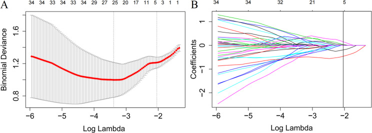
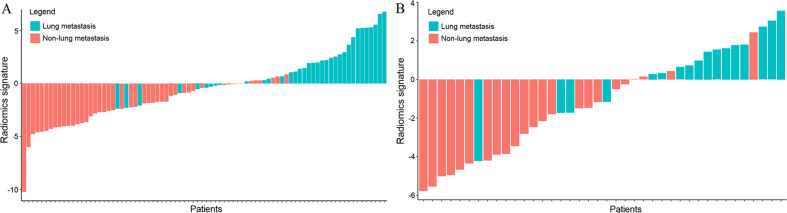
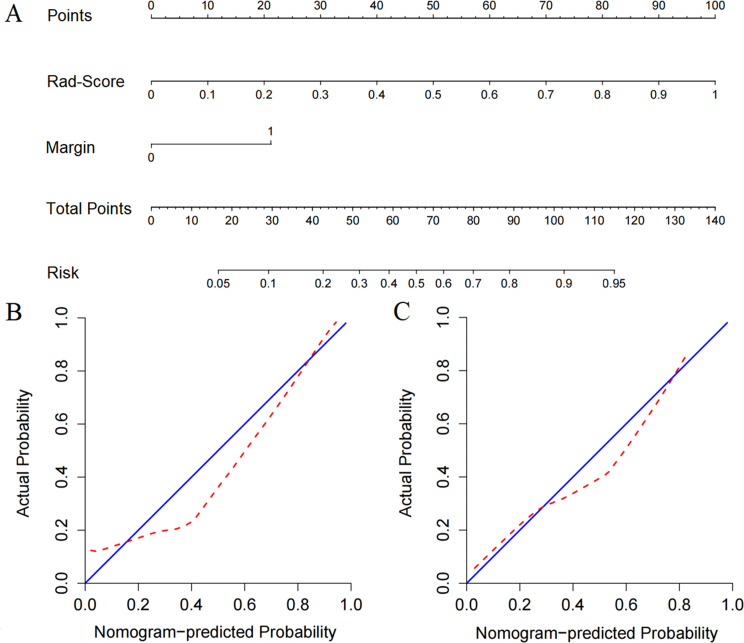
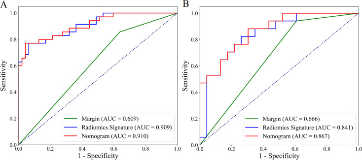
Methods: In total, 122 patients with clinicopathologically confirmed STS who underwent pretreatment T1-weighted contrast-enhanced (T1-CE) and T2-weighted fat-suppressed (T2FS) MRI scans were enrolled between Jul. 2017 and Mar. 2021. Radiomics signatures were established by calculating and selecting radiomics features from the two sequences. Clinical independent predictors were evaluated by statistical analysis. The radiomics nomogram was constructed from margin and radiomics features by multivariable logistic regression. Finally, the study used receiver operating characteristic (ROC) and calibration curves to evaluate performance of radiomics models. Decision curve analyses (DCA) were performed to evaluate clinical usefulness of the models.
Results: The margin was considered as an independent predictor (p < 0.05). A total of 4 MRI features were selected and used to develop the radiomics signature. By incorporating the margin and radiomics signature, the developed nomogram showed the best prediction performance in the training (AUCs, margin vs. radiomics signature vs. nomogram, 0.609 vs. 0.909 vs. 0.910) and validation (AUCs, margin vs. radiomics signature vs. nomogram, 0.666 vs. 0.841 vs. 0.894) cohorts. DCA indicated potential usefulness of the nomogram model.
Conclusions: This feasibility study evaluated predictive values of multi-parametric MRI for the prediction of lung metastasis, and proposed a nomogram model to potentially facilitate the individualized treatment decision-making for STSs.
Keywords: Lung metastasis; MRI; Nomogram; Soft-tissue sarcoma.
© 2024. The Author(s).
Conflict of interest statement
The authors declare that they have no competing interests.
Figures

Similar articles
-
A contrast-enhanced MRI-based nomogram to identify lung metastasis in soft-tissue sarcoma: A multi-centre study.Med Phys. 2023 May;50(5):2961-2970. doi: 10.1002/mp.16136. Epub 2022 Dec 7. Med Phys. 2023. PMID: 36433749
-
Magnetic Resonance Imaging-Based Radiomics Nomogram for Prediction of the Histopathological Grade of Soft Tissue Sarcomas: A Two-Center Study.J Magn Reson Imaging. 2021 Jun;53(6):1683-1696. doi: 10.1002/jmri.27532. Epub 2021 Feb 18. J Magn Reson Imaging. 2021. PMID: 33604955
-
Multi-parametric MRI-based radiomics for the diagnosis of malignant soft-tissue tumor.Magn Reson Imaging. 2022 Sep;91:91-99. doi: 10.1016/j.mri.2022.05.003. Epub 2022 May 4. Magn Reson Imaging. 2022. PMID: 35525523
-
Multi-institutional validation of a radiomics signature for identification of postoperative progression of soft tissue sarcoma.Cancer Imaging. 2024 May 8;24(1):59. doi: 10.1186/s40644-024-00705-8. Cancer Imaging. 2024. PMID: 38720384 Free PMC article.
-
Diagnostic Value of Magnetic Resonance Imaging Radiomics and Machine-learning in Grading Soft Tissue Sarcoma: A Mini-review on the Current State.Acad Radiol. 2025 Jan;32(1):311-315. doi: 10.1016/j.acra.2024.08.035. Epub 2024 Sep 10. Acad Radiol. 2025. PMID: 39261231 Review.
References
MeSH terms
Grants and funding
- 31770147/China National Natural Science Foundation
- 201909J033/Project of Pneumoconiosis Prevention and Control of China's coal mines foundation
- 2021-MS-205/Natural Science Foundation of Liaoning Province
- LD202029/Medical-Engineering Joint Fund for Cancer Hospital of China Medical University and Dalian University of technology
- LQNK201744/Education Department Foundation of Liaoning
LinkOut - more resources
Full Text Sources
Medical
