. 2024 Sep;12(17):e70038.
doi: 10.14814/phy2.70038.

Osteopontin deletion attenuates cyst growth but exacerbates fibrosis in mice with cystic kidney disease

Affiliations

Osteopontin deletion attenuates cyst growth but exacerbates fibrosis in mice with cystic kidney disease

Kyle P Jansson et al. Physiol Rep. 2024 Sep.

Abstract

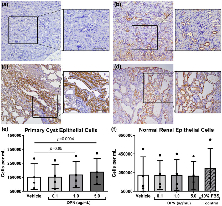
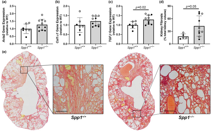
Osteopontin (OPN) is a multi-functional glycoprotein that coordinates the innate immune response, prevents nanocrystal formation in renal tubule fluid, and is a biomarker for kidney injury. OPN expression is markedly increased in cystic epithelial cells of polycystic kidney disease (PKD) kidneys; however, its role in PKD progression remains unclear. We investigated the in vitro effects of recombinant OPN on the proliferation of tubular epithelial cells from PKD and normal human kidneys and in vivo effects of OPN deletion on kidney cyst formation, fibrosis, and mineral metabolism in pcy/pcy mice, a non-orthologous model of autosomal-dominant PKD. In vitro studies revealed that OPN enhanced the proliferation of PKD cells but had no effect on normal kidney cells. Deletion of OPN in pcy/pcy mice significantly reduced kidney cyst burden; however, this was accompanied by increased fibrosis and no change in kidney function. The loss of OPN had no effect on kidney macrophage numbers, cyst epithelial cell proliferation, or apoptosis. Furthermore, there was no difference in kidney mineral deposition or mineral metabolism parameters between pcy/pcy mice with and without OPN expression. Global deletion of OPN reduced kidney cyst burden, while paradoxically exacerbating kidney fibrosis in mice with cystic kidney disease.

Keywords: Osteopontin; PKD; fibrosis; matricellular proteins; mineral metabolism.


Conflict of interest statement

DPW has received research funding from Synkine Therapeutics. All other authors have nothing to disclose.

Figures

FIGURE 1
FIGURE 1
Osteopontin (OPN) expression is increased in kidneys from mice with cystic kidney disease and stimulates cyst epithelial cell proliferation in vitro. Immunohistochemistry (IHC) of OPN protein expression (brown) in kidneys from (a) mice with Spp1 (OPN) gene deletion, (b) wild‐type controls, (c) pcy/pcy mice (non‐orthologous PKD model), and (d) RC/RC mice (orthologous PKD model) (enlarged inset is 20× magnification; scale bar = 100 μm). Further in vitro experiments were performed to test the effect of increasing concentrations of OPN on cellular proliferation in (e) human primary cyst epithelial cells and (f) primary human tubular epithelial cells from normal kidneys (analyzed by 1‐way paired ANOVA).
FIGURE 2
FIGURE 2
Kidney cyst burden is reduced in pcy/pcy mice exhibiting OPN (Spp1) deletion. (a) Gross tissue evaluation of kidneys from pcy/pcy mice with and without Spp1 expression. (b, c) H&E staining of kidney sections to assess cystic index in study mice (magnified images taken at 10x; error bar = 500 μm). Additional kidney related outcomes were conducted, including (d) two‐kidney to total body weight ratio (KW/BW), (e) BUN, and (f) serum creatinine (analyzed by Student's t‐test; closed circles = males, open circles = females).
FIGURE 3
FIGURE 3
Kidney fibrosis is increased in pcy/pcy mice in the absence of OPN. Quantitative RT‐PCR analysis to evaluate gene expression for key fibrosis mediators demonstrated increased kidney expression of genes encoding (a) α‐smooth muscle Actin (Acta2), (b) α1 subunit of type I collagen (Col1α1), and (c) transforming growth factor β (TGFβ). (d, e) Quantification of fibrosis by picrosirius red staining of kidney histology sections from pcy/pcy mice with and without OPN (Spp1) expression (analyzed by Student's t‐test; closed circles = males, open circles = females).
FIGURE 4
FIGURE 4
Osteopontin deficiency has minimal effect on mineral metabolism parameters in pcy/pcy mice. Serum measurements of mineral metabolism parameters were conducted, including (a) phosphorus, (b) calcium, (c) parathyroid hormone (PTH), and (d) intact fibroblast growth factor 23 (FGF23). (e, f) Evaluation of kidney mineral deposition revealed only sparse kidney mineral deposits in both groups. (g) Von Kossa staining of kidney sections showing mineral deposits (red arrows) attached to epithelial surfaces within cysts. (h, i) Evaluation of bone mineral content in femurs from study mice showed evidence of cortical bone porosity (black arrows) in both groups (analyzed by Student's t‐test; closed circles = males, open circles = females).
FIGURE 5
FIGURE 5
Osteopontin deficiency does not alter kidney macrophage numbers in pcy/pcy mice. Immunohistochemistry staining of kidney sections for macrophage marker CD68 in pcy/pcy mice (a) with and (b) without OPN expression (scale bar = 100 um), along with (c) subsequent quantification of CD68‐positive cells from these sections (data expressed as positive cells per kidney section). (d) Assessment of kidney gene expression by qRT‐PCR for Adgre1 (F4/80), an alternative macrophage marker (closed circles = males, open circles = females).
FIGURE 6
FIGURE 6
Kidney fibrosis is an early phenotypic finding in pcy/pcy mice with OPN deletion. Evaluation of kidney disease parameters at an earlier time point (20 weeks‐of‐age) in pcy/pcy mice with and without OPN expression, including (a, b) cyst burden by histology, (c, d) quantification of biochemical markers of kidney function, (e, f) Ki‐67 staining to assess proliferation of cyst‐lining epithelial cells, (g, h) TUNEL‐staining to quantify apoptosis of cyst lining cells (data expressed as positive cells per kidney section for both Ki‐67 and TUNEL stains), and (i, j) picrosirius red staining to evaluate kidney fibrosis (analyzed by Student's t‐test; closed circles = males, open circles = females).

Similar articles

Cited by

  • Microvascular aberrations found in human polycystic kidneys are an early feature in a Pkd1 mutant mouse model.
    Jafree DJ, Perera C, Ball M, Tolomeo D, Pomeranz G, Wilson L, Davis B, Mason WJ, Funk EM, Kolatsi-Joannou M, Polschi R, Malik S, Stewart BJ, Price KL, Mitchell H, Motallebzadeh R, Muto Y, Lees R, Needham S, Moulding D, Chandler JC, Nandanwar S, Walsh CL, Winyard PJD, Scambler PJ, Hägerling R, Clatworthy MR, Humphreys BD, Lythgoe MF, Walker-Samuel S, Woolf AS, Long DA. Jafree DJ, et al. Dis Model Mech. 2025 Apr 1;18(4):dmm052024. doi: 10.1242/dmm.052024. Epub 2025 Apr 28. Dis Model Mech. 2025. PMID: 40114603 Free PMC article.

References

    1. Anders, H. J. , Suarez‐Alvarez, B. , Grigorescu, M. , Foresto‐Neto, O. , Steiger, S. , Desai, J. , Marschner, J. A. , Honarpisheh, M. , Shi, C. , Jordan, J. , Muller, L. , Burzlaff, N. , Bauerle, T. , & Mulay, S. R. (2018). The macrophage phenotype and inflammasome component NLRP3 contributes to nephrocalcinosis‐related chronic kidney disease independent from IL‐1‐mediated tissue injury. Kidney International, 93, 656–669. - PubMed
    1. Bank N , Su, W. S. , & Aynedjian, H. S. (1978). A micropuncture study of renal phosphate transport in rats with chronic renal failure and secondary hyperparathyroidism. The Journal of Clinical Investigation, 61, 884–894. - PMC - PubMed
    1. Cowley, B. D., Jr. , Ricardo, S. D. , Nagao, S. , & Diamond, J. R. (2001). Increased renal expression of monocyte chemoattractant protein‐1 and osteopontin in ADPKD in rats. Kidney International, 60, 2087–2096. - PubMed
    1. Cui, R. , Takahashi, F. , Ohashi, R. , Gu, T. , Yoshioka, M. , Nishio, K. , Ohe, Y. , Tominaga, S. , Takagi, Y. , Sasaki, S. , Fukuchi, Y. , & Takahashi, K. (2007). Abrogation of the interaction between osteopontin and alphavbeta3 integrin reduces tumor growth of human lung cancer cells in mice. Lung Cancer, 57, 302–310. - PubMed
    1. Denhardt, D. T. , Noda, M. , O'Regan, A. W. , Pavlin, D. , & Berman, J. S. (2001). Osteopontin as a means to cope with environmental insults: Regulation of inflammation, tissue remodeling, and cell survival. The Journal of Clinical Investigation, 107, 1055–1061. - PMC - PubMed

MeSH terms

LinkOut - more resources