. 2024 Oct 1;18(39):26733-26750.
doi: 10.1021/acsnano.4c06921. Epub 2024 Sep 5.

Hydrogel Microneedle Patches Loaded with Stem Cell Mitochondria-Enriched Microvesicles Boost the Chronic Wound Healing

Affiliations

Hydrogel Microneedle Patches Loaded with Stem Cell Mitochondria-Enriched Microvesicles Boost the Chronic Wound Healing

Wen-De Yao et al. ACS Nano. .

Abstract

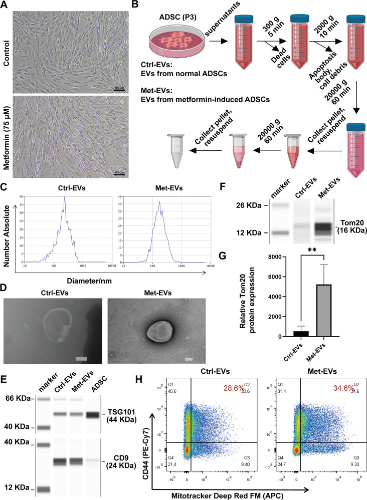
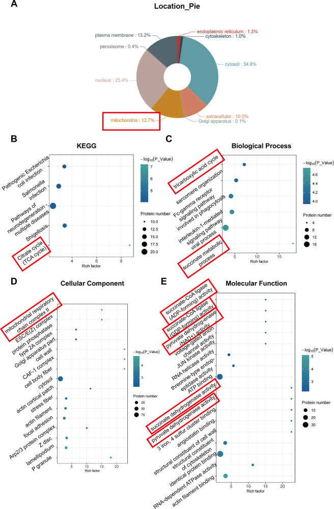
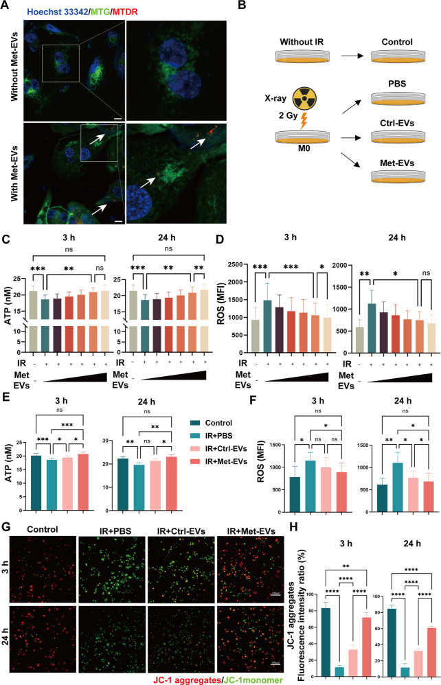
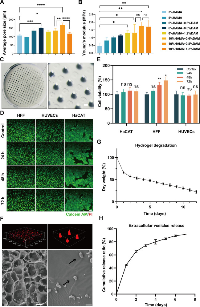
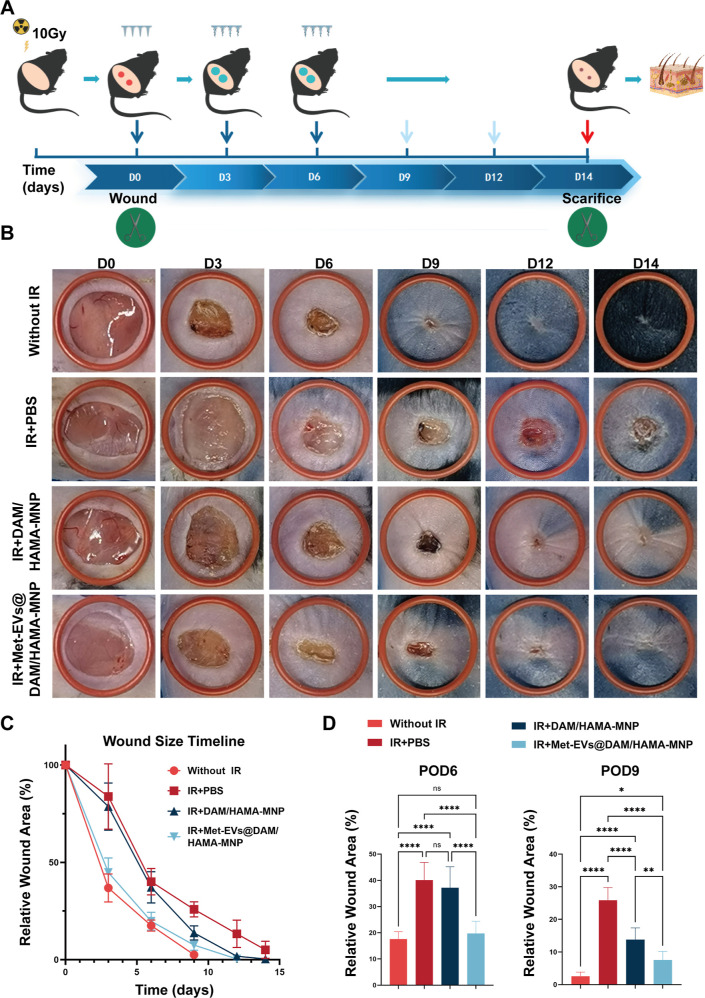
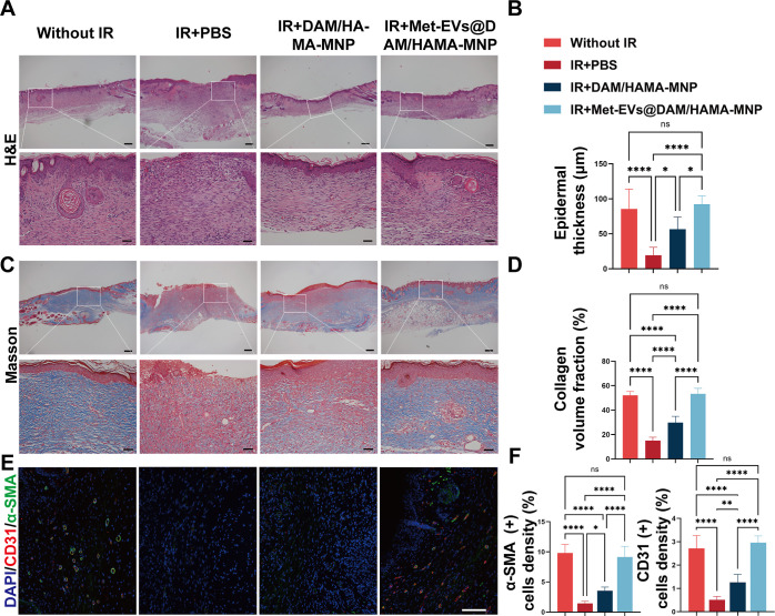
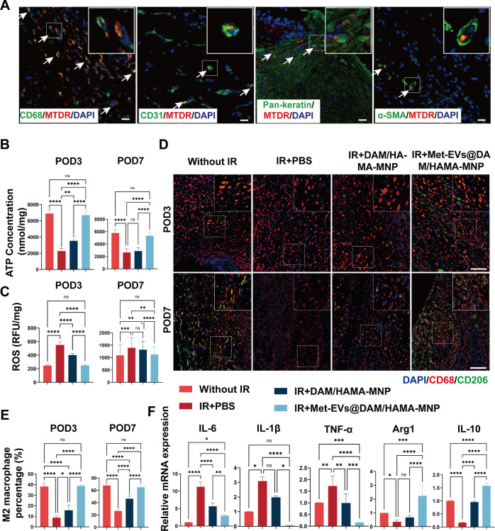
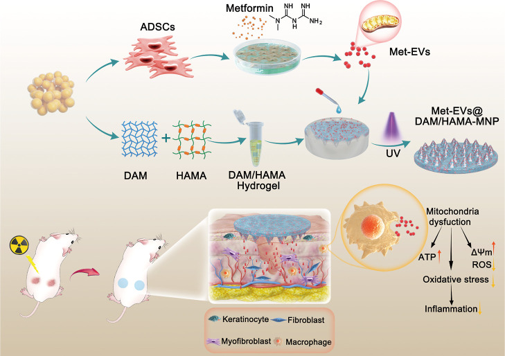
Rescuing or compensating mitochondrial function represents a promising therapeutic avenue for radiation-induced chronic wounds. Adult stem cell efficacies are primarily dependent on the paracrine secretion of mitochondria-containing extracellular vesicles (EVs). However, effective therapeutic strategies addressing the quantity of mitochondria and mitochondria-delivery system are lacking. Thus, in this study, we aimed to design an effective hydrogel microneedle patch (MNP) loaded with stem cell-derived mitochondria-rich EVs to gradually release and deliver mitochondria into the wound tissues and boost wound healing. We, first, used metformin to enhance mitochondrial biogenesis and thereby increasing the secretion of mitochondria-containing EVs (termed "Met-EVs") in adipose-derived stem cells. To verify the therapeutic effects of Met-EVs, we established an in vitro and an in vivo model of X-ray-induced mitochondrial dysfunction. The Met-EVs ameliorated the mitochondrial dysfunction by rescuing mitochondrial membrane potential, increasing adenosine 5'-triphosphate levels, and decreasing reactive oxygen species production by transferring active mitochondria. To sustain the release of EVs into damaged tissues, we constructed a Met-EVs@Decellularized Adipose Matrix (DAM)/Hyaluronic Acid Methacrylic Acid (HAMA)-MNP. Met-EVs@DAM/HAMA-MNP can load and gradually release Met-EVs and their contained mitochondria into wound tissues to alleviate mitochondrial dysfunction. Moreover, we found Met-EVs@DAM/HAMA-MNP can markedly promote macrophage polarization toward the M2 subtype with anti-inflammatory and regenerative functions, which can, in turn, enhance the healing process in mice with skin wounds combined radiation injuries. Collectively, we successfully fabricated a delivery system for EVs, Met-EVs@DAM/HAMA-MNP, to effectively deliver stem cell-derived mitochondria-rich EVs. The effectiveness of this system has been demonstrated, holding great potential for chronic wound treatments in clinic.

Keywords: hydrogel microneedle patch; metformin; mitochondria-enriched extracellular vesicle; radiation-induced injury; stem cell.


Conflict of interest statement

The authors declare no competing financial interest.

Figures

Figure 1
Figure 1
Fabrication and characterization of ADSC-EVs. (A) Morphology of control ADSCs (above) and metformin-treated ADSCs (below) by light microscopy. Scale bar, 100 μm. (B) Flowchart of the isolation of EVs. (C) Nanoparticle tracking analysis (NTA) of the range of particle size distribution of Ctrl-versus Met-EVs. (D) Transmission electron microscopy (TEM) images of Ctrl- and Met-EVs. Scale bar, 100 nm. (E) CD9 and TSG101 expression in Ctrl- and Met-EVs and ADSCs was assessed using capillary immunoassays. (F) Tom20 expression levels in Ctrl- and Met-EVs were evaluated by capillary immunoassays. (G) Capillary immunoassays detected a significant difference in Tom20 expression between Ctrl- and Met-EVs (n = 4). (H) Flow cytometric analysis of CD44+MTDR+ EVs in Ctrl-EVs (28.6%) and Met-EVs (34.6%). **p < 0.01. EVs, extracellular vesicles; Met, metformin; Ctrl, control; ADSCs, adipose-derived stem cells.
Figure 2
Figure 2
Enrichment analysis of differential proteins between Met- and Ctrl-EVs. (A) Analysis of subcellular localization of differentially expressed proteins. (B) Kyoto Encyclopedia of Genes and Genomes (KEGG) pathway enrichment analysis of the upregulated proteins. (C–E) Gene Ontology (GO) classification of the upregulated proteins in terms of biological processes, cellular components, and molecular functions. Met, metformin; Ctrl, control; EVs, extracellular vesicles; KEGG, Kyoto Encyclopedia of Genes and Genomes; GO, Gene Ontology.
Figure 3
Figure 3
Met-EVs ameliorate X-ray-induced mitochondrial dysfunction in macrophages. (A) Representative microscopic images of MTDR-labeled exogenous mitochondria (red) in macrophages after treatment with Met-EVs. Mitochondria (indicated by white arrows) within the transferred Met-EVs were observed in recipient macrophages prelabeled with MitoTracker Green FM (MTG, green). Scale bar, 10 μm. (B) Schematic diagram of the experimental groups set up for comparison with the groups Without IR, IR + phosphate-buffered saline, PBS, IR + Ctrl-EVs, and IR + Met-EVs. (C,D) ATP and ROS levels in X-ray-injured macrophages treated with Met-EVs. (0, 3.0 × 10⁶, 1.0 × 10⁷, 3.0 × 10⁷, 108 or 3.0 × 10⁸ particles/mL: wedges). Left: 3 h post-treatment. Right, 24 h post-treatment. (E,F) ATP and ROS levels in X-ray-injured macrophages treated with Met- and Ctrl-EVs at a concentration of 108 particles/mL. Left, 3 h post-treatment. Right, 24 h post-treatment. (G) Representative images of MMP determined using a JC-1 kit (JC-1 aggregates, red, bioactive mitochondria; JC-1 monomer, green, impaired mitochondria) in X-ray-injured macrophages treated with Met- or Ctrl-EVs at a concentration of 108 particles/mL. Top panel, 3 h post-treatment; bottom panel, 24 h post-treatment. Scale bars,100 μm. (H) Ratio of JC-1 aggregates (red) to the total intensity of red and green fluorescence (%). Both fluorescence intensities (red and green) were analyzed using ImageJ software. n = 4–9, in each group, ns p > 0.05, *p < 0.05, **p < 0.01, ***p < 0.001, and ****p < 0.0001. Met, metformin; MTDR, MitoTracker Deep Red; EVs, extracellular vesicles; Ctrl, control; IR, irradiation; PBS, phosphate-buffered saline.
Figure 4
Figure 4
Preparation and characterization of Met-EVs@DAM/HAMA-MNP. (A) Mean pore size of each hydrogel group at different concentration ratios (n = 3). (B) Compression moduli of each group of hydrogels at different concentrations (n = 3). (C) Stereoscopic DAM/HAMA-MNP images. The diameters of MNP tip spacing, height, and base were 700, 500, and 270 μm, respectively. Scale bar, 100 μm. Live and dead cell assays (D) and CCK-8 assays for cell viability (E) of HFF, HUVECs, and HaCAT cells treated with liquid DAM/HAMA hydrogel extracts produced at indicated time points (0, 24-, 48-, 72 h). The cells were labeled with Calcein AM (green) and PI (red) in (D). Scale bars in (D), 100 μm. (F) Top left and right: confocal laser scanning microscopy (CLSM) scanning for 3D reconstruction of the images of MNP loaded with Dil-labeled EVs (red); bottom left and bottom right: scanning electron microscopy (SEM) images of DAM/HAMA-MNPs loaded with EVs, indicated by black arrows. Scale bar, bottom left 100 μm, bottom right 1 μm. (G) In vitro degradation curve of the DAM/HAMA hydrogel (n = 3). (H) EV release curve of Met-EVs@DAM/HAMA analyzed using the bicinchoninic acid (BCA) assay (n = 5). ns, p > 0.05, *p < 0.05, **p < 0.01, ***p < 0.001, and ****p < 0.0001. Met, metformin; EVs, extracellular vesicles; DAM, decellularized adipose matrix; HAMA, hyaluronic acid methacrylic acid; MNP, microneedle patch; HFF, human foreskin fibroblast; HUVECs, human umbilical vein endothelial cells; HaCAT, human epidermal keratinocytes; CLSM, confocal laser scanning microscopy.
Figure 5
Figure 5
Met-EVs@DAM/HAMA-MNP treatment for radiation combined with skin wounds in mice. (A) Schematic of radiation combined with skin wound model and treatment strategy. (B) Representative graphs of wound healing at each stage in different groups and images of treated wounds on POD 0, 3, 6, 9, 12, and 14. (The red rubber ring has an inner diameter of 14 mm.) (C) Healing curves of the dorsal wounds of mice in each group. The healing curves reflected the relative wound area rate at each stage of the postoperative period (n = 8). (D) Statistical analysis of the relative wound areas in each group on POD6 and 9 (n = 8). ns p > 0.05, *p < 0.05, **p < 0.01, and ****p < 0.0001. Met, metformin; EVs, extracellular vesicles; DAM, decellularized adipose matrix; HAMA, hyaluronic acid methacrylic acid; MNP, microneedle patch.
Figure 6
Figure 6
Histological analysis of healed wounds in mice on POD14. (A) Representative images of H&E staining of wound tissues and comparison of wound tissues epithelial thickness (B) on POD14 between each group (n = 6). (C) Masson’s trichrome staining on POD14 showed that the IR + PBS group still had a wound with no epidermal overlying tissue and significantly less collagen deposition than those in the other groups. (D) Comparison of collagen deposition fraction on POD14 between each group (n = 6). (E,F) Immunofluorescence staining of wound tissue sections on POD14 by labeling CD31 (red) and α-smooth muscle actin (α-SMA) (green) and comparing the expression levels of CD31 and α-SMA in each group (n = 6). Scale bars: (A) and (C), top, 200 μm, bottom, 50 μm; (E), 100 μm. ns p > 0.05, *p < 0.05, **p < 0.01, and ****p < 0.0001. POD, postoperative day; H&E, hematoxylin and eosin; IR, irradiation; PBS, phosphate-buffered saline; α-SMA, α-smooth muscle actin.
Figure 7
Figure 7
Histological analysis of healed wounds in mice on POD3 and 7. (A) Met-EVs containing MTDR (red)-labeled mitochondria were delivered to the wound, and 3 days later, the tissue was excised and immunofluorescently stained to observe the delivery of mitochondria by Met-EVs to macrophages (CD68, green), vascular endothelial cells (CD31, green), epithelial cells (Pan-keratin, green), and myofibroblasts (α-SMA, green) under CLSM. MTDR-labeled mitochondria (indicated by white arrows) were captured in the wound tissues. Scale bar, 50 μm. (B) ATP content of wound tissues was measured on POD3 and 7 (n = 6). (C) ROS levels in wound tissues were measured on POD3 and 7 (n = 6). (D) Representative images of the wound tissue stained with CD68 (red) and CD206 (green) on POD3 and 7. Scale bar, 100 μm. (E) Percentage of M2-subtype macrophages (cell number ratio of CD206+ cells to CD68+ cells) in wound tissues on POD3 and 7 (n = 4–6). (F) Relative mRNA expression of IL-6, IL-1β, TNF-α, arginase 1 (Arg1), and IL-10 were examined in the wound tissues on POD7 (n = 4–6). ns p > 0.05, *p < 0.05, **p < 0.01, ***p < 0.001, and ****p < 0.0001. POD, postoperative day; Met, metformin; EVs, extracellular vesicles; MTDR, MitoTracker Deep Red; α-SMA, alpha-smooth muscle actin; CLSM, confocal laser scanning microscopy; ROS, reactive oxygen species; IL, interleukin; TNF-α, tumor necrosis factor-α.
Figure 8
Figure 8
Schematic illustration of the fabrication of Met-EVs@DAM/HAMA-MNP and skin wound treatment in a mice model. Met-EVs@DAM/HAMA-MNPs can continuously and effectively deliver EVs containing active mitochondria to irradiated wound tissues to improve mitochondrial dysfunction by increasing ATP production, decreasing ROS content and oxidative stress pressure, and promoting macrophage polarization from the pro-inflammatory M1-subtype toward the M2-subtype with anti-inflammatory and wound healing functions in skin wound tissues.

References

    1. Behroozian T.; Bonomo P.; Patel P.; Kanee L.; Finkelstein S.; van den Hurk C.; Chow E.; Wolf J. R.; Behroozian T.; Bonomo P.; et al. Multinational Association of Supportive Care in Cancer (MASCC) Clinical Practice Guidelines for the Prevention and Management of Acute Radiation Dermatitis: International Delphi Consensus-Based Recommendations. Lancet Oncol 2023, 24, e172–e185. 10.1016/s1470-2045(23)00067-0. - DOI - PubMed
    1. Du Z.; Liu H.; Huang X.; Li Y.; Wang L.; Liu J.; Long S.; Li R.; Xiang Q.; Luo S. Design and Synthesis of a Mitochondria-Targeting Radioprotectant for Promoting Skin Wound Healing Combined with Ionizing Radiation Injury. Pharmaceuticals 2022, 15, 721.10.3390/ph15060721. - DOI - PMC - PubMed
    1. Lintel H.; Abbas D. B.; Lavin C. V.; Griffin M.; Guo J. L.; Guardino N.; Churukian A.; Gurtner G. C.; Momeni A.; Longaker M. T.; Wan D. C. Transdermal Deferoxamine Administration Improves Excisional Wound Healing in Chronically Irradiated Murine Skin. J. Transl. Med. 2022, 20, 274.10.1186/s12967-022-03479-4. - DOI - PMC - PubMed
    1. Wang Z.; Chen Z.; Jiang Z.; Luo P.; Liu L.; Huang Y.; Wang H.; Wang Y.; Long L.; Tan X.; Liu D.; Jin T.; Wang Y.; Wang Y.; Liao F.; Zhang C.; Chen L.; Gan Y.; Liu Y.; Yang F.; et al. Cordycepin Prevents Radiation Ulcer by Inhibiting Cell Senescence via NRF2 and AMPK in Rodents. Nat. Commun. 2019, 10, 2538.10.1038/s41467-019-10386-8. - DOI - PMC - PubMed
    1. Yu C. Y.; Xu H.; Ji S.; Kwok R. T.; Lam J. W.; Li X.; Krishnan S.; Ding D.; Tang B. Z. Mitochondrion-Anchoring Photosensitizer with Aggregation-Induced Emission Characteristics Synergistically Boosts the Radiosensitivity of Cancer Cells to Ionizing Radiation. Adv. Mater. 2017, 29, 1606167.10.1002/adma.201606167. - DOI - PubMed

Publication types

LinkOut - more resources