Case Reports
. 2024 Sep 5;11(9):e01492.
doi: 10.14309/crj.0000000000001492. eCollection 2024 Sep.

Pneumatosis Cystoides Intestinalis Which Developed During Treatment for Mycobacterium avium Complex Lung Disease: A Case Series of 3 Patients

Affiliations
Case Reports

Pneumatosis Cystoides Intestinalis Which Developed During Treatment for Mycobacterium avium Complex Lung Disease: A Case Series of 3 Patients

Hiroyuki Uehara et al. ACG Case Rep J. .

Abstract

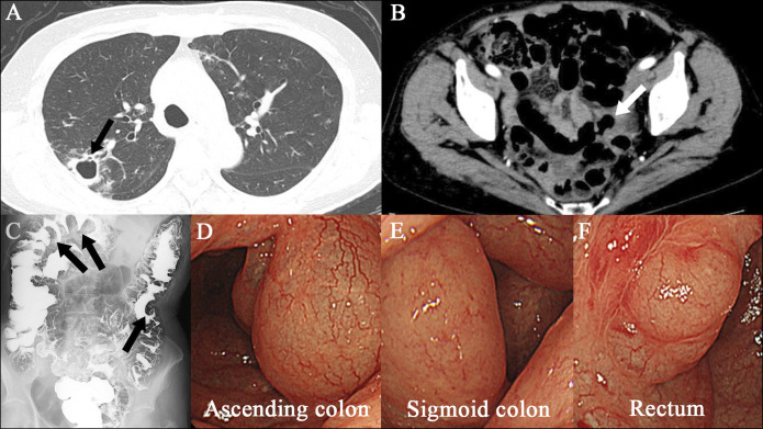
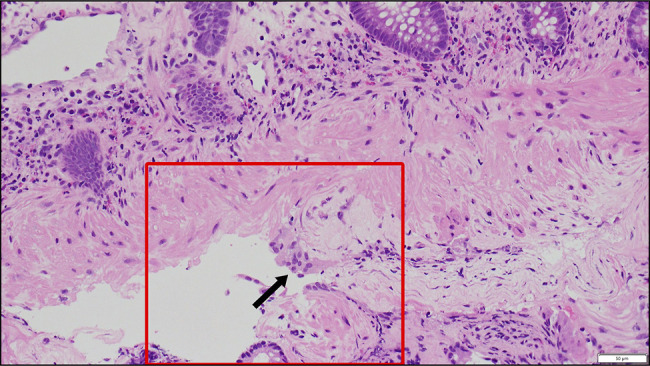
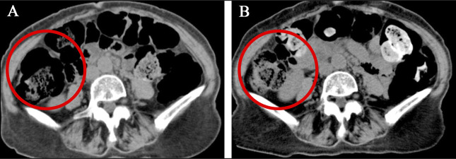
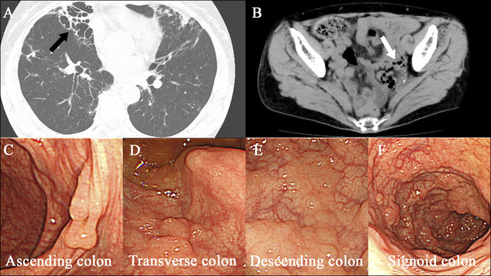
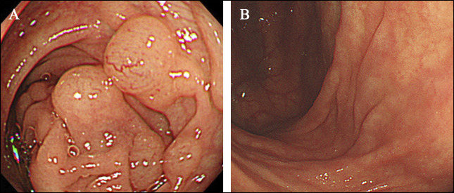
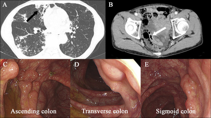
Pneumatosis cystoides intestinalis (PCI) is an uncommon condition characterized by the presence of a collection of individual gas cysts in the submucosa and subserosa of the intestine. The etiology of PCI is still unclear. We experienced 3 cases with PCI during treatment for pulmonary Mycobacterium avium complex (MAC) infection. Each case was treated conservatively. We believe our case series will highlight the importance of examining the gastrointestinal tract of patients with MAC infection and hopefully elucidate the clinical characteristics of PCI which developed during MAC treatment.

Keywords: antibiotics; colon; endoscopy; mycobacterium avium complex infection; pneumatosis cystoides intestinalis.


Figures

Figure 1.
Figure 1.
Images of case 1. An axial chest CT scan (panel A, arrow) shows right upper lobe cavitation. An axial abdominal CT scan (panel B, arrow) during antibiotic treatment for Mycobacterium avium complex pulmonary infection reveals air-filled cysts in the wall of the colon. Images from air-contrast barium enema show polypoid filling defects (panel C, arrows) consistent with pneumatosis cystoides intestinalis. Images obtained by colonoscopy (panels D, E, and F) during antibiotic treatment show multiple cystic lesions. CT, computed tomography.
Figure 2.
Figure 2.
Biopsy specimen obtained from the transverse colon of case 1. Biopsy specimen from the transverse colon reveals a submucosal cyst with a multinucleated giant cell (arrow) on 40× hematoxylin and eosin staining, consistent with pneumatosis cystoides intestinalis.
Figure 3.
Figure 3.
Abdominal CT of case 1 conducted before and after discontinuation of antibiotic treatment. An axial section of abdominal CT (panel A, circle) of case 1 conducted during antibiotic treatment for Mycobacterium avium complex pulmonary infection. It shows air-filled gas cysts in the wall of the ascending colon. After antibiotic treatment was stopped, abdominal CT (panel B, circle) shows improvement of pneumatosis cystoides intestinalis. CT, computed tomography.
Figure 4.
Figure 4.
Images of case 2. An axial chest CT scan (panel A, arrow) shows right lower lobe cavitation. An axial abdominal CT scan (panel B, arrow) during antibiotic treatment for Mycobacterium avium complex pulmonary infection reveals air-filled cysts in the wall of the colon. Images obtained by colonoscopy (panels C, D, E, and F) during antibiotic treatment show multiple cystic lesions. CT, computed tomography.
Figure 5.
Figure 5.
Colonoscopy of case 2 conducted before and after discontinuation of antibiotic treatment. A colonoscopy (panel A) reveals cystic lesions through the ascending colon to the sigmoid colon of case 2 during antibiotic treatment. After antibiotic treatment was discontinued, no cystic lesions were seen on colonoscopy (panel B).
Figure 6.
Figure 6.
Images of case 3. An axial chest CT scan (panel A, arrow) shows right lower lobe cavitation. An axial abdominal CT scan (panel B, arrow) during antibiotic treatment for Mycobacterium avium complex pulmonary infection reveals air-filled cysts in the wall of the colon. Images obtained by colonoscopy (panels C, D, and E) during antibiotic treatment show multiple cystic lesions. CT, computed tomography.

Similar articles

References

    1. Im J, Anjum F. Pneumatosis intestinalis 2023 Apr 27. In: StatPearls [Internet]. StatPearls Publishing: Treasure Island (FL), 2023.
    1. Wang YJ, Wang YM, Zheng YM, Jiang HQ, Zhang J. Pneumatosis cystoides intestinalis: Six case reports and a review of the literature. BMC Gastroenterol. 2018;18(1):100. - PMC - PubMed
    1. Azzaroli F, Turco L, Ceroni L, et al. Pneumatosis cystoides intestinalis. World J Gastroenterol. 2011;17(44):4932–6. - PMC - PubMed
    1. Hsueh KC, Tsou SS, Tan KT. Pneumatosis intestinalis and pneumoperitoneum on computed tomography: Beware of non-therapeutic laparotomy. World J Gastrointest Surg. 2011;3(6):86–8. - PMC - PubMed
    1. Morris MS, Gee AC, Cho SD, et al. Management and outcome of pneumatosis intestinalis. Am J Surg. 2008;195(5):679–83. - PubMed

Publication types

LinkOut - more resources