

Diffusion tensor imaging biomarkers and clinical assessments in amyotrophic lateral sclerosis (ALS) patients: an exploratory study
- PMID: 39239063
- PMCID: PMC11374192
- DOI: 10.1097/MS9.0000000000002332
Diffusion tensor imaging biomarkers and clinical assessments in amyotrophic lateral sclerosis (ALS) patients: an exploratory study
Abstract
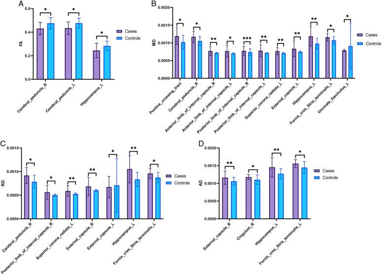
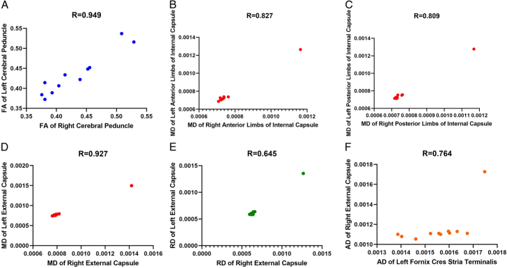
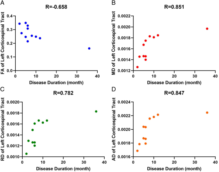
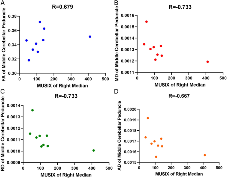
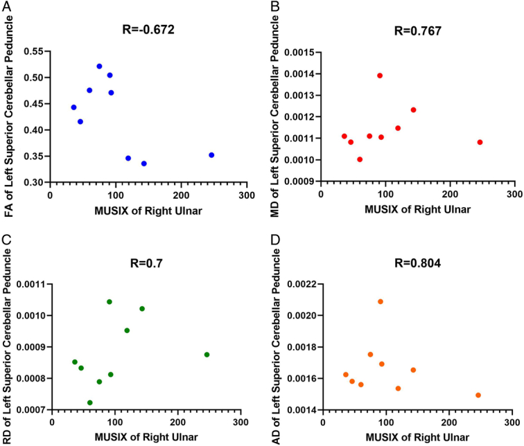
Amyotrophic lateral sclerosis (ALS) is a neurodegenerative disease characterized by progressive loss of upper and lower motor neurons. Biomarkers are needed to improve diagnosis, gauge progression, and evaluate treatment. Diffusion tensor imaging (DTI) is a promising biomarker for detecting microstructural alterations in the white matter tracts. This study aimed to assess DTI metrics as biomarkers and to examine their relationship with clinical assessments in patients with ALS. Eleven patients with ALS and 21 healthy controls (HCs) underwent 3T MRI with DTI. DTI metrics, including fractional anisotropy (FA), mean diffusivity (MD), radial diffusivity (RD), and axial diffusivity (AD), were compared between key motor and extra-motor tract groups. Group comparisons and correlations between DTI metrics also correlated with clinical scores of disability (ALSFRS-R), muscle strength (dynamometry), and motor unit loss (MUNIX). Widespread differences were found between patients with ALS and HCs in DTI metrics, including decreased FA and increased diffusivity metrics. However, MD and RD are more sensitive metrics for detecting white matter changes in patients with ALS. Significant interhemispheric correlations between the tract DTI metrics were also observed. DTI metrics showed symmetry between the hemispheres and correlated with the clinical assessments. MD, RD, and AD increases significantly correlated with lower ALSFRS-R and MUNIX scores and weaker dynamometry results. DTI reveals microstructural damage along the motor and extra-motor regions in ALS patients. DTI metrics can serve as quantitative neuroimaging biomarkers for diagnosis, prognosis, monitoring of progression, and treatment. Combined analysis of imaging, electrodiagnostic, and functional biomarkers shows potential for characterizing disease pathophysiology and progression.
Keywords: amyotrophic lateral sclerosis; biomarkers; diffusion tensor imaging; motor neuron disease; neuroimaging; prognosis.
Copyright © 2024 The Author(s). Published by Wolters Kluwer Health, Inc.
Conflict of interest statement
The authors declares no conflicts of interest.Sponsorships or competing interests that may be relevant to content are disclosed at the end of this article.
Figures
References
-
- Ghaderi S, Fatehi F, Kalra S, et al. . MRI biomarkers for memory-related impairment in amyotrophic lateral sclerosis: a systematic review. Amyotroph Later Scler Frontotemp Degener 2023;24:572–588. - PubMed
-
- Mohammadi S, Ghaderi S. Motor band sign in motor neuron diseases using magnetic resonance imaging: a systematic review. Acta Neurol Scand 2023;2023:e6677967.
LinkOut - more resources
Full Text Sources
Miscellaneous